This paper proposes a method for automatic classification of plaques in coronary arteries using convolutional neural networks (CNN) and transfer learning on intravascular optical coherence tomography (IVOCT) images. The approach aims to leverage deep feature learning to detect and classify plaques directly from high-resolution IVOCT images, enhancing early detection of atherosclerosis.
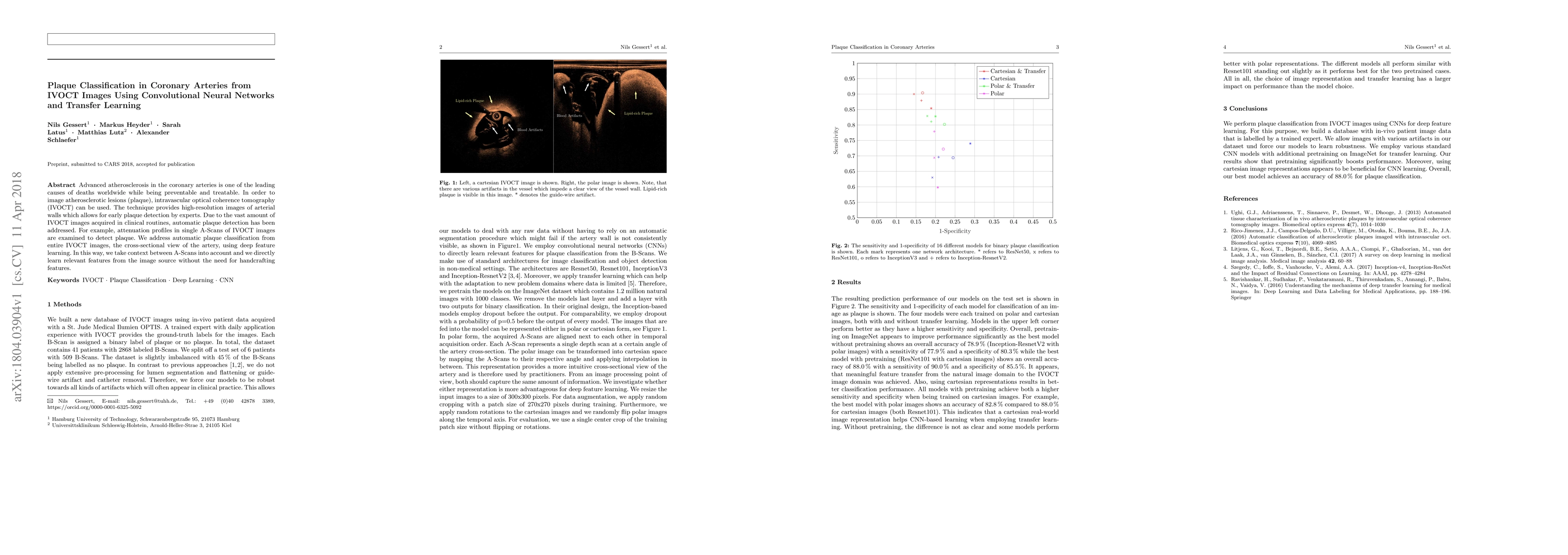
Advanced atherosclerosis in the coronary arteries is one of the leading causes of deaths worldwide while being preventable and treatable. In order to image atherosclerotic lesions (plaque), intravascular optical coherence tomography (IVOCT) can be used. The technique provides high-resolution images of arterial walls which allows for early plaque detection by experts. Due to the vast amount of IVOCT images acquired in clinical routines, automatic plaque detection has been addressed. For example, attenuation profiles in single A-Scans of IVOCT images are examined to detect plaque. We address automatic plaque classification from entire IVOCT images, the cross-sectional view of the artery, using deep feature learning. In this way, we take context between A-Scans into account and we directly learn relevant features from the image source without the need for handcrafting features.
Get AI-generated insights about this paper's methodology, results, and significance.
Current paper (gray), citations (green), references (blue)
Akira Furui, Takeshi Yoshidomi, Shinji Kume et al.
Title | Authors | Year | Actions |
---|---|---|---|
A cross-sectional study of the anatomy of the jaws of a central-European Caucasian population using cone beam computer tomography as a prerequisite for designing pre-formed calcium phosphate cement scaffolds.
|
J. Matschke, S. Farahzadi, P. Sembdner, S. Holtzhausen, Lysann M Kroschwald, Paula Korn, T. A. Schröder, Christiane Keil, Kristin Paetzold-Byhain, Günter Lauer, A. Franke
|
2024
|
View Paper |
Factors influencing the outcome of volumetry tools for pulmonary nodule analysis: a systematic review and attempted meta-analysis
|
Erique Guedes Pinto, D. Penha, Sofia Ravara, C. Monaghan, B. Hochhegger, Edson Marchiori, L. Taborda-Barata, K. Irion
|
2023
|
View Paper |
Multi-Feature Fusion Method for Identifying Carotid Artery Vulnerable Plaque
|
X. Xu, L. Huang, R. Wu, Wu Zhang, G. Ding, L. Liu, M. Chi, Jiang Xie
|
2021
|
View Paper |
Locally operated assistant manipulators with selectable connection system for robotically assisted laparoscopic solo surgery
|
Shohei Fukui, T. Kawai, Y. Nishizawa, A. Nishikawa, Tatsuo Nakamura, Noriyasu Iwamoto, Y. Horise, K. Masamune
|
2021
|
View Paper |
Generalization vs. Specificity: In Which Cases Should a Clinic Train its Own Segmentation Models?
|
J. Schreier, F. Attanasi, H. Laaksonen
|
2020
|
View Paper |
A novel automated lumen segmentation and classification algorithm for detection of irregular protrusion after stents deployment
|
Su Yang, H. Yoon, Seyed Kamaleddin Mostafavi Yazdi, Jong-Ha Lee
|
2020
|
View Paper |
Research on automatic identification based on IVOCT images of coronary plaque
|
Qin Li, Haoran Sun, Jingbo Wang, Na Qin, Wei Liu
|
2019
|
View Paper |
Inter-observer reliability of alternative diagnostic methods for proximal humerus fractures: a comparison between attending surgeons and orthopedic residents in training
|
Luiz Fernando Cocco, João Alberto Yazzigi, Eduardo Felipe Kin Ito Kawakami, Helio Jorge Fernandes Alvachian, F. B. Dos Reis, M. Luzo
|
2019
|
View Paper |
Multi-organ segmentation of the head and neck area: an efficient hierarchical neural networks approach
|
Elias Tappeiner, Samuel Pröll, M. Hönig, P. Raudaschl, P. Zaffino, M. Spadea, G. Sharp, R. Schubert, K. Fritscher
|
2019
|
View Paper |
Characterization of coronary artery pathological formations from OCT imaging using deep learning.
|
A. Abdolmanafi, Luc Duong, N. Dahdah, Ibrahim Ragui Adib, F. Cheriet
|
2018
|
View Paper |
Pathobox: The Collaborative Tele-pathology Platform with Access Management
|
R. Lebre, Rui Jesus, Pedro Nunes, C. Costa
|
2020
|
View Paper |
Comments (0)