This research introduces crossed laser phase plates (XLPP) for enhancing phase-contrast in transmission electron microscopy, employing two intersecting laser standing waves to improve image contrast and suppress ghost images. The study provides a theoretical model and simulations showing increased information transfer and reduced diffraction effects.
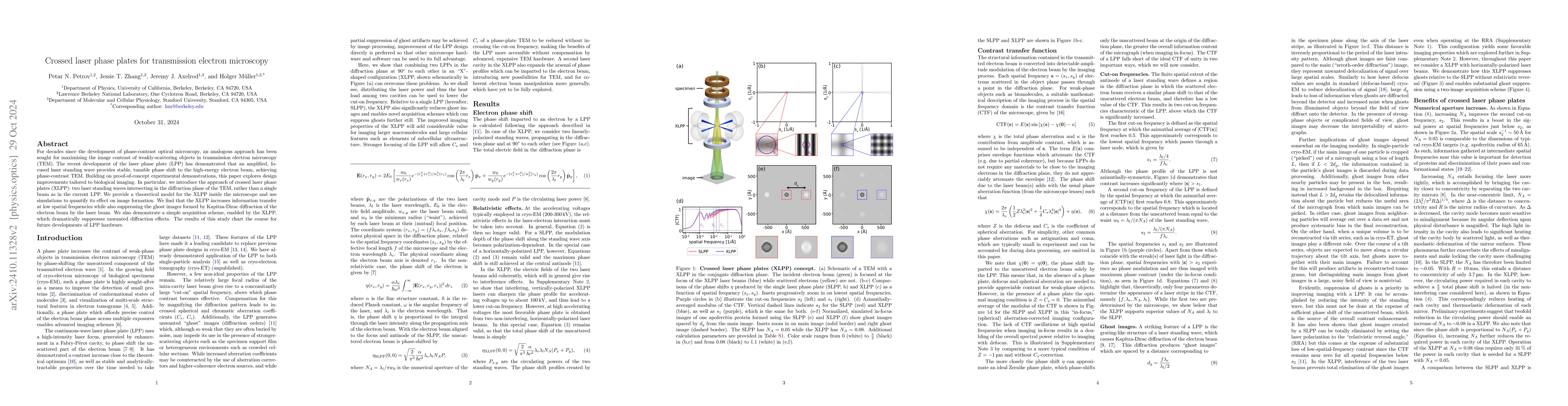
For decades since the development of phase-contrast optical microscopy, an analogous approach has been sought for maximizing the image contrast of weakly-scattering objects in transmission electron microscopy (TEM). The recent development of the laser phase plate (LPP) has demonstrated that an amplified, focused laser standing wave provides stable, tunable phase shift to the high-energy electron beam, achieving phase-contrast TEM. Building on proof-of-concept experimental demonstrations, this paper explores design improvements tailored to biological imaging. In particular, we introduce the approach of crossed laser phase plates (XLPP): two laser standing waves intersecting in the diffraction plane of the TEM, rather than a single beam as in the current LPP. We provide a theoretical model for the XLPP inside the microscope and use simulations to quantify its effect on image formation. We find that the XLPP increases information transfer at low spatial frequencies while also suppressing the ghost images formed by Kapitza-Dirac diffraction of the electron beam by the laser beam. We also demonstrate a simple acquisition scheme, enabled by the XLPP, which dramatically suppresses unwanted diffraction effects. The results of this study chart the course for future developments of LPP hardware.
Get AI-generated insights about this paper's methodology, results, and significance.
Current paper (gray), citations (green), references (blue)
No similar papers found for this research.
No citations found for this paper.
Title | Authors | Year | Actions |
---|---|---|---|
Cryo-EM phase-plate images reveal unexpected levels of apparent specimen damage
|
Jonathan Remis, Petar N. Petrov, Jessie T. Zhang, J. Axelrod, Hang Cheng, Shahar Sandhaus, Holger Mueller, R. Glaeser
|
2024
|
View Paper |
Cavity QED in a high NA resonator
|
Danial Shadmany, Aishwarya Kumar, Anna Soper, L. Palm, Chuan Yin, Henry Ando, Bowen Li, Lavanya Taneja, Matt Jaffe, Schuster David, Jon Simon
|
2024
|
View Paper |
Modern approaches to improving phase contrast electron microscopy
|
J. Axelrod, Jessie T. Zhang, Petar N. Petrov, R. Glaeser, Holger Mȕller
|
2024
|
View Paper |
OVERCOMING RESOLUTION LOSS DUE TO THERMAL MAGNETIC FIELD FLUCTUATIONS FROM PHASE PLATES IN TRANSMISSION ELECTRON MICROSCOPY
|
J. Axelrod, Petar N. Petrov, Jessie T. Zhang, J. Remis, B. Buijsse, R. Glaeser, H. Mȕller
|
2023
|
View Paper |
Estimating conformational landscapes from Cryo-EM particles by 3D Zernike polynomials
|
D. Herreros, Roy R. Lederman, J. Krieger, A. Jiménez-Moreno, M. Martínez, D. Myška, D. Střelák, J. Filipovič, C. Sorzano, J. M. Carazo
|
2023
|
View Paper |
Cryomicroscopy in situ: what is the smallest molecule that can be directly identified without labels in a cell?
|
C. Russo, Joshua L. Dickerson, K. Naydenova
|
2022
|
View Paper |
High finesse bow-tie cavity for strong atom-photon coupling in Rydberg arrays.
|
Yu-Ting Chen, M. Szurek, Beili Hu, J. de Hond, B. Braverman, V. Vuletić
|
2022
|
View Paper |
Micro-fabricated mirrors with finesse exceeding one million
|
Naijun Jin, Charlie McLemore, David Mason, J. Hendrie, Yizhi Luo, Megan L. Kelleher, P. Kharel, F. Quinlan, S. Diddams, P. Rakich
|
2022
|
View Paper |
Perspective: Emerging strategies for determining atomic-resolution structures of macromolecular complexes within cells
|
Petar N. Petrov, H. Mueller, R. Glaeser
|
2021
|
View Paper |
Conquer by cryo-EM without physically dividing.
|
G. Lander, R. Glaeser
|
2021
|
View Paper |
CryoDRGN: Reconstruction of heterogeneous cryo-EM structures using neural networks
|
Ellen D. Zhong, Tristan Bepler, B. Berger, Joseph H. Davis
|
2021
|
View Paper |
High-power near-concentric Fabry–Perot cavity for phase contrast electron microscopy
|
C. Turnbaugh, J. Axelrod, S. Campbell, J. Dioquino, Petar N. Petrov, J. Remis, O. Schwartz, Zanlin Yu, Yifan Cheng, R. Glaeser, H. Mueller
|
2020
|
View Paper |
Atomic-resolution protein structure determination by cryo-EM
|
K. M. Yip, N. Fischer, E. Paknia, A. Chari, H. Stark
|
2020
|
View Paper |
Defocus-dependent Thon-ring fading
|
R. Glaeser, W. Hagen, B. Han, R. Henderson, G. McMullan, C. Russo
|
2020
|
View Paper |
The promise and the challenges of cryo‐electron tomography
|
M. Turk, W. Baumeister
|
2020
|
View Paper |
Observation of the Relativistic Reversal of the Ponderomotive Potential.
|
J. Axelrod, S. Campbell, O. Schwartz, C. Turnbaugh, R. Glaeser, H. Mueller
|
2019
|
View Paper |
Laser phase plate for transmission electron microscopy
|
O. Schwartz, J. Axelrod, S. Campbell, C. Turnbaugh, R. Glaeser, H. Müller
|
2019
|
View Paper |
Revisiting the Structure of Hemoglobin and Myoglobin with Cryo-Electron Microscopy.
|
M. Khoshouei, R. Danev, J. Plitzko, W. Baumeister
|
2017
|
View Paper |
Near-concentric Fabry-Pérot cavity for continuous-wave laser control of electron waves.
|
O. Schwartz, J. Axelrod, D. Tuthill, P. Haslinger, C. Ophus, R. Glaeser, H. Müller
|
2016
|
View Paper |
Efficient linear phase contrast in scanning transmission electron microscopy with matched illumination and detector interferometry
|
C. Ophus, J. Ciston, Jordan S. Pierce, Tyler R. Harvey, J. Chess, B. McMorran, C. Czarnik, H. Rose, P. Ercius
|
2016
|
View Paper |
Visualizing the molecular sociology at the HeLa cell nuclear periphery
|
J. Mahamid, Stefan Pfeffer, M. Schaffer, E. Villa, R. Danev, Luis Kuhn Cuellar, F. Förster, A. Hyman, J. Plitzko, W. Baumeister
|
2016
|
View Paper |
RELION: Implementation of a Bayesian approach to cryo-EM structure determination
|
S. Scheres
|
2012
|
View Paper |
Hydration-layer models for cryo-EM image simulation.
|
Zhiguo Shang, F. Sigworth
|
2012
|
View Paper |
Tilt-Pair Analysis of Images from a Range of Different Specimens in Single-Particle Electron Cryomicroscopy
|
R. Henderson, Shaoxia Chen, James Z. Chen, N. Grigorieff, L. A. Passmore, Luciano Ciccarelli, J. Rubinstein, R. Crowther, P. Stewart, P. Rosenthal
|
2011
|
View Paper |
Object wave reconstruction by phase-plate transmission electron microscopy.
|
B. Gamm, Manuel Dries, K. Schultheiss, H. Blank, A. Rosenauer, R. Schröder, D. Gerthsen
|
2010
|
View Paper |
Design of an electron microscope phase plate using a focused continuous-wave laser
|
Holger Mueller, Jian Jin, R. Danev, John C. H. Spence, H. Padmore, R. Glaeser
|
2010
|
View Paper |
Observation of the Kapitza–Dirac effect
|
D. Freimund, K. Aflatooni, H. Batelaan
|
2001
|
View Paper |
Complex Observation in Electron Microscopy. II. Direct Visualization of Phases and Amplitudes of Exit Wave Functions
|
R. Danev, K. Nagayama
|
2001
|
View Paper |
Relation between Surface Roughness and Specular Reflectance at Normal Incidence
|
H. E. Bennett, J. O. Porteus
|
1961
|
View Paper |
Practical considerations: Defocus, stigmation, coma-free illumination, and phase plates
|
2021
|
||
Optical aberrations and ewald sphere curvature
|
2021
|
Showing first page preview. Click "Open Full PDF" to view the complete paper.
Your comment contains inappropriate language and has been rejected.
Are you sure you want to remove this tag? This action cannot be undone.
Someone just favorited a paper!
Comments (0)