Summary
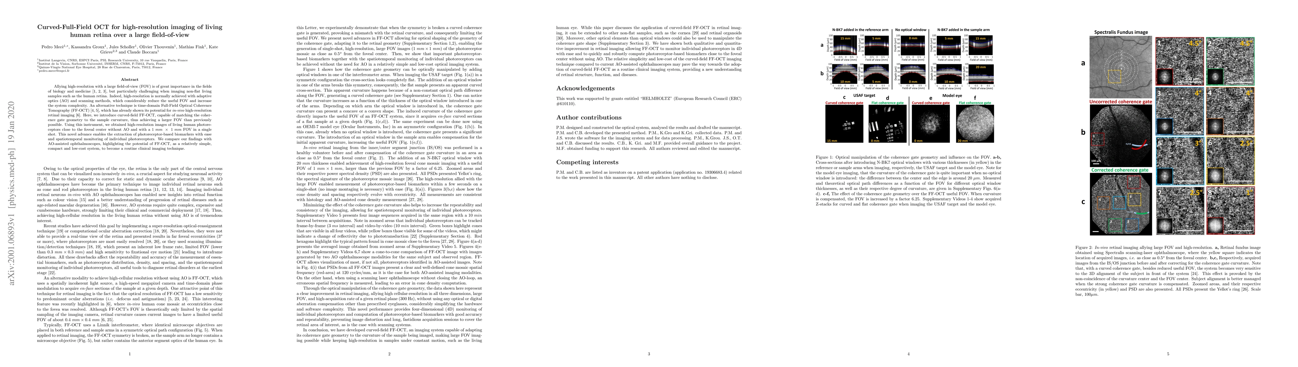
Allying high-resolution with a large field-of-view (FOV) is of great importance in the fields of biology and medicine, but particularly challenging when imaging non-flat living samples such as the human retina. Indeed, high-resolution is normally achieved with adaptive optics (AO) and scanning methods, which considerably reduce the useful FOV and increase the system complexity. An alternative technique is time-domain Full-Field Optical Coherence Tomography (FF-OCT), which has already shown its potential for in-vivo high-resolution retinal imaging. Here, we introduce curved-field FF-OCT, capable of matching the coherence gate geometry to the sample curvature, thus achieving a larger FOV than previously possible. Using this instrument, we obtained high-resolution images of living human photoreceptors close to the foveal center without AO and with a 1 mm x 1 mm FOV in a single shot. This novel advance enables the extraction of photoreceptor-based biomarkers with ease and spatiotemporal monitoring of individual photoreceptors. We compare our findings with AO-assisted ophthalmoscopes, highlighting the potential of FF-OCT, as a relatively simple, compact and low-cost system, to become a routine clinical imaging technique.
AI Key Findings
Get AI-generated insights about this paper's methodology, results, and significance.
Paper Details
PDF Preview
Key Terms
Citation Network
Current paper (gray), citations (green), references (blue)
Display is limited for performance on very large graphs.
Similar Papers
Found 4 papers| Title | Authors | Year | Actions |
|---|
Comments (0)