Publication
Metrics
AI Quick Summary
This study demonstrates high-resolution en face retinal imaging using wavefront correctionless full-field OCT (FFOCT) in vivo, combining FFOCT with spectral-domain OCT (SDOCT) for real-time optical path length matching. The technique provides diffraction-limited images of multiple retinal layers, revealing details of nerve fiber orientation, blood vessel distribution, and photoreceptor mosaic.
Paper Preview
Abstract
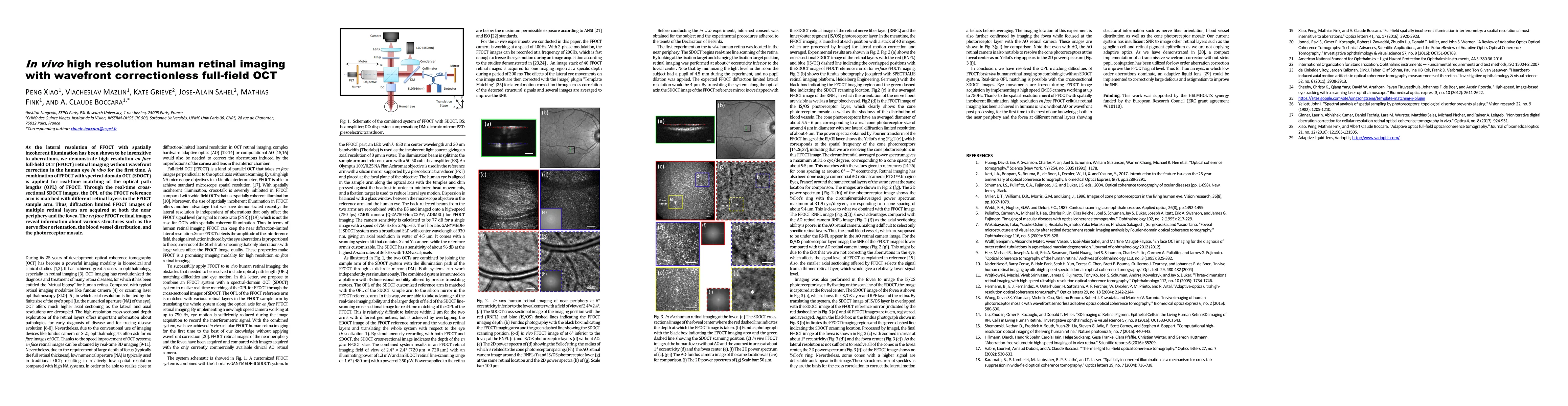
As the lateral resolution of FFOCT with spatially incoherent illumination has been shown to be insensitive to aberrations, we demonstrate high resolution en face full-field OCT (FFOCT) retinal imaging without wavefront correction in the human eye in vivo for the first time. A combination of FFOCT with spectral-domain OCT (SDOCT) is applied for real-time matching of the optical path lengths (OPL) of FFOCT. Through the real-time cross-sectional SDOCT images, the OPL of the FFOCT reference arm is matched with different retinal layers in the FFOCT sample arm. Thus, diffraction limited FFOCT images of multiple retinal layers are acquired at both the near periphery and the fovea. The en face FFOCT retinal images reveal information about various structures such as the nerve fiber orientation, the blood vessel distribution, and the photoreceptor mosaic.
AI Key Findings
Get AI-generated insights about this paper's methodology, results, significance, and more — seven facets brought into focus.
Impact
Paper Details
PDF Preview
Key Terms
Citation Network
Current paper (gray), citations (green), references (blue)
Display is limited for performance on very large graphs.
Discussion 0