DCE-Qnet: Deep Network Quantification of Dynamic Contrast Enhanced (DCE) MRI
Publication
Metrics
AI Quick Summary
**Summary:** DCE-Qnet, a deep neural network, effectively quantifies dynamic contrast-enhanced MRI data by estimating key pharmacokinetic parameters, outperforming traditional methods and demonstrating high reproducibility in both phantom and clinical settings. This approach simplifies the imaging process and enhances accuracy in parameter estimation.
Paper Preview
Abstract
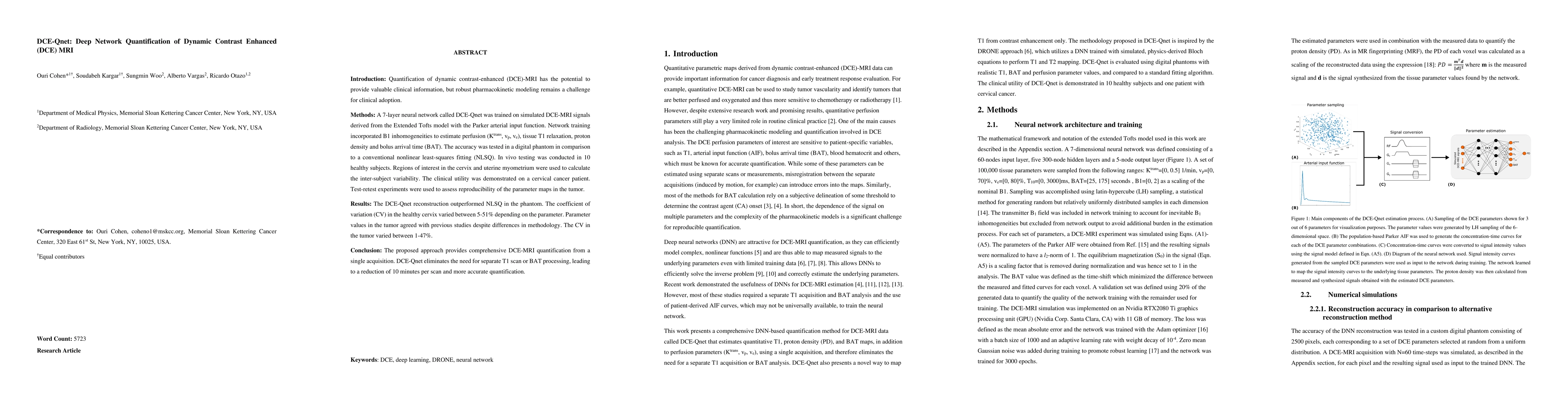
Introduction: Quantification of dynamic contrast-enhanced (DCE)-MRI has the potential to provide valuable clinical information, but robust pharmacokinetic modeling remains a challenge for clinical adoption. Methods: A 7-layer neural network called DCE-Qnet was trained on simulated DCE-MRI signals derived from the Extended Tofts model with the Parker arterial input function. Network training incorporated B1 inhomogeneities to estimate perfusion (Ktrans, vp, ve), tissue T1 relaxation, proton density and bolus arrival time (BAT). The accuracy was tested in a digital phantom in comparison to a conventional nonlinear least-squares fitting (NLSQ). In vivo testing was conducted in 10 healthy subjects. Regions of interest in the cervix and uterine myometrium were used to calculate the inter-subject variability. The clinical utility was demonstrated on a cervical cancer patient. Test-retest experiments were used to assess reproducibility of the parameter maps in the tumor. Results: The DCE-Qnet reconstruction outperformed NLSQ in the phantom. The coefficient of variation (CV) in the healthy cervix varied between 5-51% depending on the parameter. Parameter values in the tumor agreed with previous studies despite differences in methodology. The CV in the tumor varied between 1-47%. Conclusion: The proposed approach provides comprehensive DCE-MRI quantification from a single acquisition. DCE-Qnet eliminates the need for separate T1 scan or BAT processing, leading to a reduction of 10 minutes per scan and more accurate quantification.
AI Key Findings
Get AI-generated insights about this paper's methodology, results, significance, and more — seven facets brought into focus.
Impact
Paper Details
Authors
PDF Preview
Key Terms
Citation Network
Current paper (gray), citations (green), references (blue)
Display is limited for performance on very large graphs.
Discussion 0