Quick Summary:

A deep learning approach using DenseNet and SVM was found to accurately detect tissue folds in histopathology images with 96.3% accuracy, outperforming other configurations.

Summary

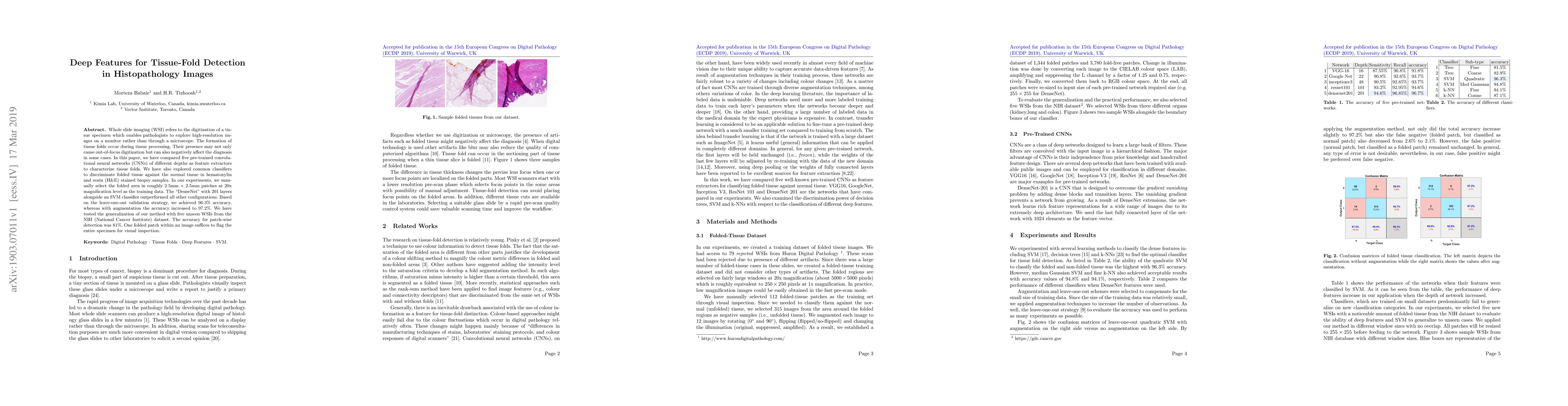
Whole slide imaging (WSI) refers to the digitization of a tissue specimen which enables pathologists to explore high-resolution images on a monitor rather than through a microscope. The formation of tissue folds occur during tissue processing. Their presence may not only cause out-of-focus digitization but can also negatively affect the diagnosis in some cases. In this paper, we have compared five pre-trained convolutional neural networks (CNNs) of different depths as feature extractors to characterize tissue folds. We have also explored common classifiers to discriminate folded tissue against the normal tissue in hematoxylin and eosin (H\&E) stained biopsy samples. In our experiments, we manually select the folded area in roughly 2.5mm $\times$ 2.5mm patches at $20$x magnification level as the training data. The ``DenseNet'' with 201 layers alongside an SVM classifier outperformed all other configurations. Based on the leave-one-out validation strategy, we achieved $96.3\%$ accuracy, whereas with augmentation the accuracy increased to $97.2\%$. We have tested the generalization of our method with five unseen WSIs from the NIH (National Cancer Institute) dataset. The accuracy for patch-wise detection was $81\%$. One folded patch within an image suffices to flag the entire specimen for visual inspection.

AI Key Findings

Get AI-generated insights about this paper's methodology, results, and significance.

Paper Details

PDF Preview

Download PDF

Key Terms

tissue (0.406)
patch (0.262)
patch (0.262)
accuracy (0.198)
accuracy (0.179)
national (0.159)
patches (0.158)
cnns (0.158)
classifier (0.156)
manually (0.155)
cancer (0.153)
inspection (0.153)
unseen (0.152)
diagnosis (0.152)
monitor (0.147)
augmentation (0.144)
wise (0.143)
cancer (0.143)
augmentation (0.143)
cnns (0.140)
Title Authors Year Actions
ArtiDiffuser: A unified framework for artifact restoration and synthesis for histology images via counterfactual diffusion model.
Chong Wang, Kaili Qu, Shuxin Li, Yi Yu, Junjun He, Chen Zhang, Yiqing Shen
2025
View Paper
Fully Automatic Content-Aware Tiling Pipeline for Pathology Whole Slide Images
Falah Jabar, L. Busund, B. Ricciuti, M. Tafavvoghi, Mette Pøhl, S. Andersen, T. Dønnem, David J. Kwiatkowski, M. Rakaee
2024
View Paper
Improving quality control of whole slide images by explicit artifact augmentation
Artur Jurgas, Marek Wodziński, Marina D'Amato, J. Laak, M. Atzori, Henning Müller
2024
View Paper
Equipping computational pathology systems with artifact processing pipelines: a showcase for computation and performance trade-offs
Neel Kanwal, Farbod Khoraminia, Umay Kiraz, Andrés Mosquera-Zamudio, Carlos Monteagudo, Emiel A. M. Janssen, T. Zuiverloon, Chunming Rong, K. Engan
2024
View Paper
Analysis and Validation of Image Search Engines in Histopathology
Isaiah Lahr, Saghir Alfasly, Peyman Nejat, Jibran A. Khan, Luke Kottom, Vaishnavi Kumbhar, Areej Alsaafin, Abubakr Shafique, S. Hemati, Ghazal Alabtah, N. Comfere, Dennis Murphree, Aaron Mangold, Saba Yasir, Chady Meroueh, Lisa Boardman, Vijay H. Shah, Joaquin J. Garcia, H. R. Tizhoosh
2024
View Paper
Are you sure it's an artifact? Artifact detection and uncertainty quantification in histological images
Neel Kanwal, M. López-Pérez, Umay Kiraz, T. Zuiverloon, Rafael Molina, K. Engan
2023
View Paper
NCL-SM: A Fully Annotated Dataset of Images from Human Skeletal Muscle Biopsies
Atif Khan, Conor Lawless, Amy E. Vincent, Charlotte Warren, Valeria Di Leo, T. Gomes, A. Mcgough
2023
View Paper
Artifact Detection and Restoration in Histology Images With Stain-Style and Structural Preservation
Jing Ke, Kai Liu, Yuxiang Sun, Yuying Xue, Jiaxuan Huang, Yizhou Lu, Jun Dai, Yaobing Chen, Xiaodan Han, Yiqing Shen, D. Shen
2023
View Paper
Development of an automated combined positive score prediction pipeline using artificial intelligence on multiplexed immunofluorescence images
Abhishek Vahadane, Shreya Sharma, Devraj Mandal, M. Dabbeeru, Josephine Jakthong, M. García-Guzmán, S. Majumdar, Chung-Wein Lee
2022
View Paper
Quality Control of Whole Slide Images using the YOLO Concept
Kimia Hemmatirad, Morteza Babaie, M. Afshari, Danial Maleki, Mahjabin Saiadi, H. Tizhoosh
2022
View Paper
Automated quality assessment of large digitised histology cohorts by artificial intelligence
Maryam Haghighat, L. Browning, K. Sirinukunwattana, S. Malacrino, Nasullah Khalid Alham, R. Colling, Ying Cui, E. Rakha, F. Hamdy, C. Verrill, J. Rittscher
2022
View Paper
PathProfiler: Automated Quality Assessment of Retrospective Histopathology Whole-Slide Image Cohorts by Artificial Intelligence, A Case Study for Prostate Cancer Research
Maryam Haghighat, L. Browning, K. Sirinukunwattana, S. Malacrino, N. K. Alham, R. Colling, Y. Cui, E. Rakha, F. Hamdy, C. Verrill, J. Rittscher
2021
View Paper
Whole-slide imaging, tissue image analysis, and artificial intelligence in veterinary pathology: An updated introduction and review
A. Zuraw, F. Aeffner
2021
View Paper
Fine-Tuning and training of densenet for histopathology image representation using TCGA diagnostic slides
Abtin Riasatian, Morteza Babaie, Danial Maleki, S. Kalra, Mojtaba Valipour, S. Hemati, Manit Zaveri, Amir Safarpoor, Sobhan Shafiei, M. Afshari, Maral Rasoolijaberi, Milad Sikaroudi, Mohammed Adnan, Sultaan Shah, C. Choi, S. Damaskinos, Clinton J. V. Campbell, P. Diamandis, L. Pantanowitz, H. Kashani, A. Ghodsi, H. Tizhoosh
2021
View Paper
Ink Marker Segmentation in Histopathology Images Using Deep Learning
Danial Maleki, M. Afshari, Morteza Babaie, H. Tizhoosh
2020
View Paper
Regional registration of whole slide image stacks containing major histological artifacts
Mahsa Paknezhad, Sheng Yang Michael Loh, Y. Choudhury, V. Koh, Timothy Tay Kwang Yong, H. S. Tan, R. Kanesvaran, P. Tan, John Yuen Shyi Peng, Weimiao Yu, Y. Tan, Yong Zhen Loy, M. Tan, H. Lee
2020
View Paper
A Comparative Study of U-Net Topologies for Background Removal in Histopathology Images
Abtin Riasatian, Maral Rasoolijaberi, Morteza Babaie, H. Tizhoosh
2020
View Paper
Multi-Task Pre-Training of Deep Neural Networks for Digital Pathology
Romain Mormont, P. Geurts, R. Marée
2020
View Paper
Brain mapping at high resolutions: Challenges and opportunities
Kyle Milligan, Aishwarya H. Balwani, Eva L. Dyer
2019
View Paper
Tissue Artifact Segmentation and Severity Assessment for Automatic Analysis Using WSI
Md Shakhawat Hossain, Galib Muhammad Shahriar, M. M. Syeed, Mohammad Faisal Uddin, Mahady Hasan, Md. Sakir Hossain, And Rubina Bari, Md. Shakhawat Hossain
2023
View Paper
The devil is in the details: Whole Slide Image acquisition and processing for artifacts detection, color variation, and data augmentation. A review.
Neel Kanwal, Fernando Pérez-Bueno, Arne Schmidt, K. Engan, R. Molina
2022
View Paper
Whole Slide Image Quality in Digital Pathology: Review and Perspectives
Romain Brixtel, Sébastien Bougleux, O. Lézoray, Yann Caillot, Benoît Lemoine, Mathieu Fontaine, Dalal Nebati, Arnaud Renouf
2022
View Paper
Class-Aware Image Search for Interpretable Cancer Identification
Arash Ebrahimian, Hossein Mohammadi, Morteza Babaie, Nima Maftoon, H. R. Tizhoosh
2020
View Paper
TUM Data Innovation Lab
View Paper

Comments (0)

This website uses cookies to ensure you get the best experience. Learn more