Authors
Publication
Metrics
Quick Actions
AI Quick Summary
This study presents a software-only method that reconstructs a 3D model of the upper jaw from just ten 2D intraoral photos, avoiding expensive hardware and painful physical impressions. Using MobileNetV2 and multi-view attention on the Dental3DS dataset, it achieves 77.49% accuracy (distance threshold 0.035) but tends to produce denser vertex clustering in certain regions, indicating uneven point distribution.
Quick Answers
What is "Deep Learning-based 3D Oral Cavity Reconstruction Using 2D Intraoral Images" about?
This study presents a software-only method that reconstructs a 3D model of the upper jaw from just ten 2D intraoral photos, avoiding expensive hardware and painful physical impressions. Using MobileNetV2 and multi-view attention on the Dental3DS dataset, it achieves 77.49% accuracy (distance threshold 0.035) but tends to produce denser vertex clustering in certain regions, indicating uneven point distribution.
What methodology did the authors use?
The approach uses ten fixed-angle 2D intraoral images (9 around the mesh plus a top-down view) to train an encoder-decoder network. MobileNetV2 serves as the image encoder per view, with positional embeddings and multi-head attention for multi-view feature fusion, followed by an MLP-based decoder that directly predicts 50,000 3D vertex coordinates. A feature modulator with a fixed scale adds diversity... More in Methodology →
What are the key results?
The model achieves 77.49% accuracy measured by nearest-neighbor matching with a distance threshold of 0.035 on the Dental3DS upper-jaw dataset. — Predicted vertices cluster in high-density regions of the ground truth, indicating uneven point distribution in the reconstructed 3D model. More in Key Results →
Why is this work significant?
Demonstrates a software-only pipeline for 3D oral cavity reconstruction directly from 2D images, potentially reducing equipment costs and patient discomfort by removing the need for physical scanners or CBCT. More in Significance →
What are the main limitations?
Chamfer distance-driven loss encourages clustering near ground-truth points, causing uneven vertex distribution across the surface. — Reconstruction quality relies on fixed viewpoints and may not generalize to unseen angles or more complex geometries. More in Limitations →
Abstract
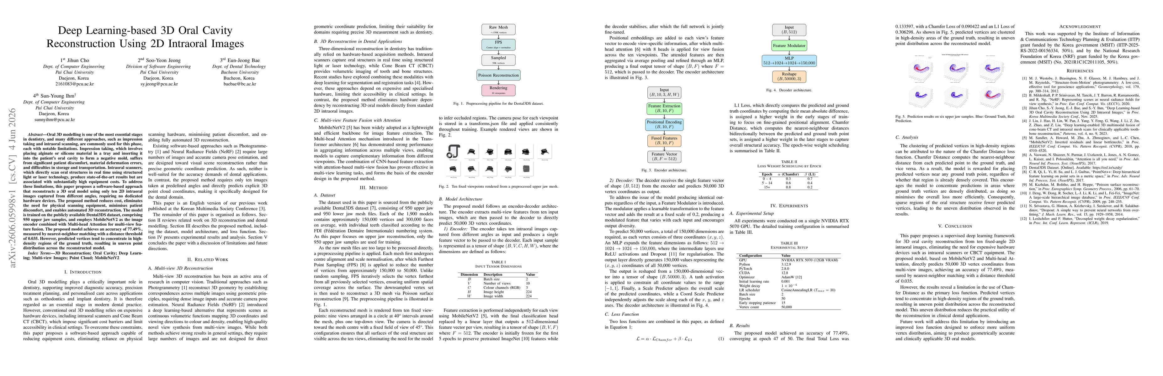
Oral 3D modelling is one of the most essential stages in dentistry, and many different approaches, such as impression taking and intraoral scanning, are commonly used for this phase, each with notable limitations. Impression taking, which involves placing alginate or silicone material in a tray and inserting it into the patient's oral cavity to form a negative mold, suffers from significant patient discomfort, material deformation errors, and difficulties in storage and transportation. Intraoral scanners, which directly scan oral structures in real time using structured light or laser technology, produce state-of-the-art results but are associated with substantially high equipment costs. To address these limitations, this paper proposes a software-based approach that reconstructs a 3D oral model using only ten 2D intraoral images captured from different angles, requiring no dedicated hardware devices. The proposed method reduces cost, eliminates the need for physical scanning equipment, minimises patient discomfort, and enables automated 3D reconstruction. The model is trained on the publicly available Dental3DS dataset, comprising 950 upper jaw samples, and employs MobileNetV2 as the image encoder combined with Multi-head Attention for multi-view feature fusion. The proposed model achieves an accuracy of 77.49%, measured by nearest-neighbor matching with a distance threshold of 0.035. However, predicted vertices tend to concentrate in high-density regions of the ground truth, resulting in uneven point distribution across the reconstructed model.
AI Key Findings
Generated Jun 05, 2026
Methodology — What approach did the authors take?
The approach uses ten fixed-angle 2D intraoral images (9 around the mesh plus a top-down view) to train an encoder-decoder network. MobileNetV2 serves as the image encoder per view, with positional embeddings and multi-head attention for multi-view feature fusion, followed by an MLP-based decoder that directly predicts 50,000 3D vertex coordinates. A feature modulator with a fixed scale adds diversity to outputs, and a two-stage loss combines Chamfer and L1 losses with epoch-based weighting to balance local and global geometry during training.
Key Results — What are the main findings?
- The model achieves 77.49% accuracy measured by nearest-neighbor matching with a distance threshold of 0.035 on the Dental3DS upper-jaw dataset.
- Predicted vertices cluster in high-density regions of the ground truth, indicating uneven point distribution in the reconstructed 3D model.
Significance — Why does this research matter?
Demonstrates a software-only pipeline for 3D oral cavity reconstruction directly from 2D images, potentially reducing equipment costs and patient discomfort by removing the need for physical scanners or CBCT.
Technical Contribution — What is the technical contribution?
Proposes a lightweight encoder-decoder framework that fuses multi-view features via multi-head attention to directly predict a dense 3D point cloud from 2D intraoral images, using a feature modulation mechanism and a combined Chamfer/L1 loss schedule.
Novelty — What is new about this work?
Introduces a ten-view fixed-angle 2D image approach with MobileNetV2 encoding and attention-based fusion to perform end-to-end 3D oral cavity reconstruction without hardware-based scanning, achieving competitive accuracy on a public dental dataset.
Limitations — What are the limitations of this study?
- Chamfer distance-driven loss encourages clustering near ground-truth points, causing uneven vertex distribution across the surface.
- Reconstruction quality relies on fixed viewpoints and may not generalize to unseen angles or more complex geometries.
- The method predicts a fixed 50,000 vertices, potentially limiting fine-grained detail in some regions.
Future Work — What did the authors propose for future work?
- Develop an improved loss function to promote more uniform vertex distribution across the surface.
- Explore adaptive or learned viewpoint configurations to enhance coverage and generalization.
- Investigate alternative decoders or refinement modules to increase local geometric accuracy without sacrificing stability.
Paper Details
How to Cite This Paper
@article{bae2026deep,
title = {Deep Learning-based 3D Oral Cavity Reconstruction Using 2D Intraoral Images},
author = {Bae, Eun-Jeong and Cho, Jihun and Jeong, Soo-Yeon and others},
year = {2026},
eprint = {2606.05998},
archivePrefix = {arXiv},
primaryClass = {cs.CV},
}
Bae, E., Cho, J., Jeong, S., & Ihm, S. (2026). Deep Learning-based 3D Oral Cavity Reconstruction Using 2D Intraoral Images. arXiv. https://arxiv.org/abs/2606.05998
Bae, Eun-Jeong, et al. "Deep Learning-based 3D Oral Cavity Reconstruction Using 2D Intraoral Images." arXiv, 2026, arxiv.org/abs/2606.05998.
PDF Preview
Similar Papers
Found 4 papers3D Structure from 2D Microscopy images using Deep Learning
QueeLim Ch'ng, Benjamin J. Blundell, Christian Sieben et al.
TerrainMesh: Metric-Semantic Terrain Reconstruction from Aerial Images Using Joint 2D-3D Learning
Qiaojun Feng, Nikolay Atanasov
Deep learning-based localization of bounded edentulous spaces in intraoral occlusal images
Shirdel, E., Azizipour, S., Magdalyanova, M.
Comments (0)