Deep Learning-Based Automated Workflow for Accurate Segmentation and Measurement of Abdominal Organs in CT Scans

Source: ArXiv

AI Quick Summary

This study develops a deep learning-based automated workflow for precise segmentation and measurement of abdominal organs in CT scans, achieving high accuracy and consistency. The workflow combines nnU-Net, U-Net++, and 3D RCNN models, demonstrating robust performance and potential for clinical adoption.

Share: Share on X LinkedIn

Summary

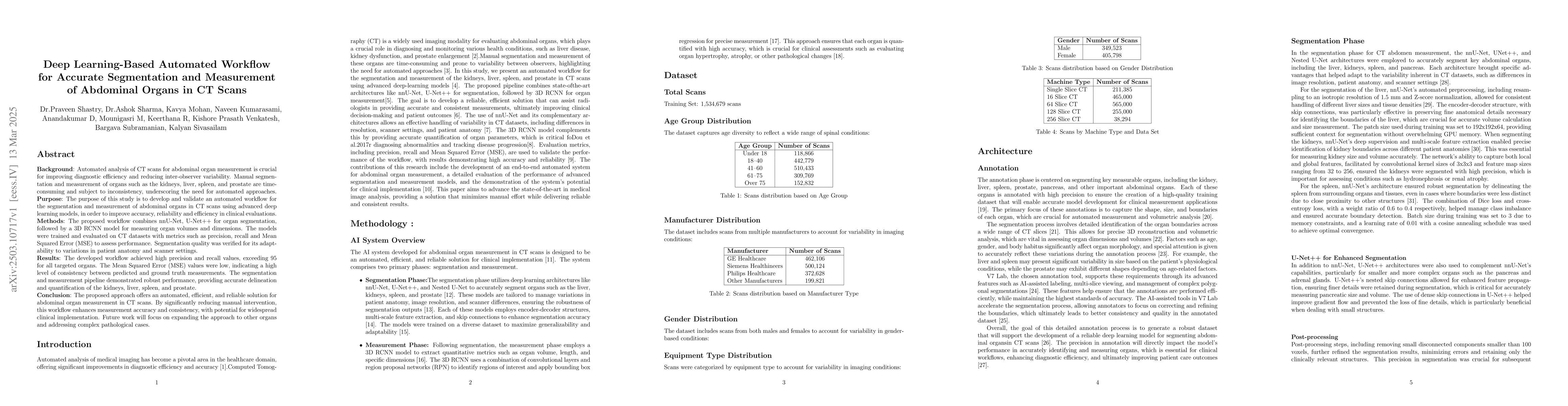
Background: Automated analysis of CT scans for abdominal organ measurement is crucial for improving diagnostic efficiency and reducing inter-observer variability. Manual segmentation and measurement of organs such as the kidneys, liver, spleen, and prostate are time-consuming and subject to inconsistency, underscoring the need for automated approaches. Purpose: The purpose of this study is to develop and validate an automated workflow for the segmentation and measurement of abdominal organs in CT scans using advanced deep learning models, in order to improve accuracy, reliability, and efficiency in clinical evaluations. Methods: The proposed workflow combines nnU-Net, U-Net++ for organ segmentation, followed by a 3D RCNN model for measuring organ volumes and dimensions. The models were trained and evaluated on CT datasets with metrics such as precision, recall, and Mean Squared Error (MSE) to assess performance. Segmentation quality was verified for its adaptability to variations in patient anatomy and scanner settings. Results: The developed workflow achieved high precision and recall values, exceeding 95 for all targeted organs. The Mean Squared Error (MSE) values were low, indicating a high level of consistency between predicted and ground truth measurements. The segmentation and measurement pipeline demonstrated robust performance, providing accurate delineation and quantification of the kidneys, liver, spleen, and prostate. Conclusion: The proposed approach offers an automated, efficient, and reliable solution for abdominal organ measurement in CT scans. By significantly reducing manual intervention, this workflow enhances measurement accuracy and consistency, with potential for widespread clinical implementation. Future work will focus on expanding the approach to other organs and addressing complex pathological cases.

AI Key Findings

Generated Jun 10, 2025

Methodology

The study employs a deep learning-based automated workflow combining nnU-Net and U-Net++ for organ segmentation, followed by a 3D RCNN model for measuring organ volumes and dimensions. The models were trained and evaluated using CT datasets with metrics such as precision, recall, and Mean Squared Error (MSE) to assess performance.

Key Results

  • The developed workflow achieved high precision and recall values, exceeding 95 for all targeted organs.
  • Mean Squared Error (MSE) values were low, indicating a high level of consistency between predicted and ground truth measurements.

Significance

This research offers an automated, efficient, and reliable solution for abdominal organ measurement in CT scans, enhancing measurement accuracy and consistency, which is crucial for improving diagnostic efficiency and reducing inter-observer variability.

Technical Contribution

The main technical contribution is the development of an integrated deep learning workflow for automated segmentation and measurement of abdominal organs in CT scans, utilizing nnU-Net, U-Net++, and a 3D RCNN model.

Novelty

This work stands out by combining multiple advanced deep learning architectures (nnU-Net, U-Net++, and 3D RCNN) into a single, robust workflow for accurate and efficient abdominal organ segmentation and measurement in CT scans, addressing the limitations of manual methods.

Limitations

  • The study did not address the computational resources required for deploying the model in real clinical settings.
  • The approach was validated on standard CT datasets; its performance on complex pathological cases remains to be explored.

Future Work

  • Expanding the approach to other organs and addressing complex pathological cases.
  • Investigating the computational requirements and potential solutions for seamless clinical implementation.

Paper Details

PDF Preview

Download PDF

Citation Network

Current paper (gray), citations (green), references (blue)

Display is limited for performance on very large graphs.

Current Paper
Citations
References
Click to view

No citations found for this paper.

Comments (0)

This website uses cookies to ensure you get the best experience. Learn more