Deep-learning-based electrode action potential mapping (DEAP Mapping) from annotation-free unipolar electrogram
Publication
Metrics
AI Quick Summary
Deep-learning-based Electrode Action Potential Mapping (DEAP Mapping) accurately reconstructs membrane potential images from unipolar ECG signals without requiring annotations, demonstrating high accuracy in ex vivo porcine heart experiments and showing potential for enhanced visualisation of atrial fibrillation substrates compared to existing methods.
Paper Preview
Abstract
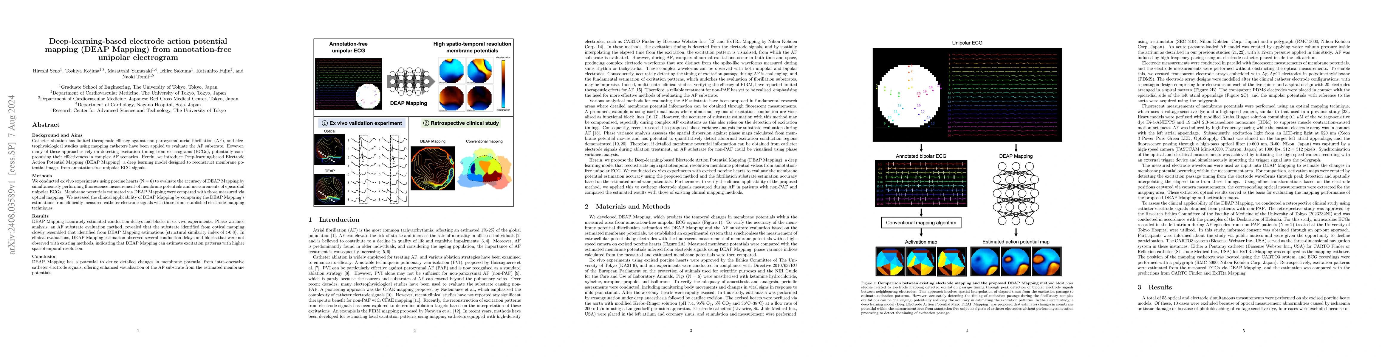
Catheter ablation has limited therapeutic efficacy against non-paroxysmal atrial fibrillation (AF), and electrophysiological studies using mapping catheters have been applied to evaluate the AF substrate. However, many of these approaches rely on detecting excitation timing from electrograms (ECGs), potentially compromising their effectiveness in complex AF scenarios. Herein, we introduce Deep-learning-based Electrode Action Potential Mapping (DEAP Mapping), a deep learning model designed to reconstruct membrane potential images from annotation-free unipolar ECG signals. We conducted ex vivo experiments using porcine hearts (N = 6) to evaluate the accuracy of DEAP Mapping by simultaneously performing fluorescence measurement of membrane potentials and measurements of epicardial unipolar ECGs. Membrane potentials estimated via DEAP Mapping were compared with those measured via optical mapping. We assessed the clinical applicability of DEAP Mapping by comparing the DEAP Mapping's estimations from clinically measured catheter electrode signals with those from established electrode-mapping techniques. DEAP Mapping accurately estimated conduction delays and blocks in ex vivo experiments. Phase variance analysis, an AF substrate evaluation method, revealed that the substrate identified from optical mapping closely resembled that identified from DEAP Mapping estimations (structural similarity index of >0.8). In clinical evaluations, DEAP Mapping estimation observed several conduction delays and blocks that were not observed with existing methods, indicating that DEAP Mapping can estimate excitation patterns with higher spatiotemporal resolution. DEAP Mapping has a potential to derive detailed changes in membrane potential from intra-operative catheter electrode signals, offering enhanced visualisation of the AF substrate from the estimated membrane potentials.
AI Key Findings
Get AI-generated insights about this paper's methodology, results, significance, and more — seven facets brought into focus.
Impact
Paper Details
Authors
PDF Preview
Citation Network
Current paper (gray), citations (green), references (blue)
Display is limited for performance on very large graphs.
Discussion 0