Deep Learning Enables Large Depth-of-Field Images for Sub-Diffraction-Limit Scanning Superlens Microscopy
Publication
Metrics
AI Quick Summary
This paper proposes a deep learning method to transform optical super-resolution images into SEM-like large depth-of-field images, eliminating the need for conductive film coatings and vacuum environments in microscopy. The method shows significant improvement in image quality, with a PSNR increase of about 0.74 dB, and has broad applications in fields like chip-level defect detection and biological sample analysis.
Paper Preview
Abstract
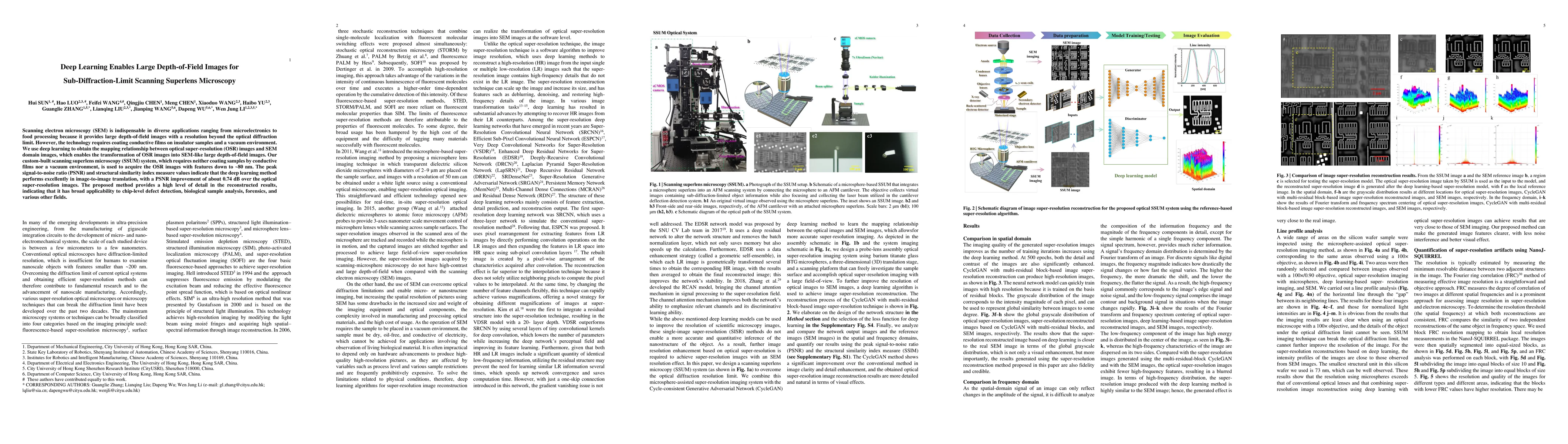
Scanning electron microscopy (SEM) is indispensable in diverse applications ranging from microelectronics to food processing because it provides large depth-of-field images with a resolution beyond the optical diffraction limit. However, the technology requires coating conductive films on insulator samples and a vacuum environment. We use deep learning to obtain the mapping relationship between optical super-resolution (OSR) images and SEM domain images, which enables the transformation of OSR images into SEM-like large depth-of-field images. Our custom-built scanning superlens microscopy (SSUM) system, which requires neither coating samples by conductive films nor a vacuum environment, is used to acquire the OSR images with features down to ~80 nm. The peak signal-to-noise ratio (PSNR) and structural similarity index measure values indicate that the deep learning method performs excellently in image-to-image translation, with a PSNR improvement of about 0.74 dB over the optical super-resolution images. The proposed method provides a high level of detail in the reconstructed results, indicating that it has broad applicability to chip-level defect detection, biological sample analysis, forensics, and various other fields.
AI Key Findings
Get AI-generated insights about this paper's methodology, results, significance, and more — seven facets brought into focus.
Impact
Paper Details
Authors
PDF Preview
Key Terms
Citation Network
Current paper (gray), citations (green), references (blue)
Display is limited for performance on very large graphs.
Discussion 0