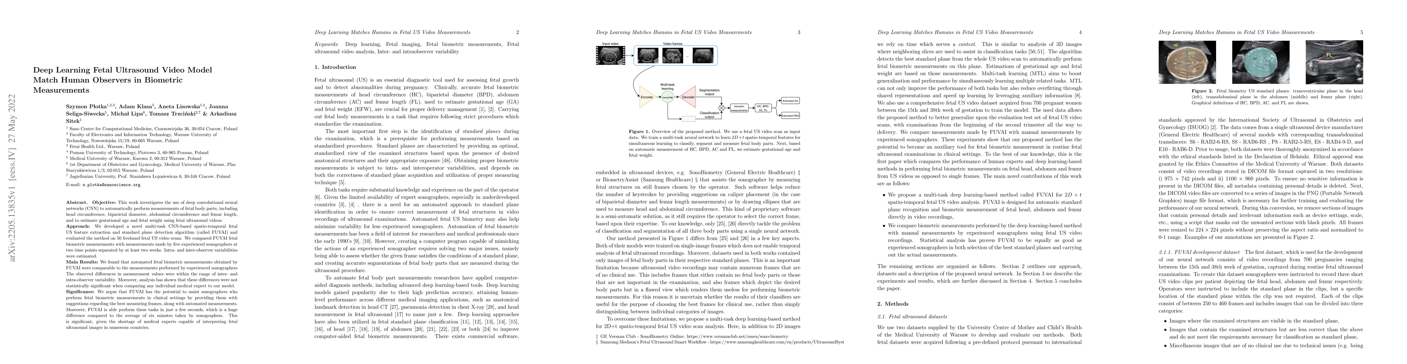
Summary
Objective. This work investigates the use of deep convolutional neural networks (CNN) to automatically perform measurements of fetal body parts, including head circumference, biparietal diameter, abdominal circumference and femur length, and to estimate gestational age and fetal weight using fetal ultrasound videos. Approach. We developed a novel multi-task CNN-based spatio-temporal fetal US feature extraction and standard plane detection algorithm (called FUVAI) and evaluated the method on 50 freehand fetal US video scans. We compared FUVAI fetal biometric measurements with measurements made by five experienced sonographers at two time points separated by at least two weeks. Intra- and inter-observer variabilities were estimated. Main results. We found that automated fetal biometric measurements obtained by FUVAI were comparable to the measurements performed by experienced sonographers The observed differences in measurement values were within the range of inter- and intra-observer variability. Moreover, analysis has shown that these differences were not statistically significant when comparing any individual medical expert to our model. Significance. We argue that FUVAI has the potential to assist sonographers who perform fetal biometric measurements in clinical settings by providing them with suggestions regarding the best measuring frames, along with automated measurements. Moreover, FUVAI is able perform these tasks in just a few seconds, which is a huge difference compared to the average of six minutes taken by sonographers. This is significant, given the shortage of medical experts capable of interpreting fetal ultrasound images in numerous countries.
AI Key Findings
Generated Sep 02, 2025
Methodology
This work investigates the use of deep convolutional neural networks (CNN) to automatically perform measurements of fetal body parts, including head circumference, biparietal diameter, abdominal circumference, and femur length, as well as estimating gestational age and fetal weight using fetal ultrasound videos. A novel multi-task CNN-based spatio-temporal fetal US feature extraction and standard plane detection algorithm, called FUVAI, was developed and evaluated on 50 freehand fetal US video scans.
Key Results
- Automated fetal biometric measurements obtained by FUVAI were comparable to measurements performed by five experienced sonographers.
- Differences in measurement values between FUVAI and sonographers were within the range of inter- and intra-observer variability and were not statistically significant.
- FUVAI can perform these tasks in just a few seconds, significantly faster than the average of six minutes taken by sonographers.
- FUVAI has the potential to assist sonographers by suggesting the best measuring frames and providing automated measurements.
Significance
FUVAI's ability to assist sonographers in clinical settings is significant, especially given the shortage of medical experts capable of interpreting fetal ultrasound images in numerous countries.
Technical Contribution
A multi-task deep learning-based framework for fetal ultrasound video analysis and interpretation, referred to as FUVAI, which can simultaneously localize standard planes in video sequences, classify and measure fetal biometric parameters, and estimate gestational age and fetal weight.
Novelty
FUVAI's success is based on its ability to automatically analyze 2D+t spatio-temporal fetal ultrasound video scans simultaneously, localizing standard planes, classifying, and measuring fetal body parts, which outperforms state-of-the-art neural networks in terms of segmentation and classification accuracy.
Limitations
- The method was trained on data from a single ultrasound device manufacturer and a single institution, raising uncertainty about its transferability to other manufacturers and institutions.
- Accuracy could likely be improved with more training cases and more diverse training examples.
Future Work
- Further validation with data from portable devices for use in underdeveloped countries.
- Improvement of the automatic method by using a more diverse training set with more examples of images with shadowing/movement.
Paper Details
PDF Preview
Key Terms
Citation Network
Current paper (gray), citations (green), references (blue)
Display is limited for performance on very large graphs.
Similar Papers
Found 4 papersFetalNet: Multi-task Deep Learning Framework for Fetal Ultrasound Biometric Measurements
Arkadiusz Sitek, Szymon Płotka, Tomasz Trzciński et al.
Uncertainty Modeling in Ultrasound Image Segmentation for Precise Fetal Biometric Measurements
Shuge Lei
MMSummary: Multimodal Summary Generation for Fetal Ultrasound Video
Xiaoqing Guo, Qianhui Men, J. Alison Noble
| Title | Authors | Year | Actions |
|---|
Comments (0)