ArxivLens

Quick Summary:

This paper proposes uncertainty modeling techniques to enhance ultrasound image segmentation for precise fetal biometric measurements, achieving an average absolute error of 8.0833mm for fetal head and 2.6163mm for fetal femur. The use of Test-Time Augmentation shows effective interpretability of data uncertainty, improving the reliability of measurements for clinical applications.

Authors:

arXiv Id: 2401.09639
Date Published: 2024-01-19
Date Updated: 2024-01-19

Categories:

Key Terms (TF-IDF):

ultrasound (0.292)
segmentation (0.241)
uncertainty (0.239)
head (0.204)
measurement (0.196)
clinical (0.190)
error (0.187)
absolute (0.176)
medical (0.173)
modeling (0.141)
relative (0.139)
average (0.134)
image (0.131)
medical image (0.130)
interpretability (0.122)
practitioners (0.119)
informed (0.119)
contributes (0.117)
computer vision (0.114)
similarly (0.114)
License: http://creativecommons.org/licenses/by/4.0/

Summary:

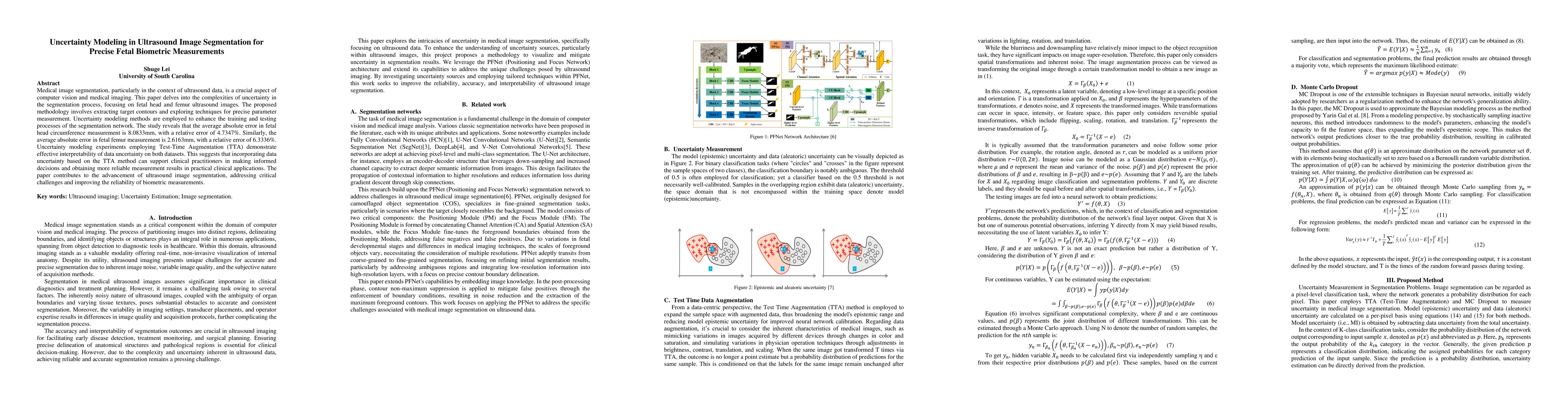
Medical image segmentation, particularly in the context of ultrasound data, is a crucial aspect of computer vision and medical imaging. This paper delves into the complexities of uncertainty in the segmentation process, focusing on fetal head and femur ultrasound images. The proposed methodology involves extracting target contours and exploring techniques for precise parameter measurement. Uncertainty modeling methods are employed to enhance the training and testing processes of the segmentation network. The study reveals that the average absolute error in fetal head circumference measurement is 8.0833mm, with a relative error of 4.7347%. Similarly, the average absolute error in fetal femur measurement is 2.6163mm, with a relative error of 6.3336%. Uncertainty modeling experiments employing Test-Time Augmentation (TTA) demonstrate effective interpretability of data uncertainty on both datasets. This suggests that incorporating data uncertainty based on the TTA method can support clinical practitioners in making informed decisions and obtaining more reliable measurement results in practical clinical applications. The paper contributes to the advancement of ultrasound image segmentation, addressing critical challenges and improving the reliability of biometric measurements.

Citation & Reference Network Visualization

This graph shows how this paper is connected to other research. The current paper is shown in dark gray, papers that cite it are in green, and papers it references are in blue. You can:

Current Paper
Papers citing this work (Citations)
Papers cited by this work (References)

• Hover over any node to see the paper title

• Click a node to view that paper's details

• Drag nodes to rearrange the graph

• Use the zoom controls (bottom right) to zoom in/out

Citations and References

No citations found for this paper.

Comments (0)

This website uses cookies to ensure you get the best experience. Learn more