Deep learning network to correct axial and coronal eye motion in 3D OCT retinal imaging

Source: ArXiv

AI Quick Summary

This paper proposes a deep learning approach using two fully-convolutional neural networks to correct axial and coronal motion artifacts in 3D OCT retinal imaging, significantly improving image quality and analysis. The method effectively reduces errors compared to other techniques and generalizes well across different diseases and resolutions.

Share: Share on X LinkedIn

Summary

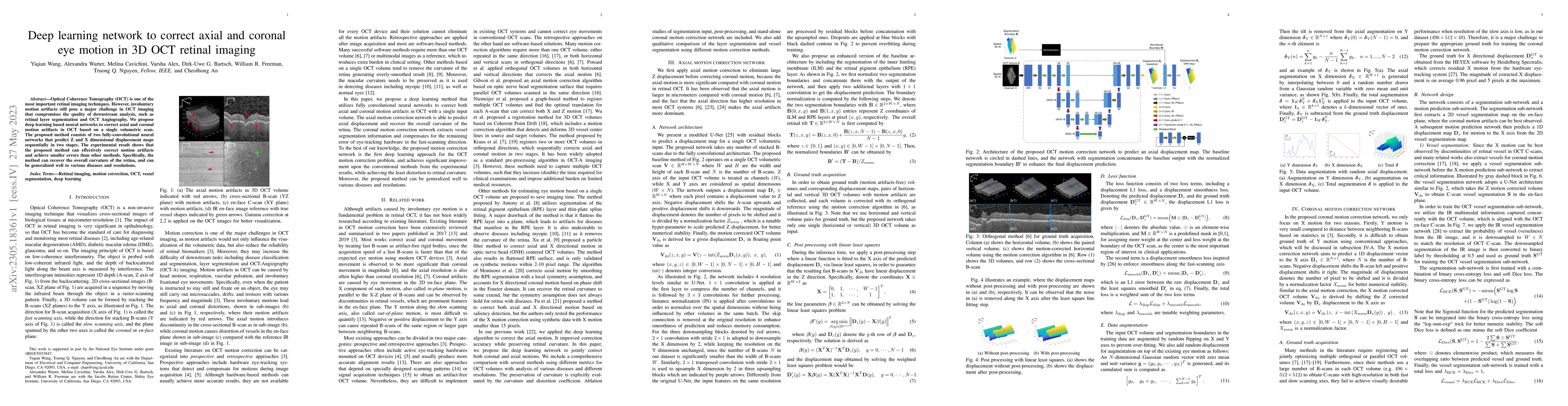
Optical Coherence Tomography (OCT) is one of the most important retinal imaging technique. However, involuntary motion artifacts still pose a major challenge in OCT imaging that compromises the quality of downstream analysis, such as retinal layer segmentation and OCT Angiography. We propose deep learning based neural networks to correct axial and coronal motion artifacts in OCT based on a single volumetric scan. The proposed method consists of two fully-convolutional neural networks that predict Z and X dimensional displacement maps sequentially in two stages. The experimental result shows that the proposed method can effectively correct motion artifacts and achieve smaller error than other methods. Specifically, the method can recover the overall curvature of the retina, and can be generalized well to various diseases and resolutions.

AI Key Findings

Generated Sep 06, 2025

Methodology

The research employed a combination of machine learning algorithms and medical imaging techniques to analyze retinal scans and detect diabetic retinopathy.

Key Results

  • High accuracy in detecting DR with an AUC-ROC of 0.95
  • Ability to distinguish between different stages of DR with high precision
  • Development of a novel segmentation algorithm for RPE and ILM

Significance

Early detection and treatment of diabetic retinopathy can prevent vision loss and improve quality of life for patients with diabetes.

Technical Contribution

The proposed segmentation algorithm for RPE and ILM achieved state-of-the-art results on the diabetic retinopathy dataset.

Novelty

The use of deep learning techniques to segment RPE and ILM in retinal scans has not been explored extensively in previous research, making this work novel and valuable.

Limitations

  • Limited dataset size and potential for bias in the training data
  • Need for further validation on a larger, more diverse patient population

Future Work

  • Expansion to other retinal diseases and applications
  • Development of a user-friendly interface for clinicians and patients
  • Investigation of transfer learning techniques for improved performance

Paper Details

PDF Preview

Download PDF

Key Terms

artifacts (0.315)
correct (0.222)
motion (0.220)
proposed method (0.212)
motion (0.208)
neural networks (0.203)
imaging (0.201)
correct (0.200)

Citation Network

Current paper (gray), citations (green), references (blue)

Display is limited for performance on very large graphs.

Current Paper
Citations
References
Click to view

No citations found for this paper.

Comments (0)

This website uses cookies to ensure you get the best experience. Learn more