Summary
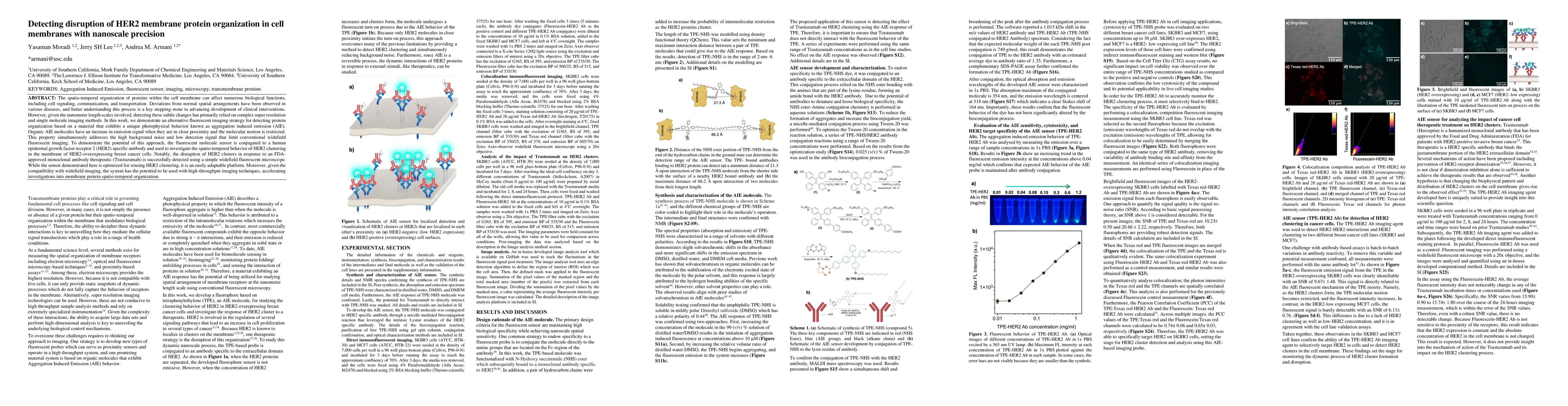
The spatio-temporal organization of proteins within the cell membrane can affect numerous biological functions, including cell signaling, communication, and transportation. Deviations from normal spatial arrangements have been observed in various diseases, and better understanding this process is a key stepping-stone to advancing development of clinical interventions. However, given the nanometer length scales involved, detecting these subtle changes has primarily relied on complex super resolution and single molecule imaging methods. In this work, we demonstrate an alternative fluorescent imaging strategy for detecting protein organization based on a material that exhibits a unique photophysical behavior known as aggregation induced emission (AIE). Organic AIE molecules have an increase in emission signal when they are in close proximity and the molecular motion is restricted. This property simultaneously addresses the high background noise and low detection signal that limit conventional widefield fluorescent imaging. To demonstrate the potential of this approach, the fluorescent molecule sensor is conjugated to a human epidermal growth factor receptor 2 (HER2) specific antibody and used to investigate the spatio-temporal behavior of HER2 clustering in the membrane of HER2-overexpressing breast cancer cells. Notably, the disruption of HER2 clusters in response to an FDA-approved monoclonal antibody therapeutic (Trastuzumab) is successfully detected using a simple widefield fluorescent microscope. While the sensor demonstrated here is optimized for sensing HER2 clustering, it is an easily adaptable platform. Moreover, given the compatibility with widefield imaging, the system has the potential to be used with high-throughput imaging techniques, accelerating investigations into membrane protein spatio-temporal organization.
AI Key Findings
Get AI-generated insights about this paper's methodology, results, and significance.
Paper Details
PDF Preview
Key Terms
Citation Network
Current paper (gray), citations (green), references (blue)
Display is limited for performance on very large graphs.
Similar Papers
Found 4 papersReal-Time Analysis of Nanoscale Dynamics in Membrane Protein Insertion via Single-Molecule Imaging
Y. Lu, C. Yang, M. Li et al.
Antibody binding reports spatial heterogeneities in cell membrane organization
Daniel P. Arnold, Sho C. Takatori, Yaxin Xu
| Title | Authors | Year | Actions |
|---|
Comments (0)