Diagnosing Glaucoma Progression with Visual Field Data Using a Spatiotemporal Boundary Detection Method
Publication
Metrics
AI Quick Summary
This paper introduces a spatiotemporal boundary detection method for diagnosing glaucoma progression using visual field data, showing improved diagnosis compared to existing spatial methods. The proposed method leverages the underlying anatomy of the optic disc to better understand vision loss, demonstrated through trials and simulations.
Paper Preview
Abstract
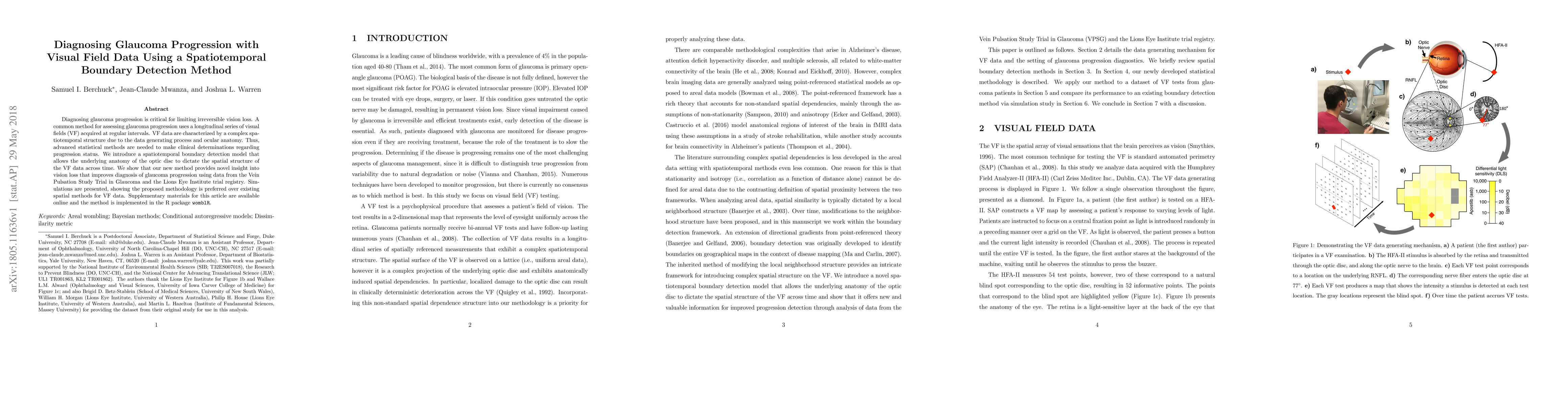
Diagnosing glaucoma progression is critical for limiting irreversible vision loss. A common method for assessing glaucoma progression uses a longitudinal series of visual fields (VF) acquired at regular intervals. VF data are characterized by a complex spatiotemporal structure due to the data generating process and ocular anatomy. Thus, advanced statistical methods are needed to make clinical determinations regarding progression status. We introduce a spatiotemporal boundary detection model that allows the underlying anatomy of the optic disc to dictate the spatial structure of the VF data across time. We show that our new method provides novel insight into vision loss that improves diagnosis of glaucoma progression using data from the Vein Pulsation Study Trial in Glaucoma and the Lions Eye Institute trial registry. Simulations are presented, showing the proposed methodology is preferred over existing spatial methods for VF data. Supplementary materials for this article are available online and the method is implemented in the R package womblR.
AI Key Findings
Get AI-generated insights about this paper's methodology, results, significance, and more — seven facets brought into focus.
Impact
Paper Details
PDF Preview
Key Terms
Citation Network
Current paper (gray), citations (green), references (blue)
Display is limited for performance on very large graphs.
Discussion 0