Diamond magnetic microscopy of malarial hemozoin nanocrystals
Publication
Metrics
Paper Preview
Abstract
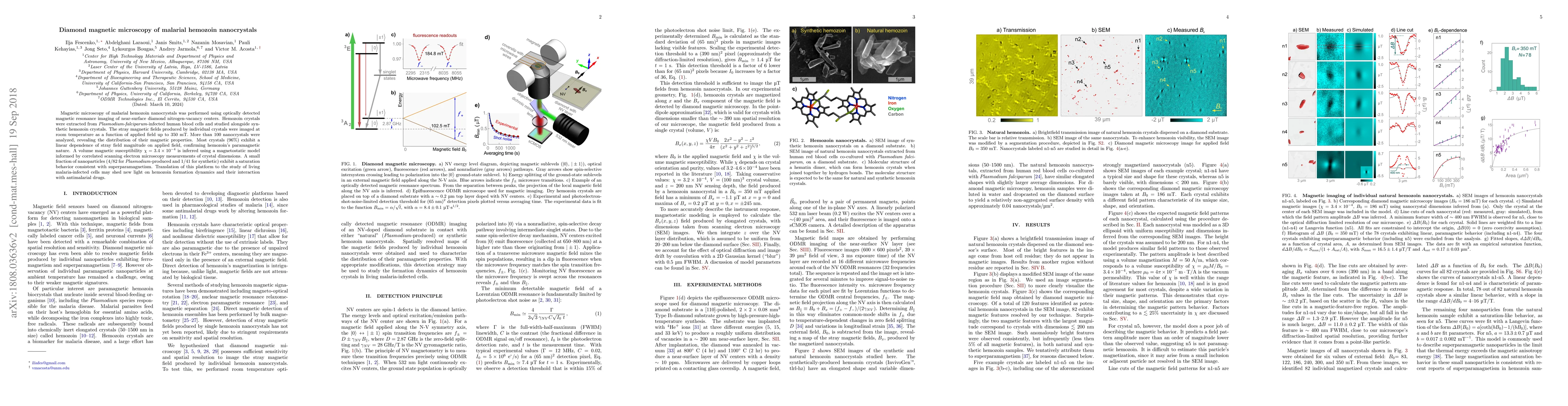
Magnetic microscopy of malarial hemozoin nanocrystals was performed using optically detected magnetic resonance imaging of near-surface diamond nitrogen-vacancy centers. Hemozoin crystals were extracted from $Plasmodium$-$falciparum$-infected human blood cells and studied alongside synthetic hemozoin crystals. The stray magnetic fields produced by individual crystals were imaged at room temperature as a function of applied field up to 350 mT. More than 100 nanocrystals were analyzed, revealing the distribution of their magnetic properties. Most crystals ($96\%$) exhibit a linear dependence of stray field magnitude on applied field, confirming hemozoin's paramagnetic nature. A volume magnetic susceptibility $\chi=3.4\times10^{-4}$ is inferred using a magnetostatic model informed by correlated scanning electron microscopy measurements of crystal dimensions. A small fraction of nanoparticles (4/82 for $Plasmodium$-produced and 1/41 for synthetic) exhibit a saturation behavior consistent with superparamagnetism. Translation of this platform to the study of living malaria-infected cells may shed new light on hemozoin formation dynamics and their interaction with antimalarial drugs.
AI Key Findings
Get AI-generated insights about this paper's methodology, results, significance, and more — seven facets brought into focus.
Impact
Paper Details
PDF Preview
Key Terms
Citation Network
Current paper (gray), citations (green), references (blue)
Display is limited for performance on very large graphs.
Discussion 0