Summary
3D-Polarized Light Imaging (3D-PLI) reconstructs nerve fibers in histological brain sections by measuring their birefringence. This study investigates another effect caused by the optical anisotropy of brain tissue - diattenuation. Based on numerical and experimental studies and a complete analytical description of the optical system, the diattenuation was determined to be below 4 % in rat brain tissue. It was demonstrated that the diattenuation effect has negligible impact on the fiber orientations derived by 3D-PLI. The diattenuation signal, however, was found to highlight different anatomical structures that cannot be distinguished with current imaging techniques, which makes Diattenuation Imaging a promising extension to 3D-PLI.
AI Key Findings
Get AI-generated insights about this paper's methodology, results, and significance.
Paper Details
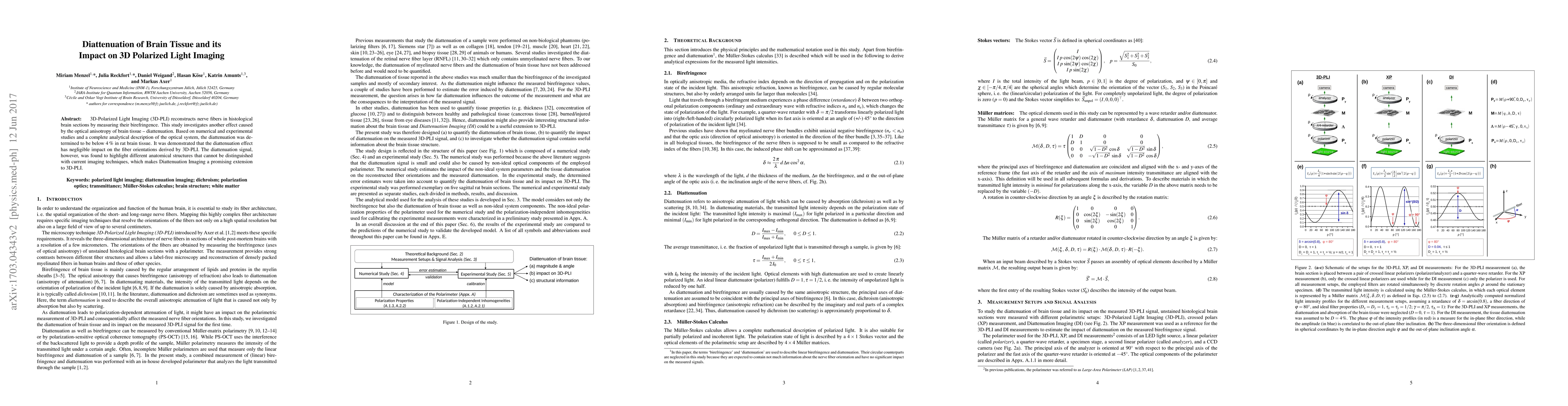
PDF Preview
Key Terms
Citation Network
Current paper (gray), citations (green), references (blue)
Display is limited for performance on very large graphs.
Similar Papers
Found 4 papers| Title | Authors | Year | Actions |
|---|
Comments (0)