ArxivLens

Quick Summary:

This study reveals that diattenuation imaging can differentiate brain tissue properties based on nerve fibre orientations and other factors like tissue homogeneity, fibre size, and myelin sheath thickness. The observed diattenuation effects are explained by anisotropic absorption and scattering, establishing diattenuation imaging as a valuable tool for distinguishing brain regions.

Journal Reference: Scientific Reports 9(1939)
DOI: 10.1038/s41598-019-38506-w
arXiv Id: 1806.07712
Comments: 18 pages, 9 figures
Date Published: 2019-02-22
Date Updated: 2019-02-22

Key Terms (TF-IDF):

brain (0.367)
referred (0.235)
light (0.209)
anisotropic (0.207)
imaging (0.183)
signal (0.172)
regions (0.170)
polarisation (0.169)
types (0.166)
observed (0.159)
minimally (0.151)
properties (0.149)
studies (0.148)
establishes (0.145)
effects (0.138)
thickness (0.135)
different (0.132)
distinguish (0.131)
valuable (0.131)
modelling (0.130)
License: http://arxiv.org/licenses/nonexclusive-distrib/1.0/

Summary:

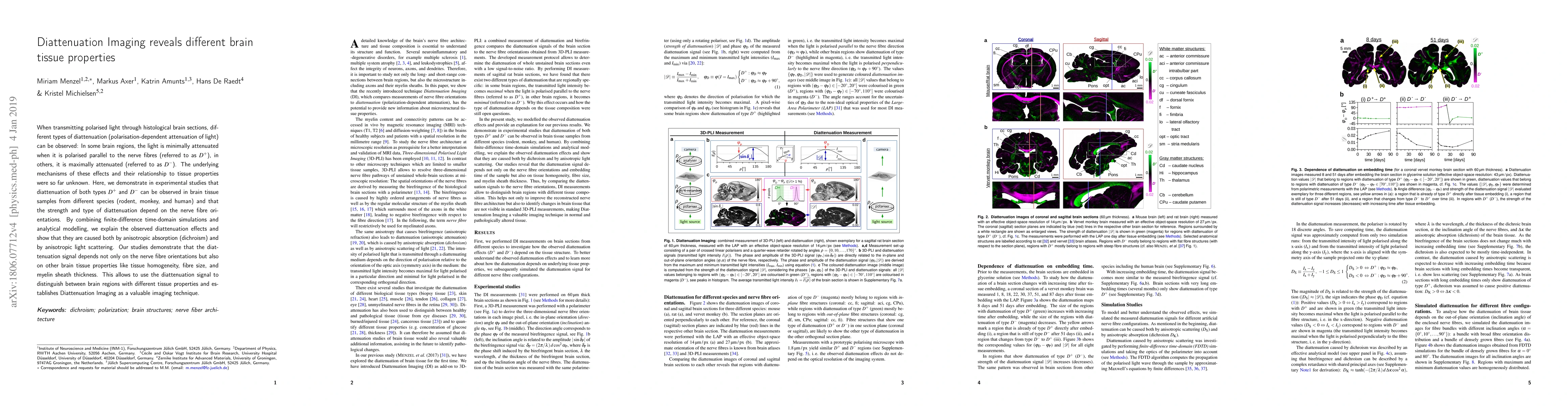
When transmitting polarised light through histological brain sections, different types of diattenuation (polarisation-dependent attenuation of light) can be observed: In some brain regions, the light is minimally attenuated when it is polarised parallel to the nerve fibres (referred to as D+), in others, it is maximally attenuated (referred to as D-). The underlying mechanisms of these effects and their relationship to tissue properties were so far unknown. Here, we demonstrate in experimental studies that diattenuation of both types D+ and D- can be observed in brain tissue samples from different species (rodent, monkey, and human) and that the strength and type of diattenuation depend on the nerve fibre orientations. By combining finite-difference time-domain simulations and analytical modelling, we explain the observed diattenuation effects and show that they are caused both by anisotropic absorption (dichroism) and by anisotropic light scattering. Our studies demonstrate that the diattenuation signal depends not only on the nerve fibre orientations but also on other brain tissue properties like tissue homogeneity, fibre size, and myelin sheath thickness. This allows to use the diattenuation signal to distinguish between brain regions with different tissue properties and establishes Diattenuation Imaging as a valuable imaging technique.

Citation & Reference Network Visualization

This graph shows how this paper is connected to other research. The current paper is shown in dark gray, papers that cite it are in green, and papers it references are in blue. You can:

Current Paper
Papers citing this work (Citations)
Papers cited by this work (References)

• Hover over any node to see the paper title

• Click a node to view that paper's details

• Drag nodes to rearrange the graph

• Use the zoom controls (bottom right) to zoom in/out

Citations and References

Title Authors Year Actions
Enhanced characterization of depolarizing samples using indices of polarization purity and polarizance–reflection–transformation spaces
Dekui Li, Ivan Montes, Mónica Canabal-Carbia, I. Estévez, O. López-Coronado, Zhongyi Guo, J. Campos, Á. Lizana
2025
View Paper
Strain alignment of conjugated polymers: Method, microstructure, and applications
Harry M. Schrickx, P. Sen, Nrup Balar, Brendan T. O'Connor
2024
View Paper
Reconstructing 3D Biomedical Architectural Order at Multiple Spatial Scales with Multimodal Stack Input
Chaojing Shi, Guocheng Sun, Kaitai Han, Mengyuan Huang, Wu Liu, Xi Liu, Zijun Wang, Qianjin Guo
2024
View Paper
Diattenuation Enhancement Induced by Tissue Staining in Multispectral Mueller Matrix Microscopy
Yuzhu Shi, Chunyan Chen, Hongtao Li, Zhiyan Liu, Chao He, Honghui He
2024
View Paper
The effect of experimental errors on optimized polarimeters
Neil C. Bruce, Iván Montes-González
2023
View Paper
A polarization image enhancement method for glioma
Yi-rong Liu, Chao-Feng Liang, H. Zhao, Yun-Mou Ou, Jian Wu
2023
View Paper
A Proposed Human Structural Brain Connectivity Matrix in the Center for Morphometric Analysis Harvard-Oxford Atlas Framework: A Historical Perspective and Future Direction for Enhancing the Precision of Human Structural Connectivity with a Novel Neuroanatomical Typology
N. Makris, R. Rushmore, J. Kaiser, Matthew D. Albaugh, M. Kubicki, Y. Rathi, Fan Zhang, L. O’Donnell, E. Yeterian, V. Caviness, D. Kennedy
2023
View Paper
Rapid and label-free histological imaging of unprocessed surgical tissues via dark-field reflectance ultraviolet microscopy
Shiwei Ye, J. Zou, Chen Huang, F. Xiang, Zonghua Wen, Nannan Wang, Jia Yu, Yuezhi He, P. Liu, Xin Mei, Hui Li, Lili Niu, Peng Gong, Wei Zheng
2022
View Paper
Spatial frequency domain Mueller matrix imaging
J. Chue-Sang, M. Litorja, A. Goldfain, T. Germer
2022
View Paper
Fast data-driven computation and intuitive visualization of fiber orientation uncertainty in 3D-polarized light imaging
D. Schmitz, Kai Benning, Nicole Schubert, M. Minnerop, K. Amunts, M. Axer
2022
View Paper
Error analysis of a Stokes imaging polarimeter based on liquid crystal variable retarders
Iván Montes-González, N. Bruce
2022
View Paper
Analyzing the Influence of Imaging Resolution on Polarization Properties of Scattering Media Obtained From Mueller Matrix
Conghui Shao, Binguo Chen, Honghui He, Chao He, Yuanxing Shen, Haoyu Zhai, Hui Ma
2022
View Paper
Polarimetric techniques for the structural studies and diagnosis of brain
O. Rodríguez-Núñez, T. Novikova
2022
View Paper
Linear retardance and diattenuation dispersion measurements of anisotropic medium with Jones calculus based spectrometric technique
F. Sanaâ, Y. Makhlouka, M. Gharbia
2022
View Paper
Linking Brain Structure, Activity, and Cognitive Function through Computation
K. Amunts, J. DeFelipe, C. Pennartz, A. Destexhe, M. Migliore, Philippe Ryvlin, S. Furber, A.C. Knoll, L. Bitsch, J. Bjaalie, Yannis E. Ioannidis, T. Lippert, M. Sanchez-Vives, R. Goebel, V. Jirsa
2022
View Paper
Roadmap on Digital Holography-Based Quantitative Phase Imaging
Vinoth Balasubramani, M. Kujawińska, C. Allier, V. Anand, Chau-Jern Cheng, C. Depeursinge, Nathaniel Hai, S. Juodkazis, J. Kalkman, A. Kuś, Moosung Lee, P. Magistretti, P. Marquet, S. Ng, Joseph Rosen, Y. Park, M. Ziemczonok
2021
View Paper
Polarimetric visualization of healthy brain fiber tracts under adverse conditions: ex vivo studies.
O. Rodríguez-Núñez, P. Schucht, E. Hewer, T. Novikova, A. Pierangelo
2021
View Paper
Refractive index of biological tissues: Review, measurement techniques, and applications.
R. Khan, B. Gul, Shamim Khan, H. Nisar, Iftikhar Ahmad
2021
View Paper
Linear diattenuation imaging of biological tissues with near infrared Mueller scanning microscopy.
M. Dubreuil, Florine Tissier, S. Rivet, Y. Grand
2020
View Paper
Cell refractive index: Models, insights, applications and future perspectives.
B. Gul, Sumara Ashraf, Shamim Khan, H. Nisar, Iftikhar Ahmad
2020
View Paper
Anatomy of nerve fiber bundles at micrometer-resolution in the vervet monkey visual system
H. Takemura, N. Palomero-Gallagher, M. Axer, D. Gräßel, M. Jorgensen, R. Woods, K. Zilles
2020
View Paper
Bringing Anatomical Information into Neuronal Network Models.
Sacha Jennifer van Albada, Aitor Morales-Gregorio, Rembrandt Bakker, G. Palm, A. Goulas, S. Bludau, Timo Dickscheid, C. Hilgetag, M. Diesmann
2020
View Paper
Axon morphology is modulated by the local environment and impacts the noninvasive investigation of its structure–function relationship
M. Andersson, H. Kjer, Jonathan Rafael-Patino, A. Pacureanu, B. Pakkenberg, J. Thiran, M. Ptito, M. Bech, A. Dahl, Vedrana Andersen Dahl, T. Dyrby
2020
View Paper
Coherent Fourier scatterometry reveals nerve fiber crossings in the brain
M. Menzel, S. Pereira
2020
View Paper
How Human Is Human Connectional Neuroanatomy?
R. Rushmore, S. Bouix, M. Kubicki, Y. Rathi, E. Yeterian, N. Makris
2020
View Paper
What we can learn from the complex architecture of single axons
K. Rockland
2020
View Paper
High-resolution 3D tractography of fibrous tissue based on polarization-sensitive optical coherence tomography
G. Yao, D. Duan
2019
View Paper
Differentiation of collagenous changes in scleroderma from unstained thin skin tissue sections using Mueller polarimetric image analysis
Mahima Sharma B. S., S. N. Unni, Leena Dennis Joseph
2019
View Paper
FAConstructor: an interactive tool for geometric modeling of nerve fiber architectures in the brain
J. Reuter, F. Matuschke, M. Menzel, Nicole Schubert, Kévin Ginsburger, C. Poupon, K. Amunts, M. Axer
2019
View Paper
Polarimetric Detection of Chemotherapy-Induced Cancer Cell Death
A. Fernández-Pérez, Olga Gutiérrez-Saiz, J. Fernández-Luna, F. Moreno, J. Saiz
2019
View Paper
Finite-Difference Time-Domain simulations of transmission microscopy enable a better interpretation of 3D nerve fiber architectures in the brain
M. Menzel, M. Axer, H. Raedt, I. Costantini, L. Silvestri, F. Pavone, K. Amunts, K. Michielsen
2018
View Paper
Coherent Fourier scatterometry reveals nerve fiber crossings in the brain
Ilvania, P. F., Ereira
2020
View Paper
Connecting the microscopic depolarizing origin of samples with macroscopic measures of the Indices of Polarimetric Purity
Mónica Canabal-Carbia, I. Estévez, Esther Nabadda, E. Garcia-Caurel, J.J. Gil, R. Ossikovski, Andrés Márquez, Ivan Moreno, J. Campos, Á. Lizana
View Paper

Comments (0)

This website uses cookies to ensure you get the best experience. Learn more