Quick Summary:

This review examines the emerging field of digital staining in optical microscopy, where deep learning techniques are used to mimic traditional biochemical staining without the need for physical dyes. It discusses current methods, challenges, and potential future advancements in this innovative approach to clinical diagnostics and research.

Summary

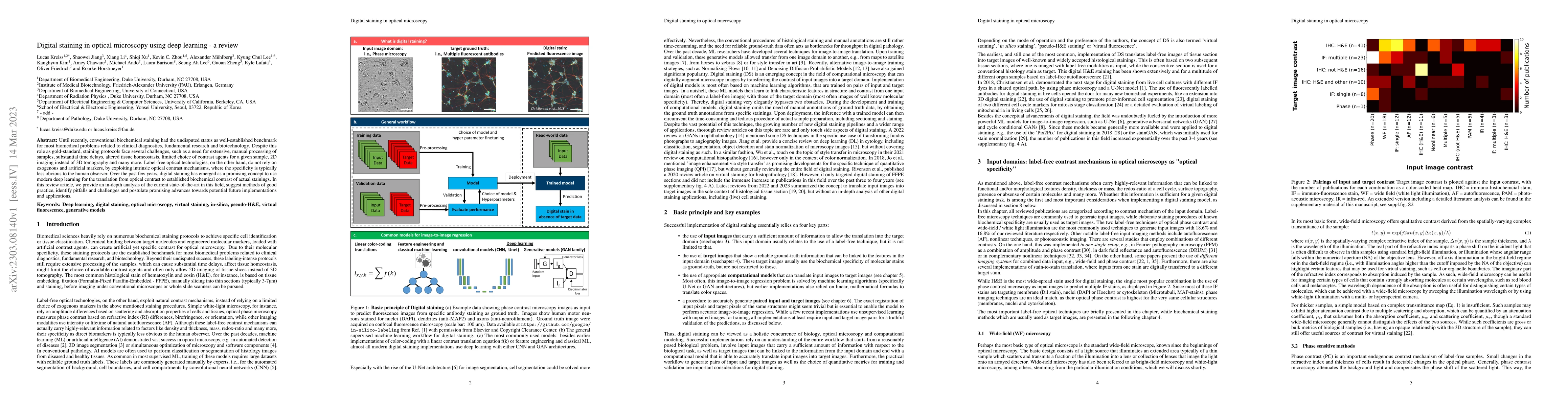
Until recently, conventional biochemical staining had the undisputed status as well-established benchmark for most biomedical problems related to clinical diagnostics, fundamental research and biotechnology. Despite this role as gold-standard, staining protocols face several challenges, such as a need for extensive, manual processing of samples, substantial time delays, altered tissue homeostasis, limited choice of contrast agents for a given sample, 2D imaging instead of 3D tomography and many more. Label-free optical technologies, on the other hand, do not rely on exogenous and artificial markers, by exploiting intrinsic optical contrast mechanisms, where the specificity is typically less obvious to the human observer. Over the past few years, digital staining has emerged as a promising concept to use modern deep learning for the translation from optical contrast to established biochemical contrast of actual stainings. In this review article, we provide an in-depth analysis of the current state-of-the-art in this field, suggest methods of good practice, identify pitfalls and challenges and postulate promising advances towards potential future implementations and applications.

AI Key Findings

Get AI-generated insights about this paper's methodology, results, and significance.

Paper Details

PDF Preview

Download PDF

Key Terms

contrast (0.229)
optical (0.182)
established (0.173)
promising (0.173)
challenges (0.164)
delays (0.151)
gold (0.144)
current state (0.138)
observer (0.137)
obvious (0.136)
manual (0.134)
tomography (0.134)
status (0.131)
clinical (0.130)
agents (0.123)
actual (0.123)
implementations (0.122)
advances (0.122)
emerged (0.122)
face (0.122)

Citation Network

Current paper (gray), citations (green), references (blue)

Current Paper
Citations
References
Click to view

Similar Papers

No similar papers found for this research.

Title Authors Year Actions
Digital Staining with Knowledge Distillation: A Unified Framework for Unpaired and Paired-But-Misaligned Data
Ziwang Xu, Lanqing Guo, Satoshi Tsutsui, Shuyan Zhang, A.C. Kot, Bihan Wen
2025
View Paper
Unpaired virtual histological staining of tissue from autofluorescence using regularized cycle-consistent adversarial networks
Zhesi Wen, Zhu-cheng Yin, Min Xu
2025
View Paper
Squamous Cell Carcinoma Margin Classification Using Vision Transformers from Digital Histopathology Images
So-yun Park, Gelan Ayana, Se-woon Choe
2025
View Paper
Vision Transformers for Low-Quality Histopathological Images: A Case Study on Squamous Cell Carcinoma Margin Classification
So-yun Park, Gelan Ayana, Beshatu Debela Wako, K. C. Jeong, Soon‐Do Yoon, Se-woon Choe
2025
View Paper
Computational staining of CD3/CD20 positive lymphocytes in human tissues with experimental confirmation in a genetically engineered mouse model
Xiang Li, Casey C. Heirman, Ashlyn G Rickard, Gina Sotolongo, Rico Castillo, Temitayo Adanlawo, Jeffery I. Everitt, Jeffery B. Hodgin, Tammara L. Watts, Andrew R. Janowczyk, Yvonne Mowery, Laura Barisoni, K. Lafata
2024
View Paper
Fourier Ptychography With Information Entropy Based No-Reference Image Quality Assessment Learning
Qijun Yang, Hujun Yin
2024
View Paper
Restoring H&E stain in faded slides via phase-to-color virtual staining in near-infrared
Hyesuk Chae, Jongho Kim, Joonsung Jeon, K. Lee, K. Lee, Ji Ung Choi, Suki Kang, Soyoung Choi, Geunbae Bang, Jong Ha Lee, E. Park, Nam Hoon Cho, Seung Ah Lee
2024
View Paper
Rapid 3D imaging at cellular resolution for digital cytopathology with a multi-camera array scanner (MCAS)
Kanghyun Kim, A. Chaware, Clare B. Cook, Shiqi Xu, Monica Abdelmalak, Colin L. Cooke, Kevin C Zhou, Mark Harfouche, Paul Reamey, Veton Saliu, Jed Doman, Clay Dugo, Gregor Horstmeyer, Richard Davis, Ian Taylor-Cho, W. Foo, Lucas Kreiss, X. Jiang, R. Horstmeyer
2024
View Paper
Impact of Loss Functions on Label-free Virtual H&E Staining
Qiang Wang, James R. Hopgood, Marta Vallejo
2024
View Paper
Autonomous Quality and Hallucination Assessment for Virtual Tissue Staining and Digital Pathology
Luzhe Huang, Yuzhu Li, N. Pillar, T. Haran, W. Wallace, Aydogan Ozcan
2024
View Paper
Virtual birefringence imaging and histological staining of amyloid deposits in label-free tissue using autofluorescence microscopy and deep learning
Xilin Yang, Bijie Bai, Yijie Zhang, Musa Aydin, Ş. Selçuk, Zhen Guo, Gregory A. Fishbein, Karine Atlan, W. Wallace, N. Pillar, Aydogan Ozcan
2024
View Paper
Self-supervised denoising for multimodal structured illumination microscopy enables long-term super-resolution live-cell imaging
Xingye Chen, Chang Qiao, Tao Jiang, Jiahao Liu, Quan Meng, Yunmin Zeng, Haoyu Chen, Hui Qiao, Dong Li, Jiamin Wu
2024
View Paper
Plug-and-play DPC-based quantitative phase microscope
Tao Peng, Zeyu Ke, Hao Wu, Jun He, Yue Sun, Shuhe Zhang, Qian Gao, Meng Shao, Shengzhao Zhang, Zhensheng Zhong, Fengya Lu, Jinhua Zhou
2024
View Paper
O RGANOID C OMPUTER V ISION : A S URVEY AND O UTLOOK
Anwaar Ulhaq
View Paper

Comments (0)

This website uses cookies to ensure you get the best experience. Learn more