Academic Profile
Statistics
Similar Authors
Papers on arXiv
We present a large-scale computational 3D topographic microscope that enables 6-gigapixel profilometric 3D imaging at micron-scale resolution across $>$110 cm$^2$ areas over multi-millimeter axial r...
We report Tensorial tomographic Fourier Ptychography (ToFu), a new non-scanning label-free tomographic microscopy method for simultaneous imaging of quantitative phase and anisotropic specimen infor...
Until recently, conventional biochemical staining had the undisputed status as well-established benchmark for most biomedical problems related to clinical diagnostics, fundamental research and biote...
To study the behavior of freely moving model organisms such as zebrafish (Danio rerio) and fruit flies (Drosophila) across multiple spatial scales, it would be ideal to use a light microscope that c...
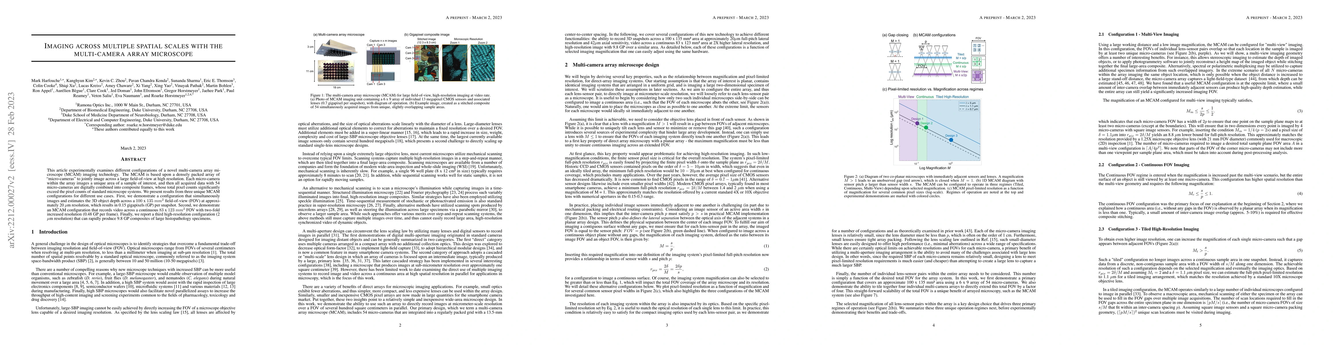
This article experimentally examines different configurations of a novel multi-camera array microscope (MCAM) imaging technology. The MCAM is based upon a densely packed array of "micro-cameras" to ...
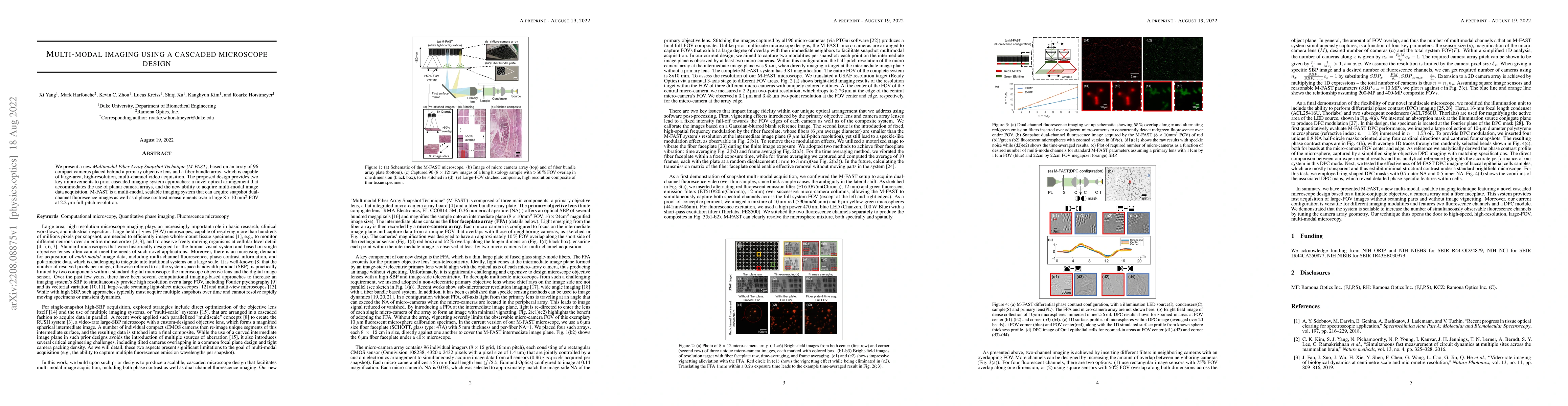
We present a new Multimodal Fiber Array Snapshot Technique (M-FAST), based on an array of 96 compact cameras placed behind a primary objective lens and a fiber bundle array. which is capable of larg...
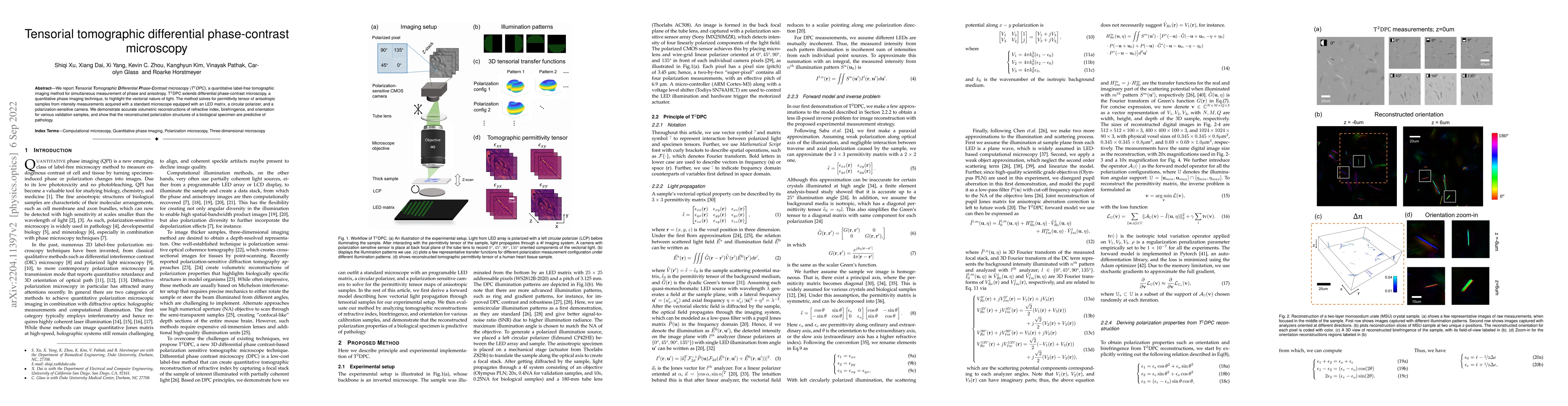
We report Tensorial Tomographic Differential Phase-Contrast microscopy (T2DPC), a quantitative label-free tomographic imaging method for simultaneous measurement of phase and anisotropy. T2DPC exten...
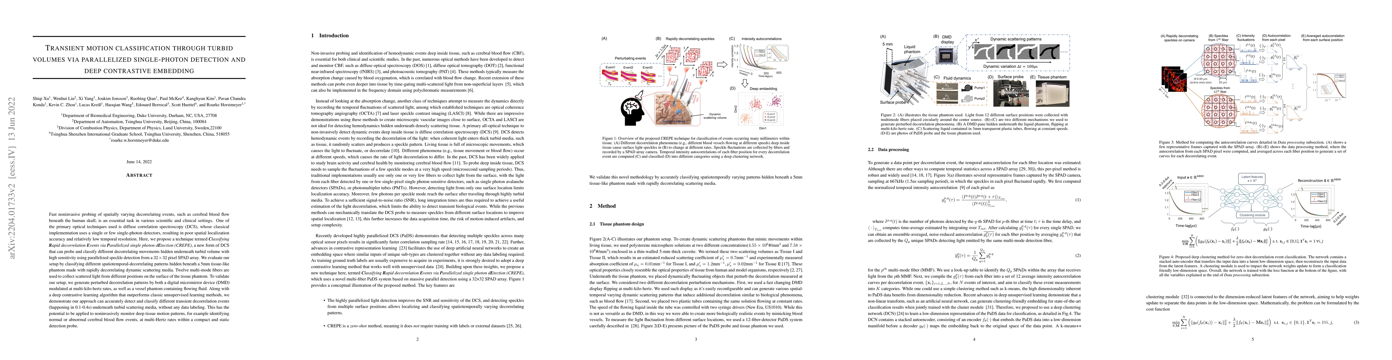
Fast noninvasive probing of spatially varying decorrelating events, such as cerebral blood flow beneath the human skull, is an essential task in various scientific and clinical settings. One of the ...
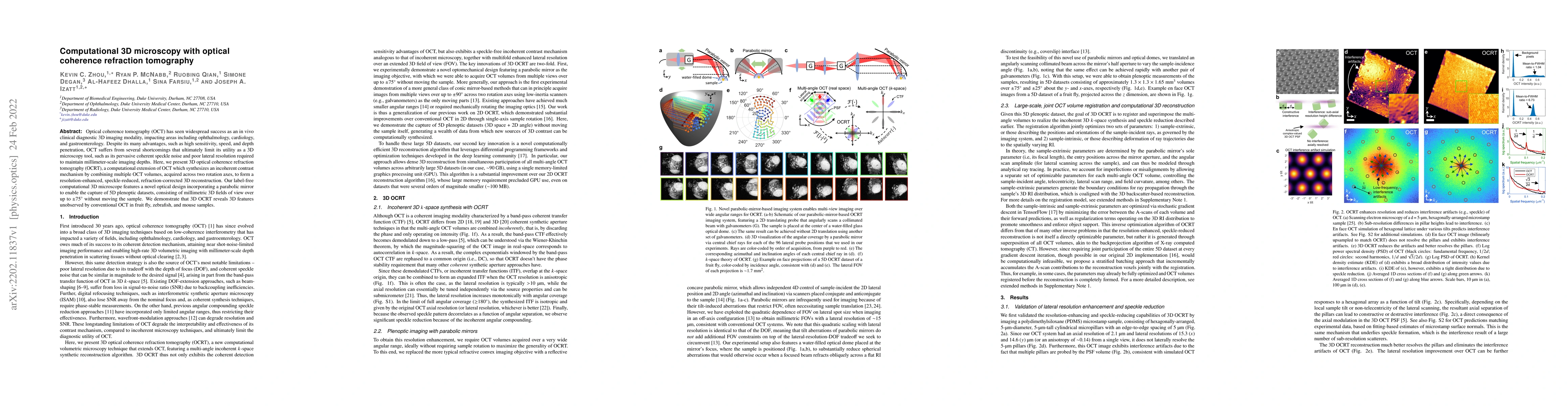
Optical coherence tomography (OCT) has seen widespread success as an in vivo clinical diagnostic 3D imaging modality, impacting areas including ophthalmology, cardiology, and gastroenterology. Despi...
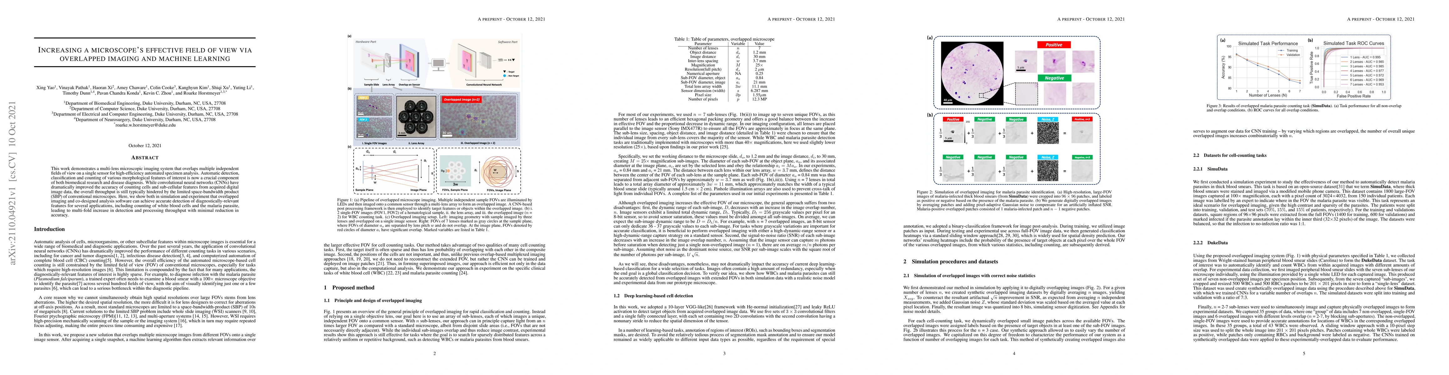
This work demonstrates a multi-lens microscopic imaging system that overlaps multiple independent fields of view on a single sensor for high-efficiency automated specimen analysis. Automatic detecti...
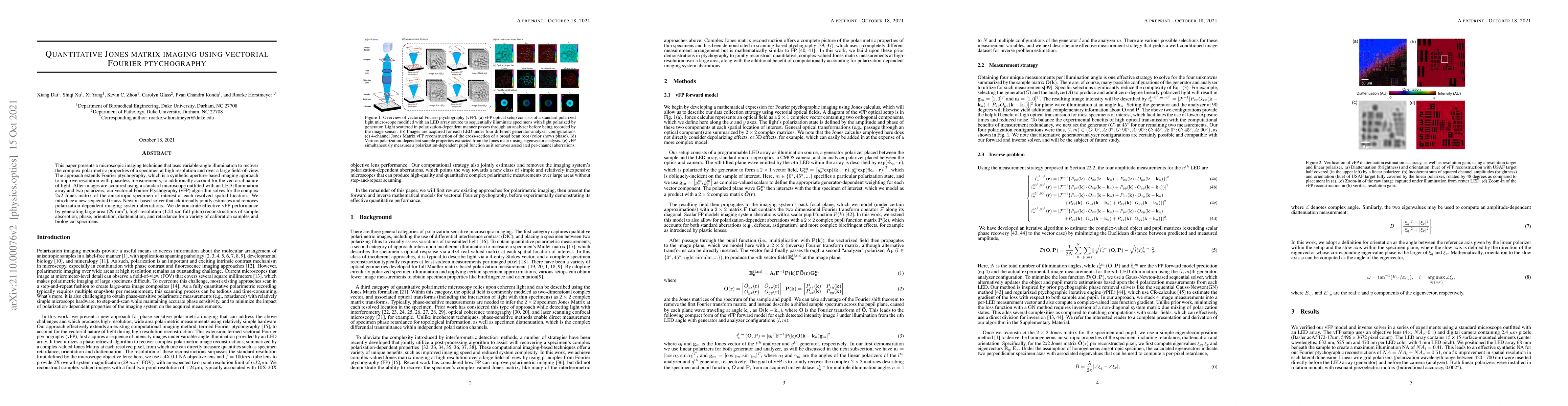
This paper presents a microscopic imaging technique that uses variable-angle illumination to recover the complex polarimetric properties of a specimen at high resolution and over a large field-of-vi...
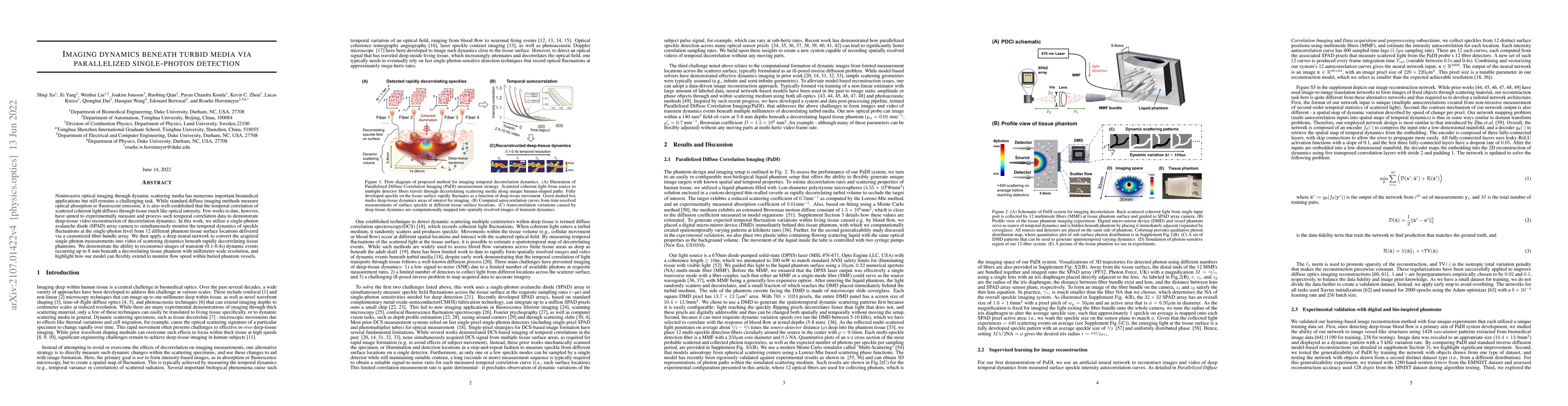
Noninvasive optical imaging through dynamic scattering media has numerous important biomedical applications but still remains a challenging task. While standard diffuse imaging methods measure optic...
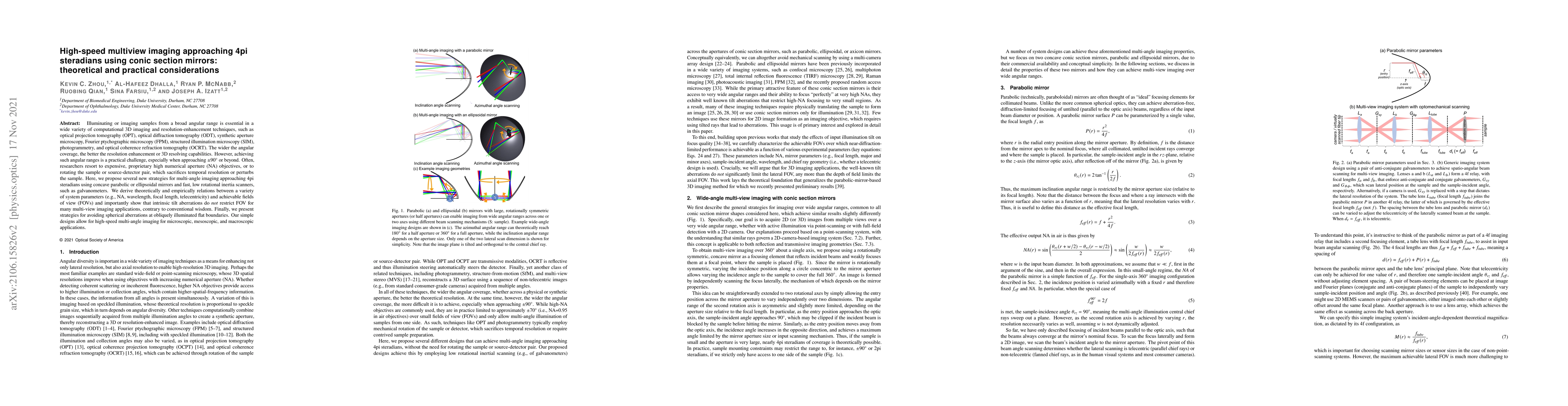
Illuminating or imaging samples from a broad angular range is essential in a wide variety of computational 3D imaging and resolution-enhancement techniques, such as optical projection tomography (OP...
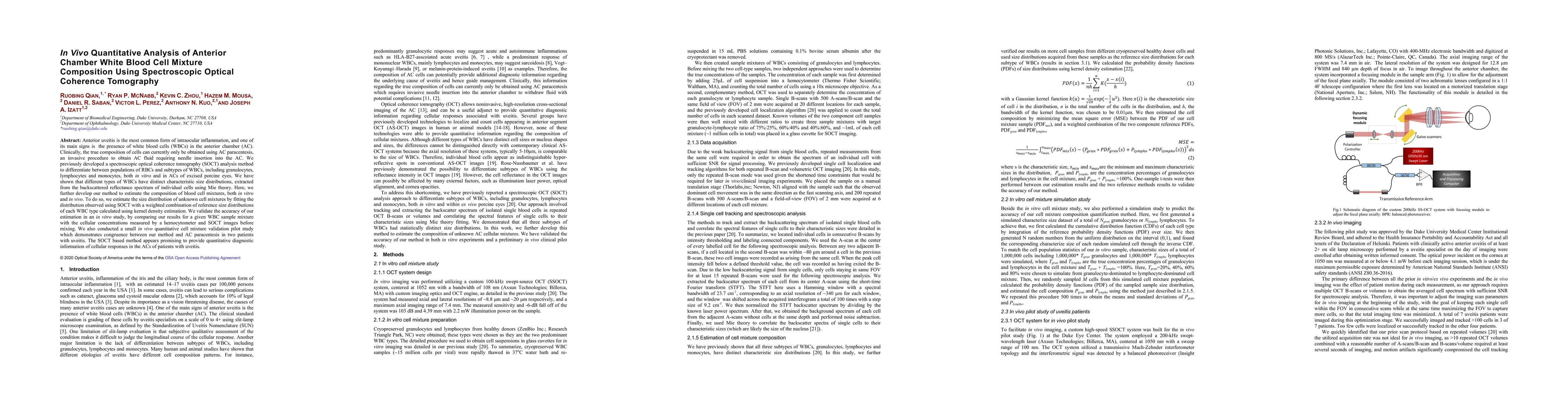
Anterior uveitis is the most common form of intraocular inflammation, and one of its main signs is the presence of white blood cells (WBCs) in the anterior chamber (AC). Clinically, the true composi...
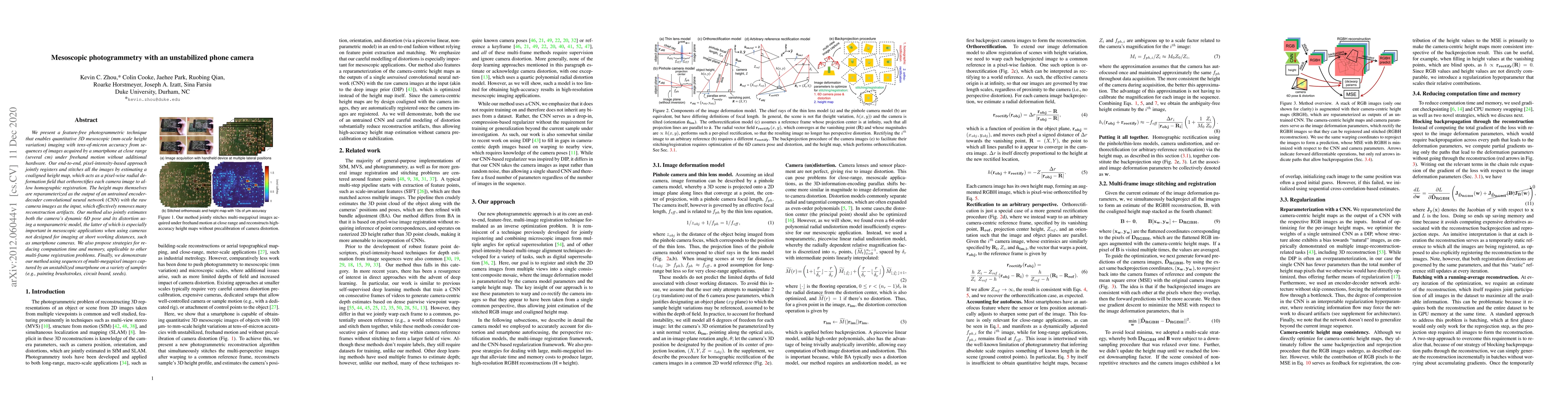
We present a feature-free photogrammetric technique that enables quantitative 3D mesoscopic (mm-scale height variation) imaging with tens-of-micron accuracy from sequences of images acquired by a sm...
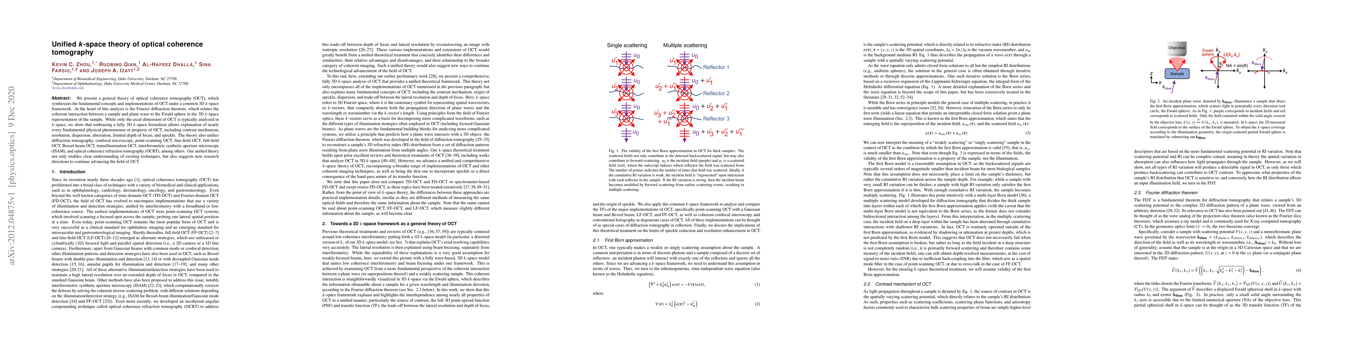
We present a general theory of optical coherence tomography (OCT), which synthesizes the fundamental concepts and implementations of OCT under a common 3D k-space framework. At the heart of this ana...
Recently, there has been growing interest and effort in developing high-speed high-precision 3D imaging technologies for a wide range of industrial, automotive and biomedical applications. Optical f...
This paper introduces a new method of data-driven microscope design for virtual fluorescence microscopy. Our results show that by including a model of illumination within the first layers of a deep ...
We present a tomographic imaging technique, termed Deep Prior Diffraction Tomography (DP-DT), to reconstruct the 3D refractive index (RI) of thick biological samples at high resolution from a sequen...
We present an approach of utilizing a multi-camera array system for capturing dynamic high-resolution videos of the human face, with improved imaging performance as compared to traditional single-came...
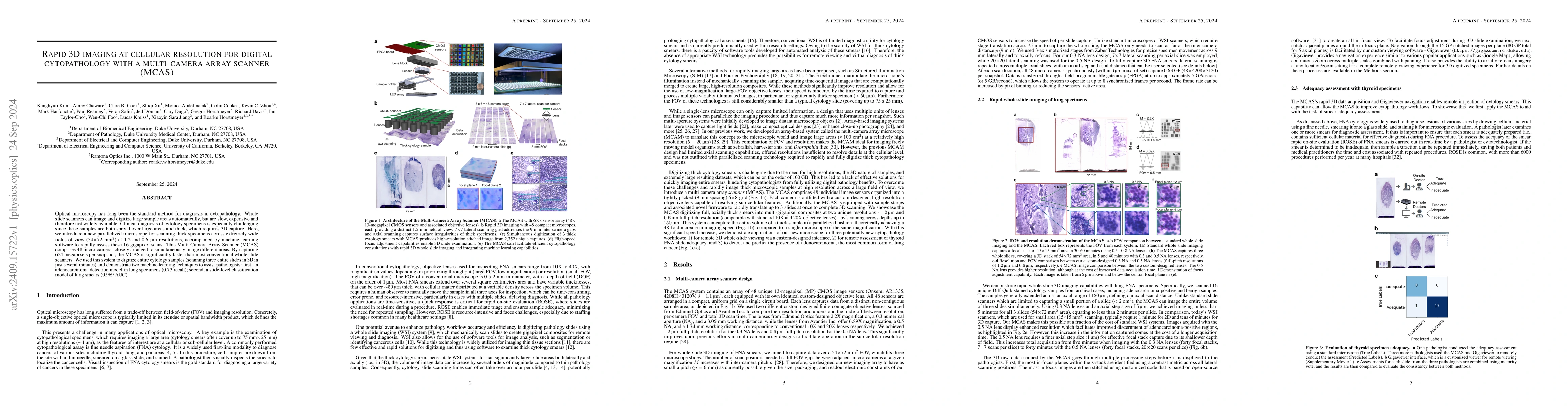
Optical microscopy has long been the standard method for diagnosis in cytopathology. Whole slide scanners can image and digitize large sample areas automatically, but are slow, expensive and therefore...
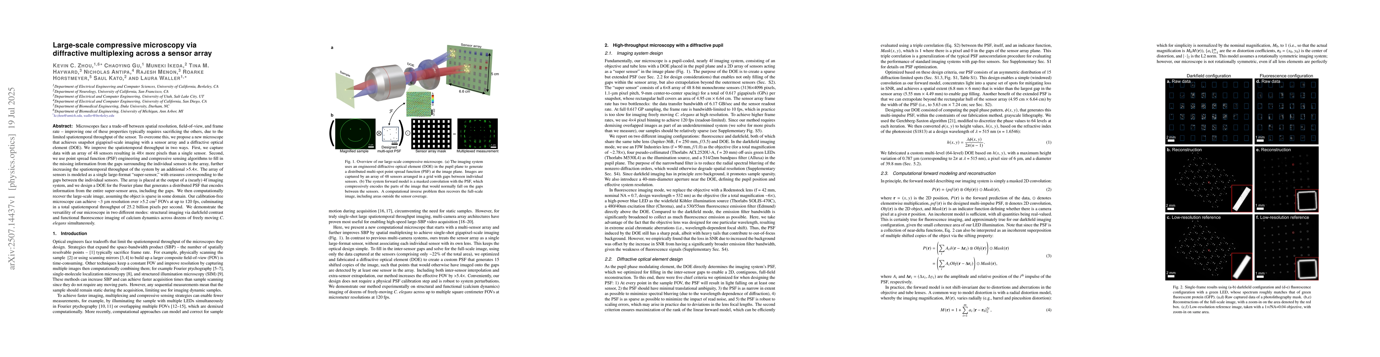
Microscopes face a trade-off between spatial resolution, field-of-view, and frame rate -- improving one of these properties typically requires sacrificing the others, due to the limited spatiotemporal...