ArxivLens

Quick Summary:

Tensorial Tomographic Differential Phase-Contrast microscopy (T2DPC) enables label-free, quantitative imaging of phase and anisotropy, reconstructing refractive index, birefringence, and orientation from intensity measurements using a standard microscope. The method demonstrates accurate volumetric reconstructions and predictive pathology in biological specimens.

arXiv Id: 2204.11397
Date Published: 2022-09-07
Date Updated: 2022-09-07

Categories:

Key Terms (TF-IDF):

microscopy (0.271)
microscopy (0.254)
quantitative (0.238)
polarization (0.224)
polarization (0.220)
phase (0.219)
quantitative (0.215)
imaging (0.213)
imaging (0.211)
phase (0.210)
samples (0.209)
contrast (0.202)
differential (0.197)
contrast (0.196)
samples (0.196)
differential (0.192)
refractive (0.179)
solves (0.165)
equipped (0.164)
reconstructed (0.163)
License: http://creativecommons.org/licenses/by/4.0/

Summary:

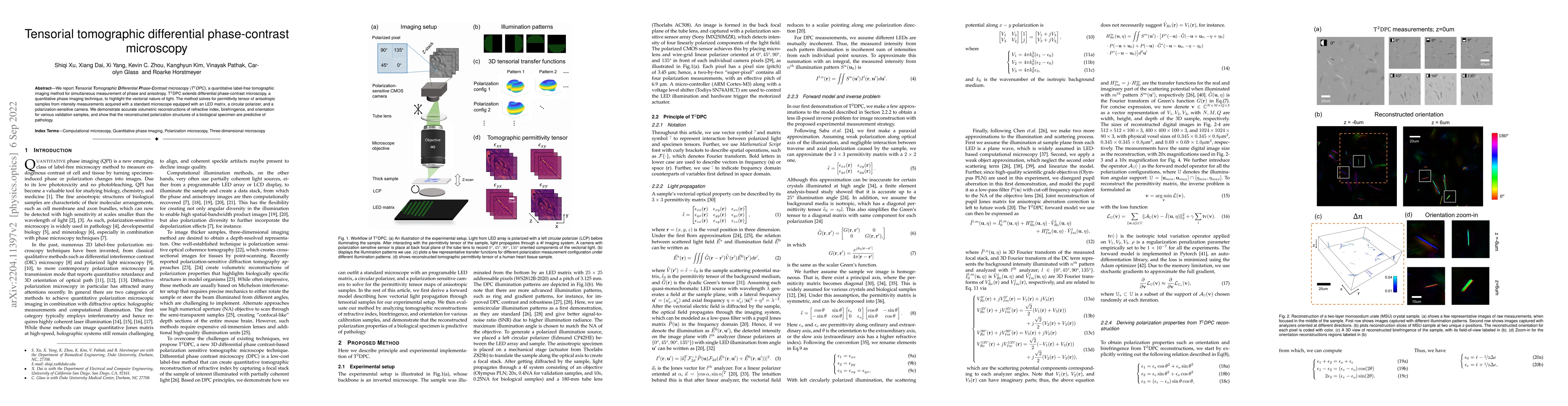
We report Tensorial Tomographic Differential Phase-Contrast microscopy (T2DPC), a quantitative label-free tomographic imaging method for simultaneous measurement of phase and anisotropy. T2DPC extends differential phase-contrast microscopy, a quantitative phase imaging technique, to highlight the vectorial nature of light. The method solves for permittivity tensor of anisotropic samples from intensity measurements acquired with a standard microscope equipped with an LED matrix, a circular polarizer, and a polarization-sensitive camera. We demonstrate accurate volumetric reconstructions of refractive index, birefringence, and orientation for various validation samples, and show that the reconstructed polarization structures of a biological specimen are predictive of pathology.

Citation & Reference Network Visualization

This graph shows how this paper is connected to other research. The current paper is shown in dark gray, papers that cite it are in green, and papers it references are in blue. You can:

Current Paper
Papers citing this work (Citations)
Papers cited by this work (References)

• Hover over any node to see the paper title

• Click a node to view that paper's details

• Drag nodes to rearrange the graph

• Use the zoom controls (bottom right) to zoom in/out

Citations and References

Title Authors Year Actions
Polarization-sensitive intensity diffraction tomography
Seungri Song, Jeongsoo Kim, T. Moon, Baekcheon Seong, Woojun Kim, Chang-Hyuk Yoo, Jun-Kyu Choi, C. Joo
2023
View Paper
Tensorial tomographic Fourier ptychography with applications to muscle tissue imaging
Shiqi Xu, Xiang Dai, P. Ritter, K. Lee, Xi Yang, L. Kreiss, Kevin C Zhou, Kanghyun Kim, A. Chaware, Jadee L. Neff, C. Glass, Seung Ah Lee, O. Friedrich, R. Horstmeyer
2023
View Paper
Multi-modal imaging using a cascaded microscope design
Xi Yang, Mark Harfouche, Kevin C Zhou, L. Kreiss, Shiqi Xu, Kanghyun Kim, R. Horstmeyer
2022
View Paper

Similar Papers

Signum phase mask differential microscopy

Sayan Ghosh, Nirmalya Ghosh, Niladri Modak, Jeeban Kumar Nayak

Published: 2023-10-11

Tensorial tomographic Fourier Ptychography with applications to muscle tissue imaging

Lucas Kreiss, Shiqi Xu, Kevin C. Zhou, Kyung Chul Lee, Kanghyun Kim, Amey Chaware, Seung Ah Lee, Oliver Friedrich, Roarke Horstmeyer, Xi Yang, Xiang Dai, Paul Ritter, Jadee Neff, Carolyn Glass

Published: 2024-03-11

Differential Dynamic Microscopy can be applied to Differential Interference Contrast images despite shadowing effects

Wolfgang Langbein, Yisu Wang, Paola Borri, Katerina Kaouri, Thomas E. Woolley, Timothy Ostler, Emily Lewis, Karl Swann

Published: 2022-12-26

Comments (0)

This website uses cookies to ensure you get the best experience. Learn more