ArxivLens

Quick Summary:

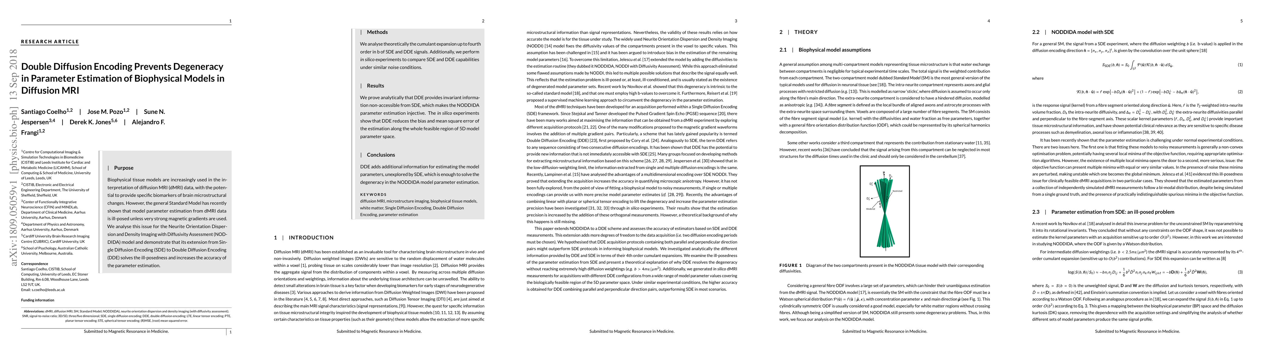
Double Diffusion Encoding (DDE) enhances the accuracy of parameter estimation in the Neurite Orientation Dispersion and Density Imaging with Diffusivity Assessment (NODDIDA) model for diffusion MRI data by providing additional invariant information. This method resolves the ill-posedness issue in Single Diffusion Encoding (SDE) and reduces estimation bias and error.

DOI: 10.1002/mrm.27714
arXiv Id: 1809.05059
Comments: 22 pages, 7 figures
Date Published: 2019-03-19
Date Updated: 2019-03-19

Key Terms (TF-IDF):

estimation (0.246)
estimation (0.238)
ill (0.227)
diffusion (0.210)
analyse (0.208)
diffusion (0.206)
encoding (0.206)
analyse (0.203)
encoding (0.200)
parameter (0.198)
parameter (0.197)
mri (0.144)
mean square (0.140)
tissue (0.139)
injective (0.137)
posed (0.136)
unless (0.136)
model (0.136)
degeneracy (0.135)
fourth (0.133)
License: http://arxiv.org/licenses/nonexclusive-distrib/1.0/

Summary:

Purpose: Biophysical tissue models are increasingly used in the interpretation of diffusion MRI (dMRI) data, with the potential to provide specific biomarkers of brain microstructural changes. However, the general Standard Model has recently shown that model parameter estimation from dMRI data is ill-posed unless very strong magnetic gradients are used. We analyse this issue for the Neurite Orientation Dispersion and Density Imaging with Diffusivity Assessment (NODDIDA) model and demonstrate that its extension from Single Diffusion Encoding (SDE) to Double Diffusion Encoding (DDE) solves the ill-posedness and increases the accuracy of the parameter estimation. Methods: We analyse theoretically the cumulant expansion up to fourth order in b of SDE and DDE signals. Additionally, we perform in silico experiments to compare SDE and DDE capabilities under similar noise conditions. Results: We prove analytically that DDE provides invariant information non-accessible from SDE, which makes the NODDIDA parameter estimation injective. The in silico experiments show that DDE reduces the bias and mean square error of the estimation along the whole feasible region of 5D model parameter space. Conclusions: DDE adds additional information for estimating the model parameters, unexplored by SDE, which is enough to solve the degeneracy in the NODDIDA model parameter estimation.

Citation & Reference Network Visualization

This graph shows how this paper is connected to other research. The current paper is shown in dark gray, papers that cite it are in green, and papers it references are in blue. You can:

Current Paper
Papers citing this work (Citations)
Papers cited by this work (References)

• Hover over any node to see the paper title

• Click a node to view that paper's details

• Drag nodes to rearrange the graph

• Use the zoom controls (bottom right) to zoom in/out

Citations and References

Title Authors Year Actions
Linear rotationally invariant kurtosis measures from double diffusion encoding MRI.
H. Moss, Thorsten Feiweier, Andreana Benitez, Jens H Jensen
2025
View Paper
3D Anatomical Structure-guided Deep Learning for Accurate Diffusion Microstructure Imaging
Xinrui Ma, Jian Cheng, Wen-Jie Fan, Ruo-Nan Wu, Yongquan Ye, Shanshan Wang
2025
View Paper
Characterization of neurite and soma organization in the brain and spinal cord with diffusion MRI
K. Schilling, Marco Palombo, Atlee Witt, K. O’Grady, Marco Pizzolato, Bennett A. Landman, Seth A. Smith
2025
View Paper
Isotropic sampling of tensor-encoded diffusion MRI.
S. Jespersen
2024
View Paper
Radiofrequency Enhancer to Recover Signal Dropouts in 7 Tesla Diffusion MRI
Varun Subramaniam, Andrew Frankini, Ameen Al Qadi, Mackenzie T. Herb, G. Verma, Bradley Delman, P. Balchandani, Akbar Alipour
2024
View Paper
The Diffusion Exchange Ratio (DEXR): A minimal sampling of diffusion exchange spectroscopy to probe exchange, restriction, and time-dependence
Teddy X. Cai, Nathan H. Williamson, R. Ravin, P. Basser
2024
View Paper
Assessment of precision and accuracy of brain white matter microstructure using combined diffusion MRI and relaxometry
S. Coelho, Ying Liao, Filip Szczepankiewicz, J. Veraart, Sohae Chung, Y. Lui, Dmitry S. Novikov, E. Fieremans
2024
View Paper
Assessment of Precision and Accuracy of Brain White Matter Microstructure using Combined Diffusion MRI and Relaxometry
S. Coelho, Ying Liao, Filip Szczepankiewicz, J. Veraart, Sohae Chung, Y. Lui, Dmitry S. Novikov, E. Fieremans
2024
View Paper
Insights and improvements in correspondence between axonal volume fraction measured with diffusion‐weighted MRI and electron microscopy
Sebastian Papazoglou, Mohammad Ashtarayeh, J. M. Oeschger, M. F. Callaghan, M. Does, S. Mohammadi
2023
View Paper
Comparison of different neurite density metrics with brain asymmetry evaluation.
I. Maximov, L. Westlye
2023
View Paper
Probing brain tissue microstructure with MRI: principles, challenges, and the role of multidimensional diffusion-relaxation encoding
Björn Lampinen, Filip Szczepankiewicz, J. Lätt, Linda Knutsson, J. Mårtensson, I. Björkman-Burtscher, D. Westen, P. Sundgren, F. Ståhlberg, M. Nilsson
2023
View Paper
Mapping tissue microstructure of brain white matter in vivo in health and disease using diffusion MRI
Yingtao Liao, S. Coelho, Jenny Chen, B. Ades-aron, Michelle M. Pang, Valentin Stepanov, R. Osorio, T. Shepherd, Y. Lui, D. Novikov, E. Fieremans
2023
View Paper
Measuring cardiomyocyte cellular characteristics in cardiac hypertrophy using diffusion‐weighted MRI
M. Farzi, Sam Coveney, M. Afzali, M. Zdora, C. Lygate, C. Rau, Alejandro F Frangi, E. Dall’Armellina, I. Teh, J. Schneider
2023
View Paper
Optimized rectification of fiber orientation density function with background threshold.
H. Moss, A. Benitez, J. Jensen
2022
View Paper
Axisymmetric diffusion kurtosis imaging with Rician bias correction: A simulation study
J. M. Oeschger, K. Tabelow, S. Mohammadi
2022
View Paper
Parameter estimation for WMTI‐Watson model of white matter using encoder–decoder recurrent neural network
Yujian Diao, I. Jelescu
2022
View Paper
Reproducibility of the Standard Model of diffusion in white matter on clinical MRI systems
S. Coelho, S. Baete, G. Lemberskiy, B. Ades-aron, Geneviève Barro, J. Veraart, D. Novikov, E. Fieremans
2022
View Paper
Multi-Contrast Magnetic Resonance Imaging of Visual White Matter Pathways in Patients With Glaucoma
S. Ogawa, H. Takemura, H. Horiguchi, A. Miyazaki, Kenji Matsumoto, Y. Masuda, K. Yoshikawa, T. Nakano
2022
View Paper
Multidimensional Diffusion MRI Methods With Confined Subdomains
Deneb Boito, Cem Yolcu, E. Özarslan
2021
View Paper
In vivo Correlation Tensor MRI reveals microscopic kurtosis in the human brain on a clinical 3T scanner
Lisa Novello, R. Henriques, A. Ianuş, T. Feiweier, N. Shemesh, J. Jovicich
2021
View Paper
Identifying microstructural changes in diffusion MRI; How to circumvent parameter degeneracy
Hossein Rafipoor, Ying-Qiu Zheng, L. Griffanti, S. Jbabdi, M. Cottaar
2021
View Paper
Diffusion time dependence, power-law scaling, and exchange in gray matter
J. Olesen, L. Østergaard, N. Shemesh, S. Jespersen
2021
View Paper
Neurite Exchange Imaging (NEXI): A minimal model of diffusion in gray matter with inter-compartment water exchange
Ileana O. Jelescu, Alexandre de Skowronski, F. Geffroy, M. Palombo, D. Novikov
2021
View Paper
On the potential for mapping apparent neural soma density via a clinically viable diffusion MRI protocol
Noemi G. Gyori, C. Clark, D. Alexander, Enrico Kaden
2021
View Paper
SPHERIOUSLY? The challenges of estimating sphere radius non-invasively in the human brain from diffusion MRI
M. Afzali, M. Nilsson, M. Palombo, Derek K. Jones
2021
View Paper
Efficient whole‐brain tract‐specific T1 mapping at 3T with slice‐shuffled inversion‐recovery diffusion‐weighted imaging
I. Leppert, Daniel A. Andrews, Jennifer S. W. Campbell, Daniel J. Park, G. Pike, J. Polimeni, Christine L. Tardif
2021
View Paper
Beyond the diffusion standard model in fixed rat spinal cord with combined linear and planar encoding
J. Olesen, L. Østergaard, N. Shemesh, S. Jespersen
2021
View Paper
Toward nonparametric diffusion‐T1 characterization of crossing fibers in the human brain
A. Reymbaut, J. Critchley, G. Durighel, T. Sprenger, M. Sughrue, K. Bryskhe, D. Topgaard
2020
View Paper
Toward Quantification: Microstructure and Magnetic Resonance Fingerprinting.
V. Kiselev, Gregor Körzdörfer, P. Gall
2020
View Paper
SPHERIOUSLY? The challenges of estimating spherical pore size non-invasively in the human brain from diffusion MRI
M. Afzali, M. Nilsson, M. Palombo, Derek K. Jones
2020
View Paper
Double diffusion encoding and applications for biomedical imaging
R. Henriques, M. Palombo, S. Jespersen, N. Shemesh, H. Lundell, A. Ianuş
2020
View Paper
The present and the future of microstructure MRI: From a paradigm shift to normal science
D. Novikov
2020
View Paper
A Signal Peak Separation Index for Axisymmetric B-Tensor Encoding
Gaëtan Rensonnet, Jonathan Rafael-Patino, B. Macq, J. Thiran, G. Girard, Marco Pizzolato Signal Processing Lab, 'Ecole polytechnique f'ed'erale de Lausanne, Lausanne, Switzerland., Icteam Institute, Universit'e catholique de Louvain, Louvain-la-Neuve, Belgium, Center for Computational Imaging, Radiology Department, C. Vaudois, University of Lausanne, D. O. Mathematics, Computer Science, T. Denmark, Kongens Lyngby, Denmark
2020
View Paper
The impact of edema and fiber crossing on diffusion MRI metrics assessed in an ex vivo nerve phantom: Multi‐tensor model vs. diffusion orientation distribution function
Zezhong Ye, Sam E. Gary, P. Sun, S. Mustafi, G. Glenn, F. Yeh, H. Merisaari, Chunyu Song, Ruimeng Yang, guo-sHu Huang, Hung-Wen Kao, C. Lin, Yu-chien Wu, J. Jensen, Sheng-Kwei Song
2020
View Paper
The sensitivity of diffusion MRI to microstructural properties and experimental factors
M. Afzali, Tomasz Pieciak, Sharlene D. Newman, E. Garyfallidis, E. Özarslan, Hu Cheng, Derek K. Jones
2020
View Paper
Axon diameter index estimation independent of fiber orientation distribution using high-gradient diffusion MRI
Q. Fan, A. Nummenmaa, T. Witzel, S. Huang
2020
View Paper
Challenges for biophysical modeling of microstructure
I. Jelescu, M. Palombo, F. Bagnato, K. Schilling
2020
View Paper
Towards in vivo g-ratio mapping using MRI: Unifying myelin and diffusion imaging
S. Mohammadi, M. Callaghan
2020
View Paper
A 4D Basis and Sampling Scheme for the Tensor Encoded Multi-Dimensional Diffusion MRI Signal
Alice P. Bates, Alessandro Daducci, P. Sadeghi, E. Caruyer
2020
View Paper
Magic DIAMOND: Multi-fascicle diffusion compartment imaging with tensor distribution modeling and tensor-valued diffusion encoding
A. Reymbaut, A. Caron, G. Gilbert, F. Szczepankiewicz, M. Nilsson, S. Warfield, M. Descoteaux, B. Scherrer
2020
View Paper
Insights into tissue microstructure using a double diffusion encoding sequence on a clinical scanner: Validation and application to experimental tumor models
Gaëtan Duchêne, J. Abarca‐Quinones, I. Leclercq, T. Duprez, F. Peeters
2020
View Paper
Towards unconstrained compartment modeling in white matter using diffusion‐relaxation MRI with tensor‐valued diffusion encoding
Björn Lampinen, Filip Szczepankiewicz, J. Mårtensson, D. van Westen, O. Hansson, C. Westin, M. Nilsson
2020
View Paper
Feasibility of axon density metrics for brain asymmetry evaluation in the UK Biobank subsample
I. Maximov, L. Westlye
2020
View Paper
Direction‐averaged diffusion‐weighted MRI signal using different axisymmetric B‐tensor encoding schemes
M. Afzali, S. Aja‐Fernández, Derek K. Jones
2020
View Paper
Accuracy and precision of statistical descriptors obtained from multidimensional diffusion signal inversion algorithms
A. Reymbaut, Paolo Mezzani, João P. de Almeida Martins, D. Topgaard
2020
View Paper
Diffusion model comparison identifies distinct tumor sub‐regions and tracks treatment response
D. McHugh, G. Lipowska-Bhalla, M. Babur, Y. Watson, I. Peset, H. Mistry, P. L. Hubbard Cristinacce, J. Naish, J. Honeychurch, K. Williams, J. O’Connor, G. Parker
2020
View Paper
The Dmipy Toolbox: Diffusion MRI Multi-Compartment Modeling and Microstructure Recovery Made Easy
Rutger Fick, D. Wassermann, R. Deriche
2019
View Paper
Generalized Richardson-Lucy (GRL) for analyzing multi-shell diffusion MRI data
Fenghua Guo, A. Leemans, M. Viergever, F. Dell’Acqua, A. Luca
2019
View Paper
Single‐ and double‐Diffusion encoding MRI for studying ex vivo apparent axon diameter distribution in spinal cord white matter
D. Anaby, D. Morozov, Inbar Seroussi, S. Hametner, N. Sochen, Y. Cohen
2019
View Paper
Traumatic and nontraumatic spinal cord injury: pathological insights from neuroimaging
Gergely Dávid, S. Mohammadi, A. Martin, J. Cohen-Adad, N. Weiskopf, Alan J. Thompson, P. Freund
2019
View Paper
Advanced encoding methods in diffusion MRI
A. Reymbaut, M. Descoteaux
2019
View Paper
Direction-Averaged Diffusion-Weighted MRI Signal using different Axisymmetric B-tensor Encoding Schemes “Submitted to Magnetic Resonance in Medicine”
M. Afzali, S. Aja‐Fernández, Derek K. Jones
2019
View Paper
Optimal Experimental Design for Biophysical Modelling in Multidimensional Diffusion MRI
S. Coelho, J. Pozo, S. Jespersen, Alejandro F Frangi
2019
View Paper
SANDI: A compartment-based model for non-invasive apparent soma and neurite imaging by diffusion MRI
M. Palombo, A. Ianuş, Michele Guerreri, Daniel Nunes, D. Alexander, N. Shemesh, Hui Zhang
2019
View Paper
A data-driven approach to optimising the encoding for multi-shell diffusion MRI with application to neonatal imaging
J. Tournier, D. Christiaens, J. Hutter, A. Price, L. Cordero-Grande, E. Hughes, M. Bastiani, M. Bastiani, S. Sotiropoulos, S. Sotiropoulos, Stephen M. Smith, D. Rueckert, S. Counsell, A. Edwards, J. Hajnal
2019
View Paper
Improved fibre dispersion estimation using b-tensor encoding
M. Cottaar, Filip Szczepankiewicz, M. Bastiani, Moisés Hernández-Fernández, S. Sotiropoulos, M. Nilsson, S. Jbabdi
2019
View Paper
Diffusion Kurtosis Imaging maps neural damage in the EAE model of multiple sclerosis
A. Chuhutin, B. Hansen, A. Wlodarczyk, T. Owens, N. Shemesh, S. Jespersen
2018
View Paper
A unique analytical solution of the white matter standard model using linear and planar encodings
M. Reisert, V. Kiselev, B. Dhital
2018
View Paper

Comments (0)

This website uses cookies to ensure you get the best experience. Learn more