ArxivLens

Quick Summary:

This study evaluates the accuracy of microscopic fractional anisotropy (μFA) estimation using spherical mean technique (SMT) in single diffusion encoding MRI, comparing it to double diffusion encoding MRI. The results show significant deviations in μFA estimates from ground truth, indicating that SMT and standard model assumptions fail to accurately capture microstructural details due to unconsidered factors like kurtosis.

Journal Reference: Magn Reson Med. 2019 May;81(5):3245-3261
DOI: 10.1002/mrm.27606
arXiv Id: 1903.08455
Date Published: 2019-03-21
Date Updated: 2019-03-21

Categories:

Key Terms (TF-IDF):

powder (0.295)
averaged (0.267)
mu (0.266)
averaged (0.250)
sm (0.246)
mu (0.242)
sm (0.222)
signals (0.201)
delta (0.196)
ms (0.189)
mri (0.189)
signals (0.184)
delta (0.179)
diffusion (0.177)
ms (0.176)
encoding (0.173)
diffusion (0.160)
encoding (0.155)
constraints (0.153)
constraints (0.142)
License: http://arxiv.org/licenses/nonexclusive-distrib/1.0/

Summary:

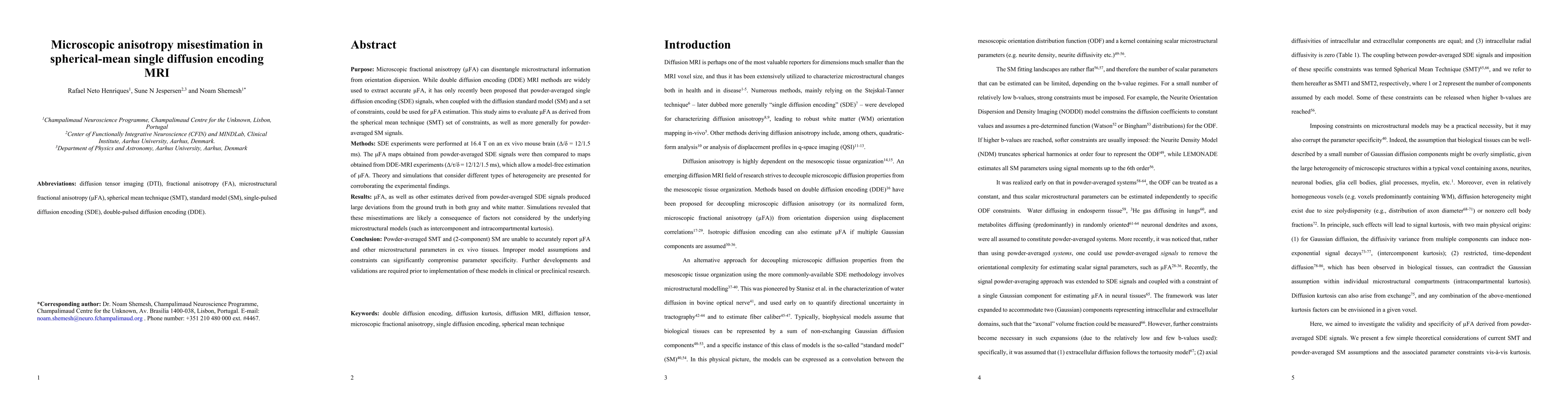
Purpose: Microscopic fractional anisotropy ({\mu}FA) can disentangle microstructural information from orientation dispersion. While double diffusion encoding (DDE) MRI methods are widely used to extract accurate {\mu}FA, it has only recently been proposed that powder-averaged single diffusion encoding (SDE) signals, when coupled with the diffusion standard model (SM) and a set of constraints, could be used for {\mu}FA estimation. This study aims to evaluate {\mu}FA as derived from the spherical mean technique (SMT) set of constraints, as well as more generally for powder-averaged SM signals. Methods: SDE experiments were performed at 16.4 T on an ex vivo mouse brain ({\Delta}/{\delta} = 12/1.5 ms). The {\mu}FA maps obtained from powder-averaged SDE signals were then compared to maps obtained from DDE-MRI experiments ({\Delta}/{\tau}/{\delta} = 12/12/1.5 ms), which allow a model-free estimation of {\mu}FA. Theory and simulations that consider different types of heterogeneity are presented for corroborating the experimental findings. Results: {\mu}FA, as well as other estimates derived from powder-averaged SDE signals produced large deviations from the ground truth in both gray and white matter. Simulations revealed that these misestimations are likely a consequence of factors not considered by the underlying microstructural models (such as intercomponent and intracompartmental kurtosis). Conclusion: Powder-averaged SMT and (2-component) SM are unable to accurately report {\mu}FA and other microstructural parameters in ex vivo tissues. Improper model assumptions and constraints can significantly compromise parameter specificity. Further developments and validations are required prior to implementation of these models in clinical or preclinical research.

Citation & Reference Network Visualization

This graph shows how this paper is connected to other research. The current paper is shown in dark gray, papers that cite it are in green, and papers it references are in blue. You can:

Current Paper
Papers citing this work (Citations)
Papers cited by this work (References)

• Hover over any node to see the paper title

• Click a node to view that paper's details

• Drag nodes to rearrange the graph

• Use the zoom controls (bottom right) to zoom in/out

Citations and References

Title Authors Year Actions
IDH Status in Brain Gliomas Can Be Predicted by the Spherical Mean MRI Technique.
V. Sedlák, Milan Němý, M. Májovský, Adéla Bubeníková, L. Nordin, Tomáš Moravec, Jana Engelová, Dalibor Sila, D. Konečná, Tomáš Belšan, E. Westman, D. Netuka
2024
View Paper
MRI Parameter Mapping via Gaussian Mixture VAE: Breaking the Assumption of Independent Pixels
Moucheng Xu, Yukun Zhou, Tobias Goodwin-Allcock, Kimia Firoozabadi, Joseph Jacob, Daniel C. Alexander, Paddy J. Slator
2024
View Paper
Human gray matter microstructure mapped using Neurite Exchange Imaging (NEXI) on a clinical scanner
Quentin Uhl, Tommaso Pavan, Thorsten Feiweier, G. Piredda, I. Jelescu
2024
View Paper
Tutorial: a guide to diffusion MRI and structural connectomics.
I. Shamir, Yaniv Assaf
2024
View Paper
Comparison of different neurite density metrics with brain asymmetry evaluation.
I. Maximov, L. Westlye
2023
View Paper
Quantifying human gray matter microstructure using neurite exchange imaging (NEXI) and 300 mT/m gradients
Quentin Uhl, Tommaso Pavan, Malwina Molendowska, Derek K. Jones, M. Palombo, Ileana Jelescu Department of Radiology, Lausanne University Hospital, Lausanne, Switzerland., School of Biology, Medicine, University of Lausanne, Cardiff University Brain Research Imaging Centre, C. University, Cardiff, United Kingdom., School of Materials Science, Informatics, Uk, joint last authorship
2023
View Paper
Mapping tissue microstructure across the human brain on a clinical scanner with soma and neurite density image metrics
S. Schiavi, M. Palombo, D. Zacà, F. Tazza, C. Lapucci, L. Castellan, M. Costagli, M. Inglese
2023
View Paper
Unique information from common diffusion MRI models about white-matter differences across the human adult lifespan
R. N. Henriques, R. Henson, M. Correia
2023
View Paper
Multi-tensor diffusion abnormalities of gray matter in an animal model of cortical dysplasia
Paulina J. Villasenõr, David Cortés-Servín, Aylín Pérez-Moriel, A. Aquiles, H. Luna-Munguía, Alonso Ramírez-Manzanares, Ricardo Coronado-Leija, J. Larriva-Sahd, L. Concha
2023
View Paper
Cardiac q‐space trajectory imaging by motion‐compensated tensor‐valued diffusion encoding in human heart in vivo
I. Teh, D. Shelley, Jordan H Boyle, Feng-lei Zhou, A. Poenar, N. Sharrack, R. Foster, N. Yuldasheva, G. Parker, E. Dall’Armellina, S. Plein, J. Schneider, Filip Szczepankiewicz
2023
View Paper
Extra-axonal contribution to double diffusion encoding-based pore size estimates in the corticospinal tract
Patricia Ulloa, Vincent Méthot, Viktor Wottschel, M. Koch
2023
View Paper
Spherical convolutional neural networks can improve brain microstructure estimation from diffusion MRI data
Leevi Kerkelä, K. Seunarine, Filip Szczepankiewicz, C. Clark
2022
View Paper
Comparison of two types of microscopic diffusion anisotropy in mouse brain
J. Jensen, Joshua Voltin, Xingju Nie, Siddhartha Dhiman, Emile T McKinnon, M. F. Falangola
2022
View Paper
Differentiating False Positive Lesions from Clinically Significant Cancer and Normal Prostate Tissue Using VERDICT MRI and Other Diffusion Models
S. Sen, V. Valindria, Paddy J. Slator, H. Pye, A. Grey, A. Freeman, C. Moore, H. Whitaker, S. Punwani, Saurabh Singh, Eleftheria Panagiotaki
2022
View Paper
Diffusion Kurtosis Imaging of Neonatal Spinal Cord in Clinical Routine
R. Trò, M. Roascio, D. Tortora, M. Severino, A. Rossi, J. Cohen-Adad, M. Fato, G. Arnulfo
2022
View Paper
Measurement of Full Diffusion Tensor Distribution Using High-Gradient Diffusion MRI and Applications in Diffuse Gliomas
Yi-Qiao Song, Ina Ly, Q. Fan, A. Nummenmaa, M. Martinez-Lage, W. Curry, J. Dietrich, D. Forst, B. Rosen, S. Huang, E. Gerstner
2022
View Paper
Diffusion MRI signal cumulants and hepatocyte microstructure at fixed diffusion time: Insights from simulations, 9.4T imaging, and histology
Francesco Grussu, K. Bernatowicz, I. Casanova-Salas, Natalia Castro, P. Nuciforo, J. Mateo, I. Barba, R. Pérez-López
2022
View Paper
Quantification of Tissue Microstructure Using Tensor-Valued Diffusion Encoding: Brain and Body
M. Afzali, Lars Mueller, Filip Szczepankiewicz, Derek K. Jones, J. Schneider
2022
View Paper
Resolution and b value dependent structural connectome in ex vivo mouse brain
Stephanie Crater, Surendra Maharjan, Y. Qi, Qi Zhao, G. Cofer, James J. Cook, G., Allan Johnson, Nian Wang
2022
View Paper
In vivo Correlation Tensor MRI reveals microscopic kurtosis in the human brain on a clinical 3T scanner
Lisa Novello, R. Henriques, A. Ianuş, T. Feiweier, N. Shemesh, J. Jovicich
2021
View Paper
Diffusion time dependence, power-law scaling, and exchange in gray matter
J. Olesen, L. Østergaard, N. Shemesh, S. Jespersen
2021
View Paper
Soma and Neurite Density MRI (SANDI) of the in-vivo mouse brain and comparison with the Allen Brain Atlas
A. Ianuş, J. Carvalho, Francisca F. Fernandes, Renata Cruz, C. Chavarrías, M. Palombo, N. Shemesh
2021
View Paper
Comparative analysis of signal models for microscopic fractional anisotropy estimation using q-space trajectory encoding
Leevi Kerkelä, F. Nery, Ross Callaghan, F. Zhou, Noemi G. Gyori, Filip Szczepankiewicz, M. Palombo, G. Parker, Hui Zhang, M. Hall, C. Clark
2021
View Paper
Test-retest reproducibility of in vivo oscillating gradient and microscopic anisotropy diffusion MRI in mice at 9.4 Tesla
Naila Rahman, Kathy Xu, M. Omer, M. Budde, Arthur Brown, C. Baron
2021
View Paper
Connectome 2.0: Developing the next-generation ultra-high gradient strength human MRI scanner for bridging studies of the micro-, meso- and macro-connectome
S. Huang, T. Witzel, B. Keil, Alina Scholz, Mathias Davids, P. Dietz, E. Rummert, Rebecca Ramb, J. Kirsch, A. Yendiki, Q. Fan, Q. Tian, Gabriel Ramos-Llordén, Hong-Hsi Lee, A. Nummenmaa, B. Bilgiç, K. Setsompop, Fuyixue Wang, A. Avram, M. Komlosh, D. Benjamini, K. N. Magdoom, S. Pathak, W. Schneider, D. Novikov, E. Fieremans, Slimane Tounekti, C. Mekkaoui, J. Augustinack, Daniel Berger, Alexander Shapson-Coe, Jeffrey M. Lichtman, P. Basser, L. Wald, B. Rosen
2021
View Paper
Diffusion-Weighted Imaging: Recent Advances and Applications.
E. Martínez-Heras, Francesco Grussu, F. Prados, E. Solana, S. Llufriu
2021
View Paper
Comparison of Neurite Orientation Dispersion and Density Imaging and Two-Compartment Spherical Mean Technique Parameter Maps in Multiple Sclerosis
Daniel Johnson, Antonio Ricciardi, W. Brownlee, B. Kanber, F. Prados, S. Collorone, Enrico Kaden, A. Toosy, D. Alexander, C. G. Gandini Wheeler-Kingshott, O. Ciccarelli, Francesco Grussu
2021
View Paper
SPHERIOUSLY? The challenges of estimating sphere radius non-invasively in the human brain from diffusion MRI
M. Afzali, M. Nilsson, M. Palombo, Derek K. Jones
2021
View Paper
Rapid microscopic fractional anisotropy imaging via an optimized linear regression formulation.
N. Arezza, D. Tse, C. Baron
2021
View Paper
Does powder averaging remove dispersion bias in diffusion MRI diameter estimates within real 3D axonal architectures?
M. Andersson, M. Pizzolato, H. Kjer, Katrine Forum Skodborg, H. Lundell, T. Dyrby
2021
View Paper
Toward more robust and reproducible diffusion kurtosis imaging
R. Henriques, S. Jespersen, Derek K. Jones, J. Veraart
2021
View Paper
Diffusional Kurtosis Imaging in the Diffusion Imaging in Python Project
R. Neto Henriques, Marta Correa, Maurizio Maralle, Elizabeth Huber, J. Kruper, S. Koudoro, J. Yeatman, E. Garyfallidis, Ariel S. Rokem
2021
View Paper
Evidence for microscopic kurtosis in neural tissue revealed by correlation tensor MRI
R. Henriques, S. Jespersen, N. Shemesh
2021
View Paper
Beyond the diffusion standard model in fixed rat spinal cord with combined linear and planar encoding
J. Olesen, L. Østergaard, N. Shemesh, S. Jespersen
2021
View Paper
Probing tissue microstructure by diffusion skewness tensor imaging
L. Ning, Filip Szczepankiewicz, M. Nilsson, Y. Rathi, C. Westin
2021
View Paper
Compartmental diffusion and microstructural properties of human brain gray and white matter studied with double diffusion encoding magnetic resonance spectroscopy of metabolites and water
H. Lundell, C. Najac, Marjolein Bulk, H. Kan, A. Webb, I. Ronen
2020
View Paper
Rapid Microscopic Fractional Anisotropy Imaging via an Optimized Kurtosis Formulation
N. Arezza, D. H. Y. Tse, C. A. Baron
2020
View Paper
SPHERIOUSLY? The challenges of estimating spherical pore size non-invasively in the human brain from diffusion MRI
M. Afzali, M. Nilsson, M. Palombo, Derek K. Jones
2020
View Paper
Double diffusion encoding and applications for biomedical imaging
R. Henriques, M. Palombo, S. Jespersen, N. Shemesh, H. Lundell, A. Ianuş
2020
View Paper
Brain White-Matter Degeneration Due to Aging and Parkinson Disease as Revealed by Double Diffusion Encoding
K. Kamiya, K. Kamagata, K. Ogaki, T. Hatano, T. Ogawa, H. Takeshige-Amano, Syo Murata, C. Andica, K. Murata, T. Feiweier, M. Hori, N. Hattori, S. Aoki
2020
View Paper
Gradient waveform design for tensor-valued encoding in diffusion MRI
Filip Szczepankiewicz, C. Westin, M. Nilsson
2020
View Paper
Tensor-valued diffusion MRI differentiates cortex and white matter in malformations of cortical development associated with epilepsy
Björn Lampinen, Ariadne Zampeli, I. Björkman-Burtscher, Filip Szczepankiewicz, K. Källén, Maria Compagno Strandberg, M. Nilsson
2020
View Paper
Free‐water DTI estimates from single b‐value data might seem plausible but must be interpreted with care
Marc Golub, R. Neto Henriques, Rita Gouveia Nunes
2020
View Paper
Disentangling white-matter damage from physiological fibre orientation dispersion in multiple sclerosis
Kasper Winther Andersen, S. Lasič, H. Lundell, M. Nilsson, D. Topgaard, F. Sellebjerg, Filip Szczepankiewicz, H. Siebner, M. Blinkenberg, T. Dyrby
2020
View Paper
Towards in vivo g-ratio mapping using MRI: Unifying myelin and diffusion imaging
S. Mohammadi, M. Callaghan
2020
View Paper
Cytosolic diffusivity and microscopic anisotropy of N‐acetyl aspartate in human white matter with diffusion‐weighted MRS at 7 T
H. Lundell, C. Ingo, T. Dyrby, I. Ronen
2020
View Paper
Feasibility of axon density metrics for brain asymmetry evaluation in the UK Biobank subsample
I. Maximov, L. Westlye
2020
View Paper
Accuracy and precision of statistical descriptors obtained from multidimensional diffusion signal inversion algorithms
A. Reymbaut, Paolo Mezzani, João P. de Almeida Martins, D. Topgaard
2020
View Paper
In vivo diffusion‐weighted MRS using semi‐LASER in the human brain at 3 T: Methodological aspects and clinical feasibility
Guglielmo Genovese, M. Marjańska, E. Auerbach, Lydia Yahia Cherif, I. Ronen, S. Lehéricy, F. Branzoli
2020
View Paper
Diffusion dispersion imaging: Mapping oscillating gradient spin‐echo frequency dependence in the human brain
A. Arbabi, J. Kai, Ali R. Khan, C. Baron
2019
View Paper
Evaluation of white matter microstructure in patients with Parkinson’s disease using microscopic fractional anisotropy
Yutaka Ikenouchi, K. Kamagata, C. Andica, T. Hatano, T. Ogawa, H. Takeshige-Amano, K. Kamiya, A. Wada, Michimasa Suzuki, S. Fujita, A. Hagiwara, Ryusuke Irie, M. Hori, G. Oyama, Y. Shimo, A. Umemura, N. Hattori, S. Aoki
2019
View Paper
Validation and noise robustness assessment of microscopic anisotropy estimation with clinically feasible double diffusion encoding MRI
Leevi Kerkelä, R. Henriques, M. Hall, C. Clark, N. Shemesh
2019
View Paper
Diffusion MRI simulation of realistic neurons with SpinDoctor and the Neuron Module
Chengran Fang, Van-Dang Nguyen, D. Wassermann, Jing-Rebecca Li
2019
View Paper
Advanced encoding methods in diffusion MRI
A. Reymbaut, M. Descoteaux
2019
View Paper
Portable simulation framework for diffusion MRI
Van-Dang Nguyen, M. Leoni, Tamara Dancheva, J. Jansson, J. Hoffman, D. Wassermann, Jing-Rebecca Li
2019
View Paper
Correlation tensor magnetic resonance imaging
R. Henriques, S. Jespersen, N. Shemesh
2019
View Paper
SANDI: A compartment-based model for non-invasive apparent soma and neurite imaging by diffusion MRI
M. Palombo, A. Ianuş, Michele Guerreri, Daniel Nunes, D. Alexander, N. Shemesh, Hui Zhang
2019
View Paper
Diffusion Dispersion Imaging: Mapping OGSE Frequency Dependence in the Human Brain
A. Arbabi, J. Kai, Ali R. Khan, C. Baron
2019
View Paper
Tensor‐valued diffusion MRI in under 3 minutes: an initial survey of microscopic anisotropy and tissue heterogeneity in intracranial tumors
M. Nilsson, Filip Szczepankiewicz, J. Brabec, Marie Taylor, C. Westin, A. Golby, D. van Westen, P. Sundgren
2019
View Paper
SpinDoctor: A MATLAB toolbox for diffusion MRI simulation
Jing-Rebecca Li, Van-Dang Nguyen, Try Nguyen Tran, J. Valdman, Cong-Bang Trang, Khieu-Van Nguyen, Duc Thach Son Vu, H. Tran, H. Tran, Thi Minh Phuong Nguyen
2019
View Paper
Tensor-valued diffusion encoding for diffusional variance decomposition (DIVIDE): Technical feasibility in clinical MRI systems
Filip Szczepankiewicz, Jens Sjölund, F. Ståhlberg, J. Lätt, M. Nilsson
2016
View Paper
Diffusional exchange versus microscopic kurtosis from CTI: Two conflicting interpretations of the same data
Arthur Chakwizira, Filip Szczepankiewicz, Markus Nilsson
2023
View Paper
Microstructural neuroimaging using spherical convolutional neural networks
Leevi Kerkelä, K. Seunarine, Filip Szczepankiewicz, C. Clark
2022
View Paper
Enhancing Diffusion Signal Augmentation Using Spherical Convolutions
Simon Koppers, D. Merhof
2021
View Paper
Measuring Microstructural Features Using Diffusion MRI
N. Shemesh
2020
View Paper

Comments (0)

This website uses cookies to ensure you get the best experience. Learn more