This study validates a simplified double diffusion encoding MRI protocol against the standard DDE 5-design, demonstrating comparable accuracy in estimating microscopic fractional anisotropy. It also highlights the method's noise robustness, revealing precision challenges in quantifying microscopic anisotropy under typical clinical signal-to-noise ratios.
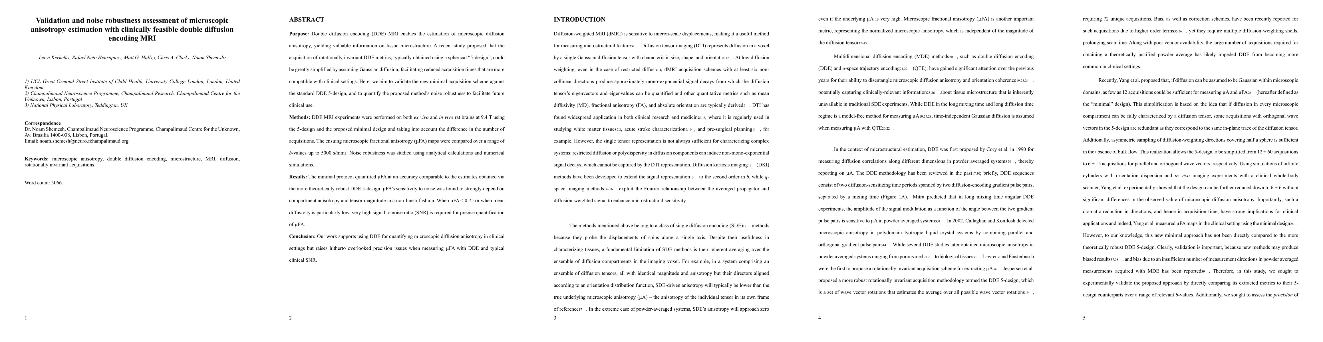
Purpose: Double diffusion encoding (DDE) MRI enables the estimation of microscopic diffusion anisotropy, yielding valuable information on tissue microstructure. A recent study proposed that the acquisition of rotationally invariant DDE metrics, typically obtained using a spherical "5-design", could be greatly simplified by assuming Gaussian diffusion, facilitating reduced acquisition times that are more compatible with clinical settings. Here, we aim to validate the new minimal acquisition scheme against the standard DDE 5-design, and to quantify the proposed method's noise robustness to facilitate future clinical use. Methods: DDE MRI experiments were performed on both ex vivo and in vivo rat brains at 9.4 T using the 5-design and the proposed minimal design and taking into account the difference in the number of acquisitions. The ensuing microscopic fractional anisotropy ({\mu}FA) maps were compared over a range of b-values up to 5000 s/mm2. Noise robustness was studied using analytical calculations and numerical simulations. Results: The minimal protocol quantified {\mu}FA at an accuracy comparable to the estimates obtained via the more theoretically robust DDE 5-design. {\mu}FA's sensitivity to noise was found to strongly depend on compartment anisotropy and tensor magnitude in a non-linear fashion. When {\mu}FA < 0.75 or when mean diffusivity is particularly low, very high signal to noise ratio (SNR) is required for precise quantification of {\mu}FA. Conclusion: Our work supports using DDE for quantifying microscopic diffusion anisotropy in clinical settings but raises hitherto overlooked precision issues when measuring {\mu}FA with DDE and typical clinical SNR.
Get AI-generated insights about this paper's methodology, results, and significance.
Current paper (gray), citations (green), references (blue)
Qi Liu, Jian Xu, Terrence Chen et al.
Title | Authors | Year | Actions |
---|---|---|---|
Self-supervised learning for denoising of multidimensional MRI data.
|
Beomgu Kang, Wonil Lee, Hyunseok Seo, H. Heo, HyunWook Park
|
2024
|
View Paper |
Unique information from common diffusion MRI models about white-matter differences across the human adult lifespan
|
R. N. Henriques, R. Henson, M. Correia
|
2023
|
View Paper |
Comparison of two types of microscopic diffusion anisotropy in mouse brain
|
J. Jensen, Joshua Voltin, Xingju Nie, Siddhartha Dhiman, Emile T McKinnon, M. F. Falangola
|
2022
|
View Paper |
In vivo Correlation Tensor MRI reveals microscopic kurtosis in the human brain on a clinical 3T scanner
|
Lisa Novello, R. Henriques, A. Ianuş, T. Feiweier, N. Shemesh, J. Jovicich
|
2021
|
View Paper |
Comparative analysis of signal models for microscopic fractional anisotropy estimation using q-space trajectory encoding
|
Leevi Kerkelä, F. Nery, Ross Callaghan, F. Zhou, Noemi G. Gyori, Filip Szczepankiewicz, M. Palombo, G. Parker, Hui Zhang, M. Hall, C. Clark
|
2021
|
View Paper |
Rapid microscopic fractional anisotropy imaging via an optimized linear regression formulation.
|
N. Arezza, D. Tse, C. Baron
|
2021
|
View Paper |
In vivo tensor-valued diffusion MRI of focal demyelination in white and deep grey matter of rodents
|
Yi He, S. Aznar, H. Siebner, T. Dyrby
|
2021
|
View Paper |
Evidence for microscopic kurtosis in neural tissue revealed by correlation tensor MRI
|
R. Henriques, S. Jespersen, N. Shemesh
|
2021
|
View Paper |
Unraveling micro-architectural modulations in neural tissue upon ischemia by Correlation Tensor MRI
|
Rita Alves, R. Henriques, Leevi Kerkelä, C. Chavarrías, S. Jespersen, N. Shemesh
|
2021
|
View Paper |
Compartmental diffusion and microstructural properties of human brain gray and white matter studied with double diffusion encoding magnetic resonance spectroscopy of metabolites and water
|
H. Lundell, C. Najac, Marjolein Bulk, H. Kan, A. Webb, I. Ronen
|
2020
|
View Paper |
Double diffusion encoding and applications for biomedical imaging
|
R. Henriques, M. Palombo, S. Jespersen, N. Shemesh, H. Lundell, A. Ianuş
|
2020
|
View Paper |
Brain White-Matter Degeneration Due to Aging and Parkinson Disease as Revealed by Double Diffusion Encoding
|
K. Kamiya, K. Kamagata, K. Ogaki, T. Hatano, T. Ogawa, H. Takeshige-Amano, Syo Murata, C. Andica, K. Murata, T. Feiweier, M. Hori, N. Hattori, S. Aoki
|
2020
|
View Paper |
Challenges for biophysical modeling of microstructure
|
I. Jelescu, M. Palombo, F. Bagnato, K. Schilling
|
2020
|
View Paper |
Correlation tensor magnetic resonance imaging
|
R. Henriques, S. Jespersen, N. Shemesh
|
2019
|
View Paper |
Improving stroke detection with Correlation Tensor MRI
|
Rita Alves, R. Henriques, Leevi Kerkelä, C. Chavarrías, N. Suñé, Jespersen, N. Shemesh
|
2021
|
View Paper |
Comments (0)