ArxivLens

Quick Summary:

This paper presents a novel graphene-based wet cell enabling high-resolution SEM imaging and EDX analysis of Au-nanoparticles in liquid environments, achieving < 5 nm resolution and surpassing conventional silicon nitride devices. The graphene membrane supports SEM requirements while maintaining imaging clarity.

Journal Reference: 2015 IOP Nanotechnology 26 315703
DOI: 10.1088/0957-4484/26/31/315703
arXiv Id: 1506.03420
Comments: 17 Pages, 6 Figures, * Authors contributed equally
Date Published: 2015-07-20
Date Updated: 2015-07-20

Key Terms (TF-IDF):

liquid (0.244)
graphene (0.242)
high resolution (0.216)
resolution (0.216)
nanoscale (0.214)
requirements (0.176)
electron (0.176)
inside (0.175)
cell (0.172)
vacuum (0.169)
imaging (0.161)
high (0.156)
membrane (0.137)
dispersive (0.135)
gold (0.132)
nanoparticles (0.131)
brownian motion (0.125)
scanning (0.122)
silicon (0.118)
biological (0.116)
License: http://arxiv.org/licenses/nonexclusive-distrib/1.0/

Summary:

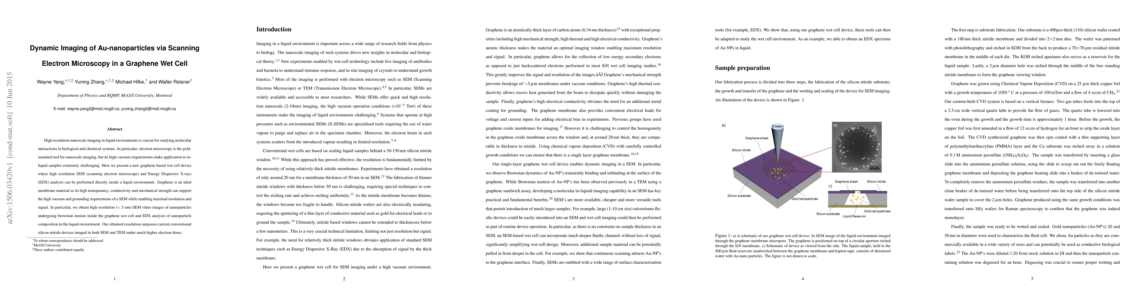
High resolution nanoscale imaging in liquid environments is crucial for studying molecular interactions in biological and chemical systems. In particular, electron microscopy is the gold-standard tool for nanoscale imaging, but its high-vacuum requirements make application to in-liquid samples extremely challenging. Here we present a new graphene based wet cell device where high resolution SEM (scanning electron microscope) and Energy Dispersive X-rays (EDX) analysis can be performed directly inside a liquid environment. Graphene is an ideal membrane material as its high transparancy, conductivity and mechanical strength can support the high vacuum and grounding requirements of a SEM while enabling maximal resolution and signal. In particular, we obtain high resolution (< 5 nm) SEM video images of nanoparticles undergoing brownian motion inside the graphene wet cell and EDX analysis of nanoparticle composition in the liquid enviornment. Our obtained resolution surpasses current conventional silicon nitride devices imaged in both SEM and TEM under much higher electron doses.

Citation & Reference Network Visualization

This graph shows how this paper is connected to other research. The current paper is shown in dark gray, papers that cite it are in green, and papers it references are in blue. You can:

Current Paper
Papers citing this work (Citations)
Papers cited by this work (References)

• Hover over any node to see the paper title

• Click a node to view that paper's details

• Drag nodes to rearrange the graph

• Use the zoom controls (bottom right) to zoom in/out

Citations and References

Title Authors Year Actions
Recent Notable Approaches to Study Self‐Assembly of Nanoparticles with X‐Ray Scattering and Electron Microscopy
F. Schulz, I. Lokteva, W. Parak, F. Lehmkühler
2021
View Paper
In situ liquid SEM imaging analysis revealing particle dispersity in aqueous solutions
Jennifer Yao, Angela Ossana Jaehun Chun, Xiao‐Ying Yu
2020
View Paper
Improving in situ liquid SEM imaging of particles
Xiao‐Ying Yu, B. Arey, S. Chatterjee, J. Chun
2019
View Paper
Study of Nanoparticle Size Limitation in Aqueous Environment by SEM
C. L. Chiang, C. W. Yeh, S. C. Fan
2018
View Paper
Past, Present, and Future Electron Microscopy of Liquid Specimens
N. Jonge, F. Ross
2016
View Paper

Comments (0)

This website uses cookies to ensure you get the best experience. Learn more