Publication

Published: Oct 21, 2025
Categories: cs.CV, cs.AI, cs.LG

Metrics

Source: ArXiv

Quick Actions

View PDF

Quick Answers

What methodology did the authors use?

The research employs a sparsely supervised semantic segmentation approach for microscopy data, combining a Gaussian Mixture Model (GMM) prior with contrastive learning and entropy loss to enhance feature representation and class discrimination. More in Methodology →

What are the key results?

Achieved state-of-the-art performance on the BetaSeg dataset with 0.88 average DSC under 0.0025% label sparsity — Demonstrated robustness to extreme label scarcity compared to traditional U-Net approaches More in Key Results →

Why is this work significant?

This work addresses the critical challenge of limited labeled data in biomedical imaging, enabling more efficient annotation processes and improving the feasibility of automated analysis for complex cellular structures. More in Significance →

What are the main limitations?

Limited to 2D microscopy data — Requires careful selection of training patches for effective sparsely supervised learning More in Limitations →

Paper Preview

Abstract

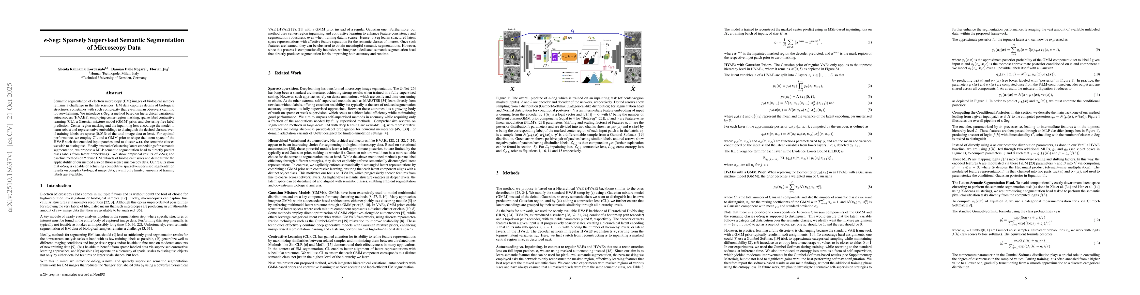
Semantic segmentation of electron microscopy (EM) images of biological samples remains a challenge in the life sciences. EM data captures details of biological structures, sometimes with such complexity that even human observers can find it overwhelming. We introduce {\epsilon}-Seg, a method based on hierarchical variational autoencoders (HVAEs), employing center-region masking, sparse label contrastive learning (CL), a Gaussian mixture model (GMM) prior, and clustering-free label prediction. Center-region masking and the inpainting loss encourage the model to learn robust and representative embeddings to distinguish the desired classes, even if training labels are sparse (0.05% of the total image data or less). For optimal performance, we employ CL and a GMM prior to shape the latent space of the HVAE such that encoded input patches tend to cluster wrt. the semantic classes we wish to distinguish. Finally, instead of clustering latent embeddings for semantic segmentation, we propose a MLP semantic segmentation head to directly predict class labels from latent embeddings. We show empirical results of {\epsilon}-Seg and baseline methods on 2 dense EM datasets of biological tissues and demonstrate the applicability of our method also on fluorescence microscopy data. Our results show that {\epsilon}-Seg is capable of achieving competitive sparsely-supervised segmentation results on complex biological image data, even if only limited amounts of training labels are available.

AI Key Findings

Generated Oct 23, 2025

Methodology — What approach did the authors take?

The research employs a sparsely supervised semantic segmentation approach for microscopy data, combining a Gaussian Mixture Model (GMM) prior with contrastive learning and entropy loss to enhance feature representation and class discrimination.

Key Results — What are the main findings?

  • Achieved state-of-the-art performance on the BetaSeg dataset with 0.88 average DSC under 0.0025% label sparsity
  • Demonstrated robustness to extreme label scarcity compared to traditional U-Net approaches
  • Enabled semantically meaningful distinction between cell structures through class-aware latent modeling

Significance — Why does this research matter?

This work addresses the critical challenge of limited labeled data in biomedical imaging, enabling more efficient annotation processes and improving the feasibility of automated analysis for complex cellular structures.

Technical Contribution — What is the technical contribution?

Development of a novel sparsely supervised framework combining GMM priors, contrastive learning, and entropy regularization for improved semantic segmentation in low-label scenarios

Novelty — What is new about this work?

Introduces a class-aware latent modeling approach through GMM priors combined with contrastive learning, providing a new paradigm for sparse supervision in biomedical image analysis

Limitations — What are the limitations of this study?

  • Limited to 2D microscopy data
  • Requires careful selection of training patches for effective sparsely supervised learning

Future Work — What did the authors propose for future work?

  • Extension to 3D volumetric data
  • Integration with active learning for adaptive data sampling
  • Exploration of multi-modal data fusion for enhanced feature representation

Paper Details

Paper ID: 2510.18637
Comments: 10 pages main text, 17 pages total
Categories:
cs.CV cs.AI cs.LG

How to Cite This Paper

BibTeX
@article{nogare2025ε,
  title         = {ε-Seg: Sparsely Supervised Semantic Segmentation of Microscopy
  Data},
  author        = {Nogare, Damian Dalle and Jug, Florian and Kordasiabi, Sheida Rahnamai},
  year          = {2025},
  eprint        = {2510.18637},
  archivePrefix = {arXiv},
  primaryClass  = {cs.CV},
}
APA 7
Nogare, D., Jug, F., & Kordasiabi, S. (2025). ε-Seg: Sparsely Supervised Semantic Segmentation of Microscopy
  Data. arXiv. https://arxiv.org/abs/2510.18637
MLA 9
Nogare, Damian Dalle, et al. "ε-Seg: Sparsely Supervised Semantic Segmentation of Microscopy
  Data." arXiv, 2025, arxiv.org/abs/2510.18637.

Comments (0)

This website uses cookies to ensure you get the best experience. Learn more