Academic Profile
Statistics
Similar Authors
Papers on arXiv
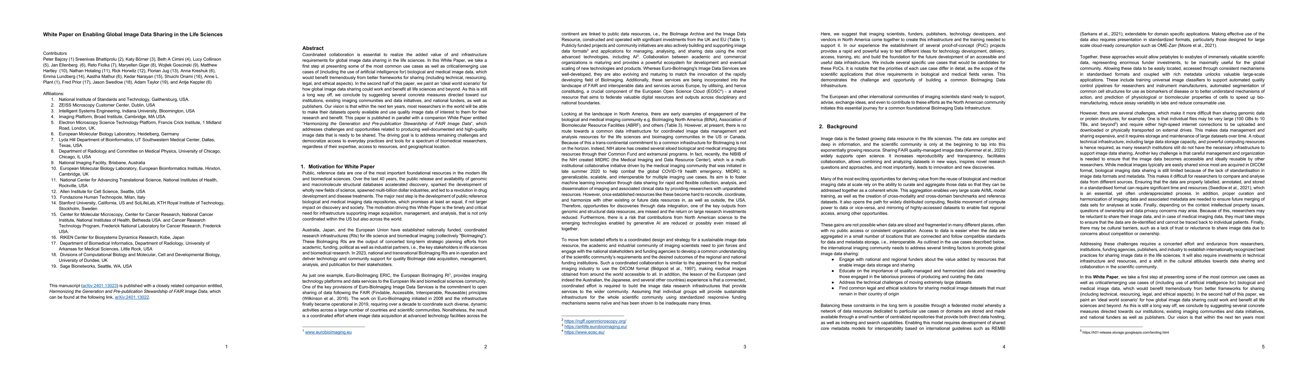
Coordinated collaboration is essential to realize the added value of and infrastructure requirements for global image data sharing in the life sciences. In this White Paper, we take a first step at ...
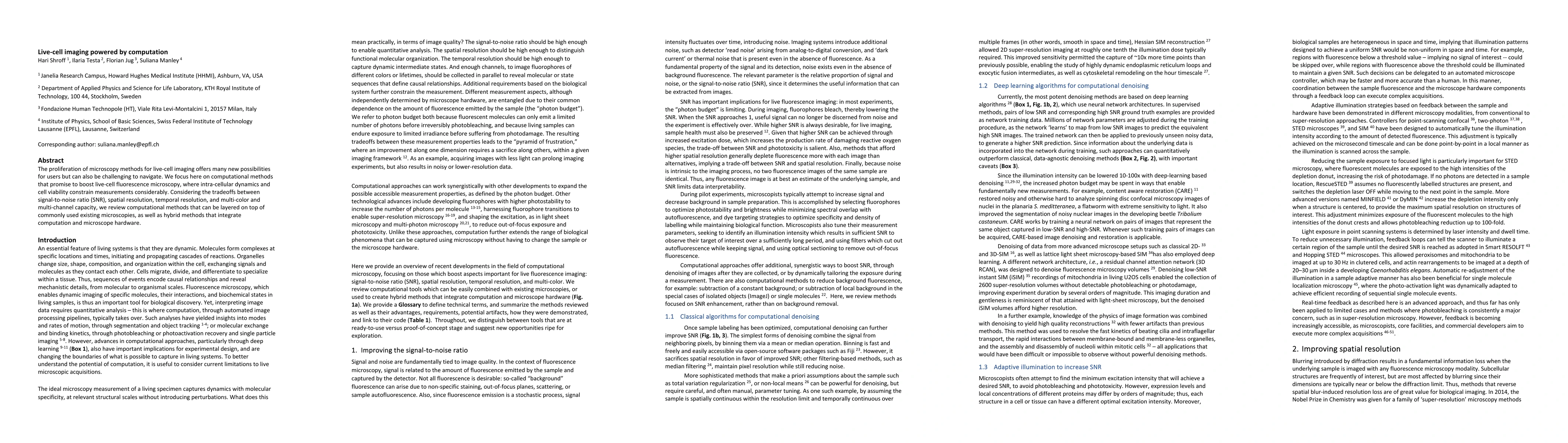
The proliferation of microscopy methods for live-cell imaging offers many new possibilities for users but can also be challenging to navigate. We focus here on computational methods that promise to ...
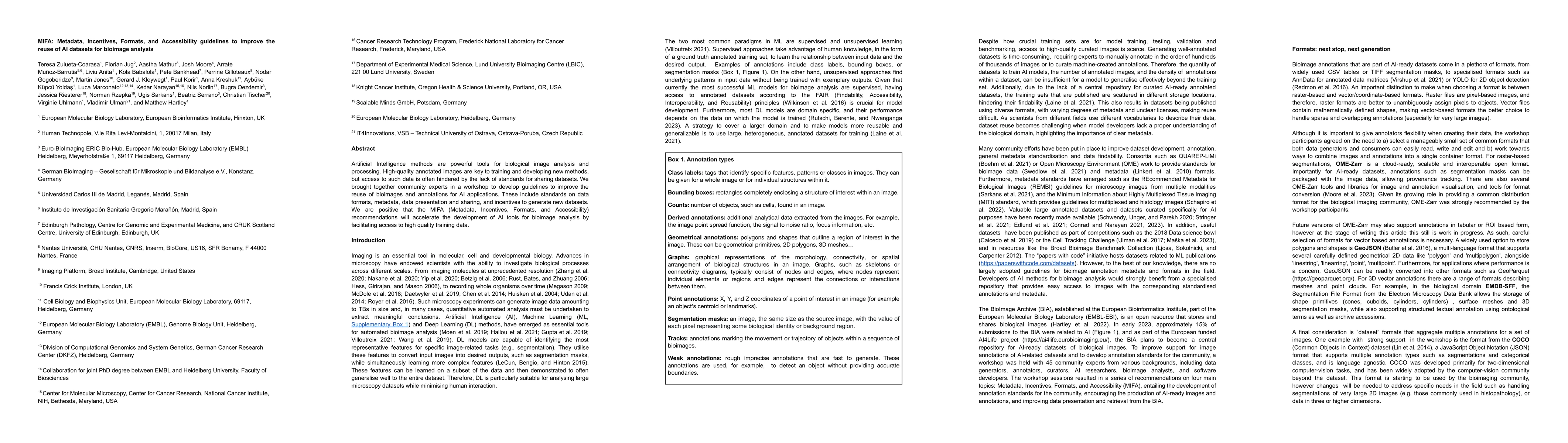
Artificial Intelligence methods are powerful tools for biological image analysis and processing. High-quality annotated images are key to training and developing new methods, but access to such data...
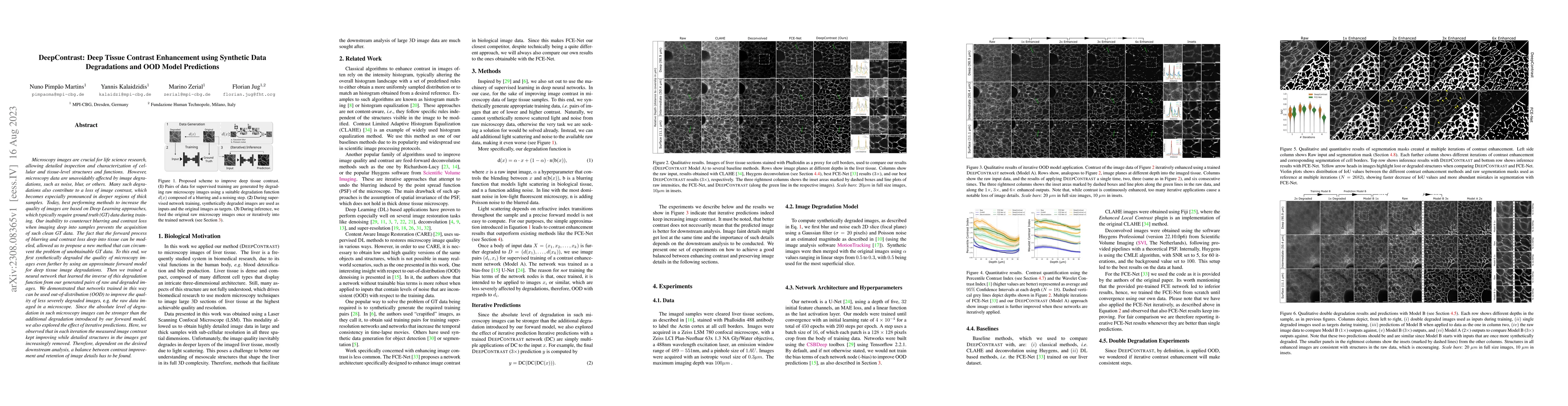
Microscopy images are crucial for life science research, allowing detailed inspection and characterization of cellular and tissue-level structures and functions. However, microscopy data are unavoid...
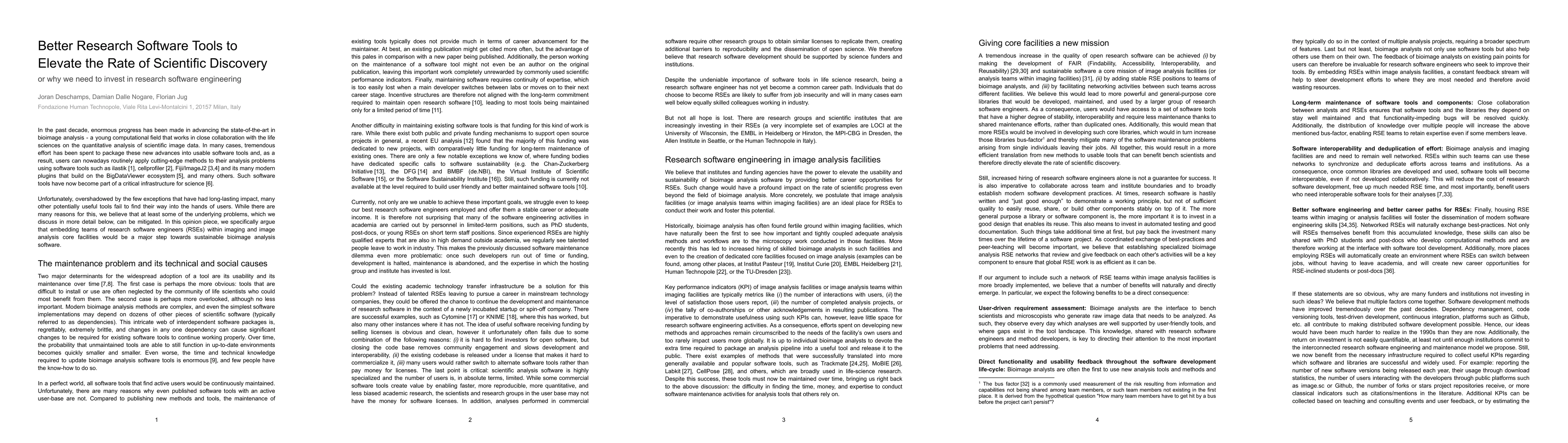
In the past decade, enormous progress has been made in advancing the state-of-the-art in bioimage analysis - a young computational field that works in close collaboration with the life sciences on t...
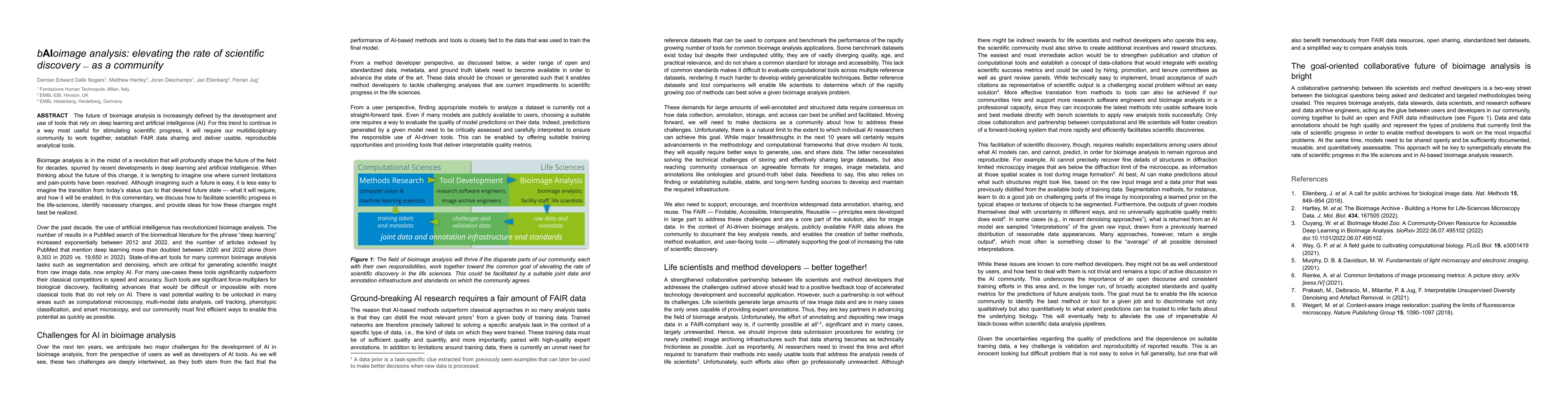
The future of bioimage analysis is increasingly defined by the development and use of tools that rely on deep learning and artificial intelligence (AI). For this trend to continue in a way most usef...
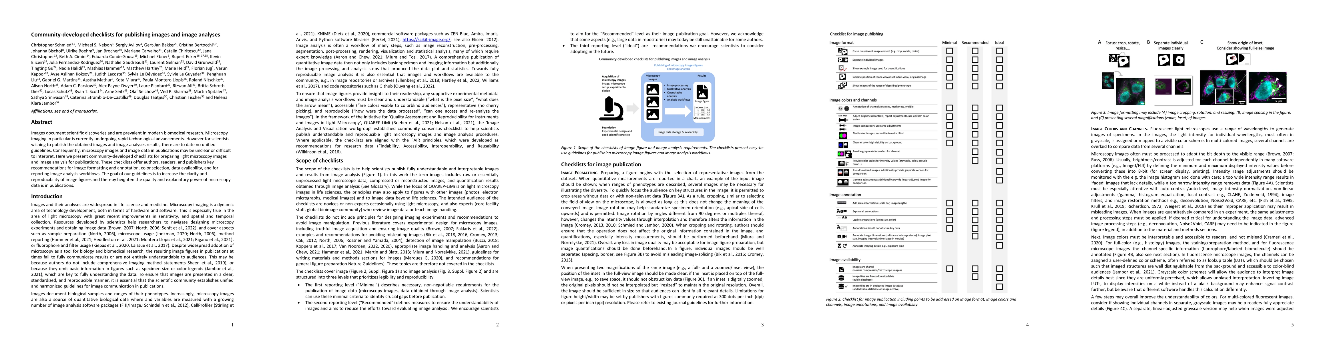
Images document scientific discoveries and are prevalent in modern biomedical research. Microscopy imaging in particular is currently undergoing rapid technological advancements. However for scienti...
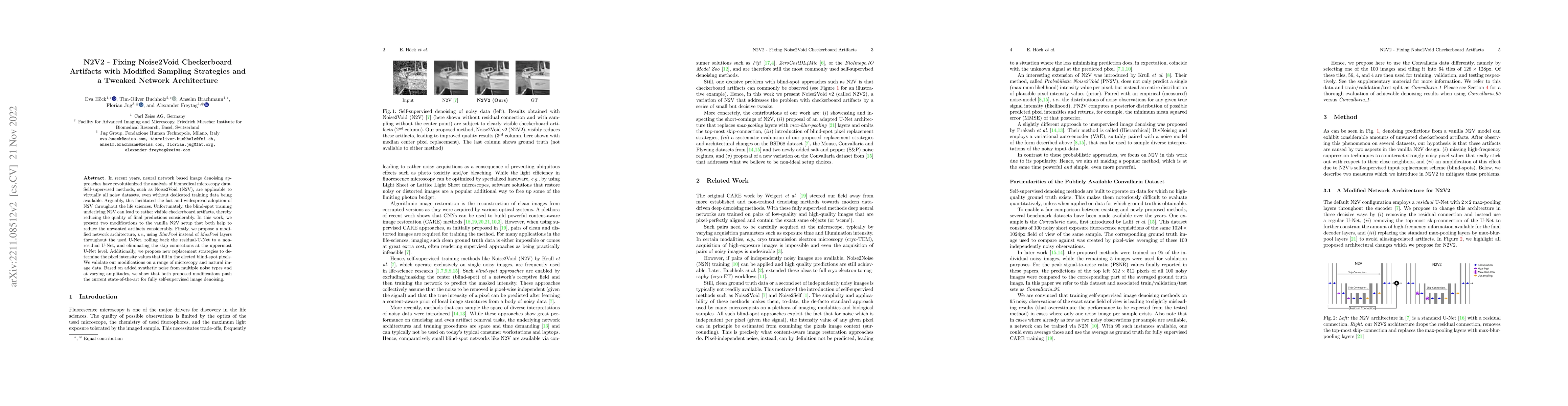
In recent years, neural network based image denoising approaches have revolutionized the analysis of biomedical microscopy data. Self-supervised methods, such as Noise2Void (N2V), are applicable to ...
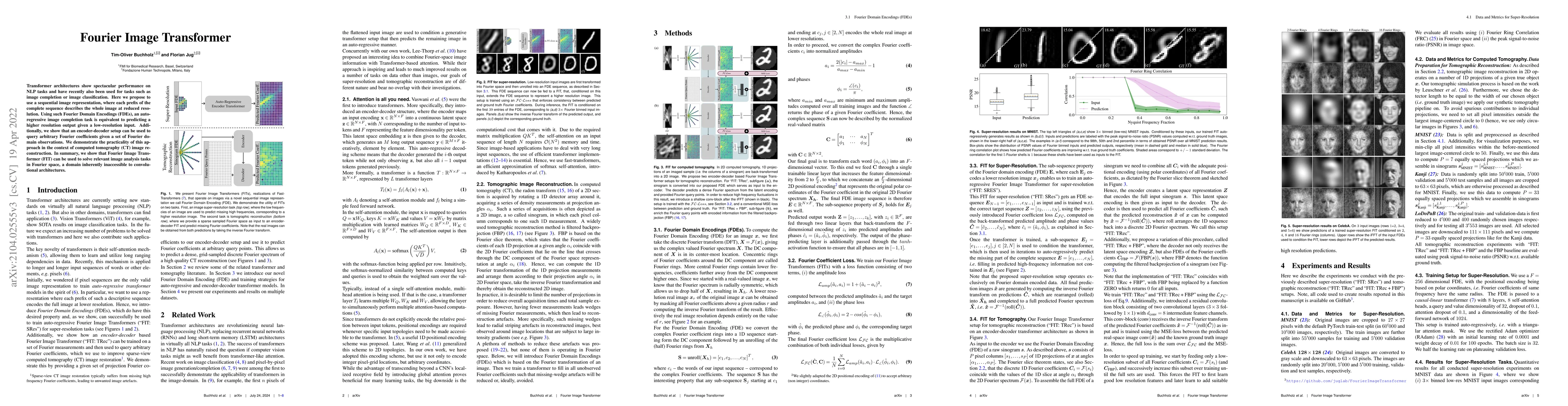
Transformer architectures show spectacular performance on NLP tasks and have recently also been used for tasks such as image completion or image classification. Here we propose to use a sequential i...
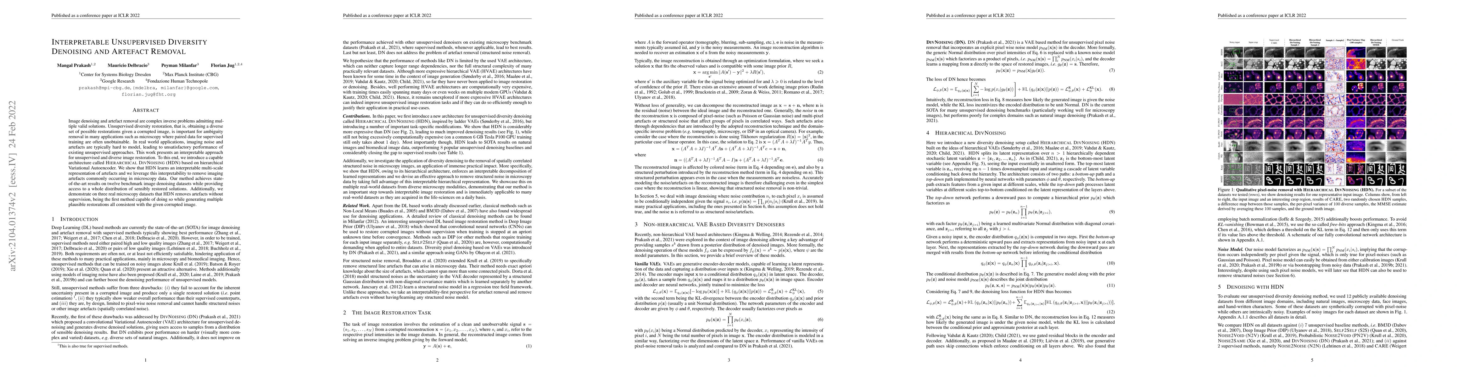
Image denoising and artefact removal are complex inverse problems admitting multiple valid solutions. Unsupervised diversity restoration, that is, obtaining a diverse set of possible restorations gi...
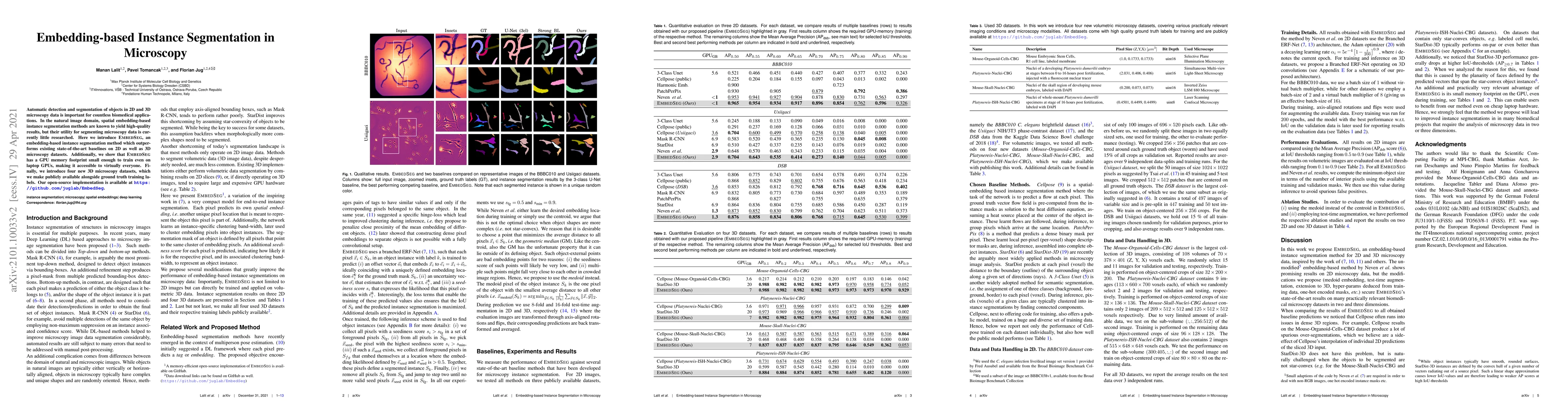
Automatic detection and segmentation of objects in 2D and 3D microscopy data is important for countless biomedical applications. In the natural image domain, spatial embedding-based instance segment...
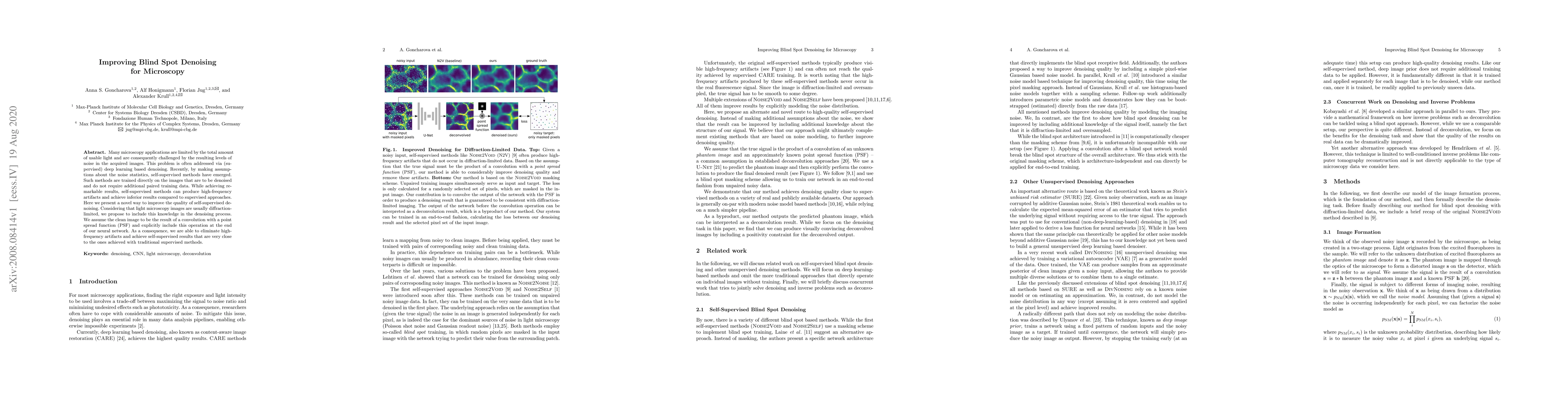
Many microscopy applications are limited by the total amount of usable light and are consequently challenged by the resulting levels of noise in the acquired images. This problem is often addressed ...
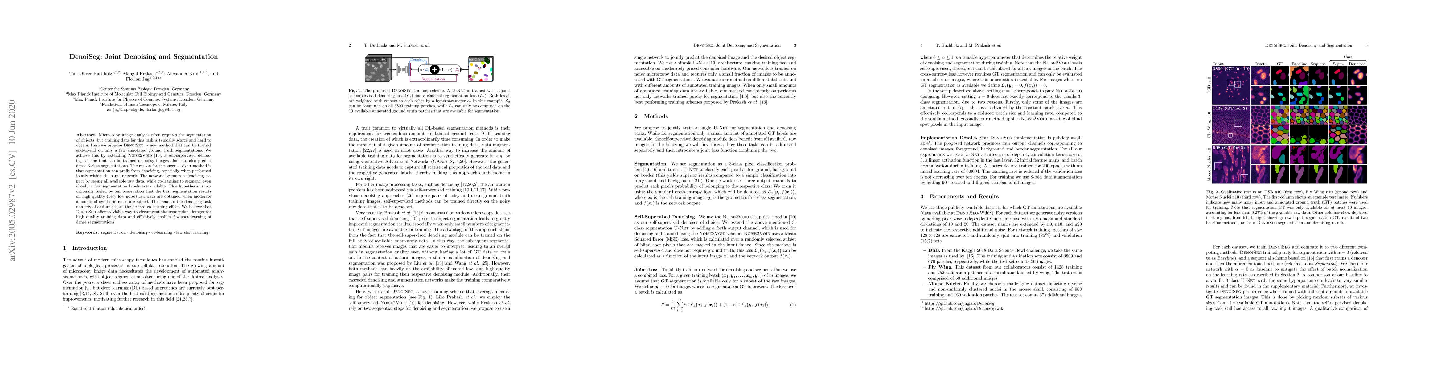
Microscopy image analysis often requires the segmentation of objects, but training data for this task is typically scarce and hard to obtain. Here we propose DenoiSeg, a new method that can be train...
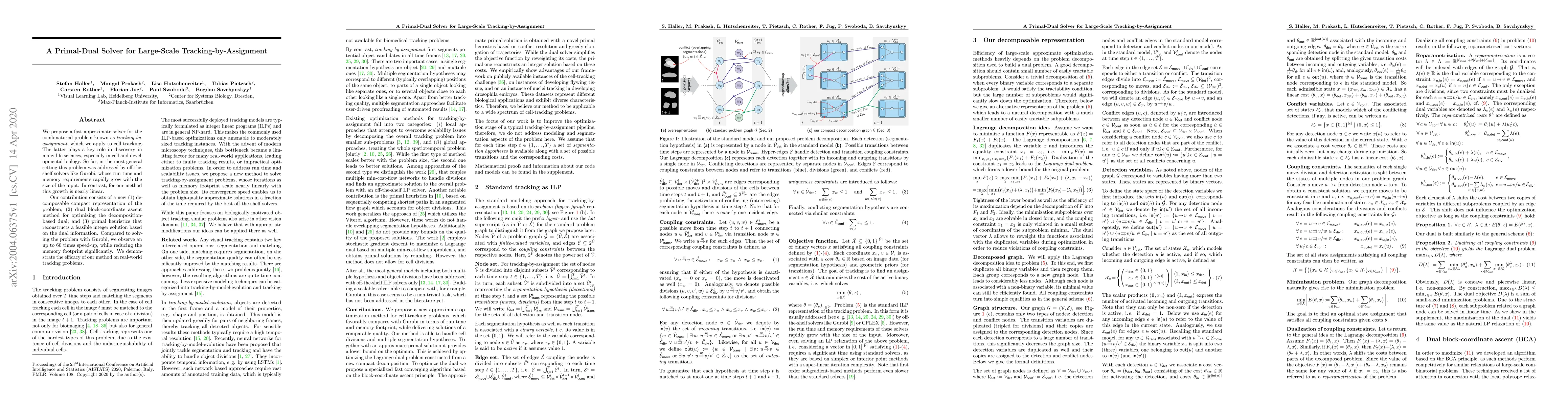
We propose a fast approximate solver for the combinatorial problem known as tracking-by-assignment, which we apply to cell tracking. The latter plays a key role in discovery in many life sciences, e...
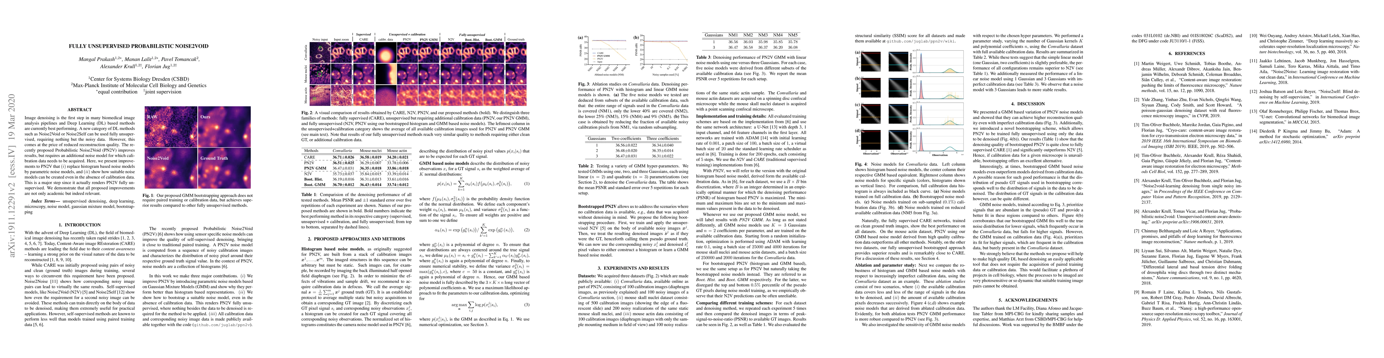
Image denoising is the first step in many biomedical image analysis pipelines and Deep Learning (DL) based methods are currently best performing. A new category of DL methods such as Noise2Void or N...
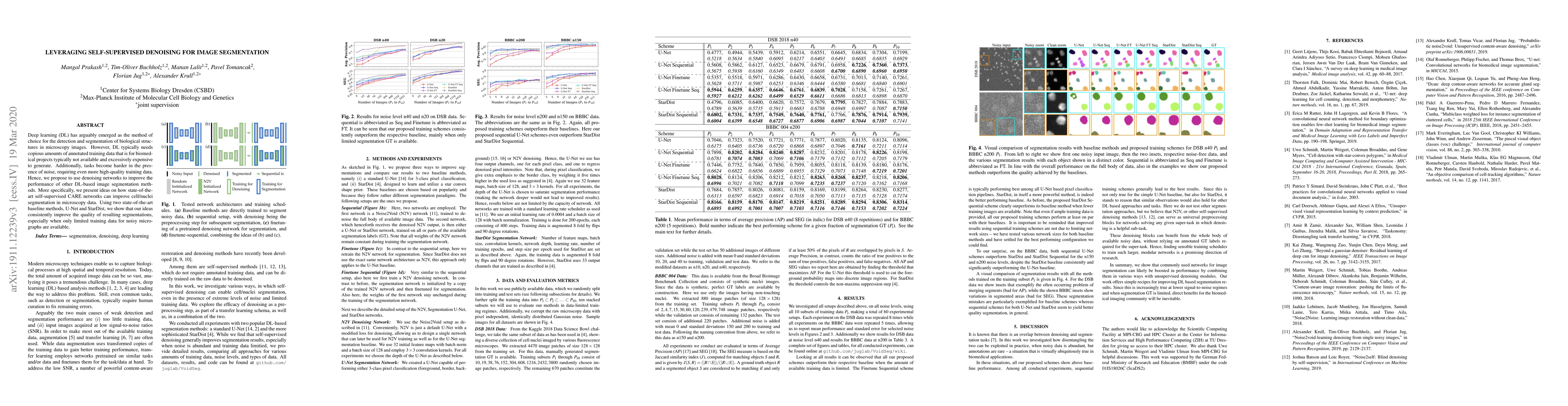
Deep learning (DL) has arguably emerged as the method of choice for the detection and segmentation of biological structures in microscopy images. However, DL typically needs copious amounts of annot...
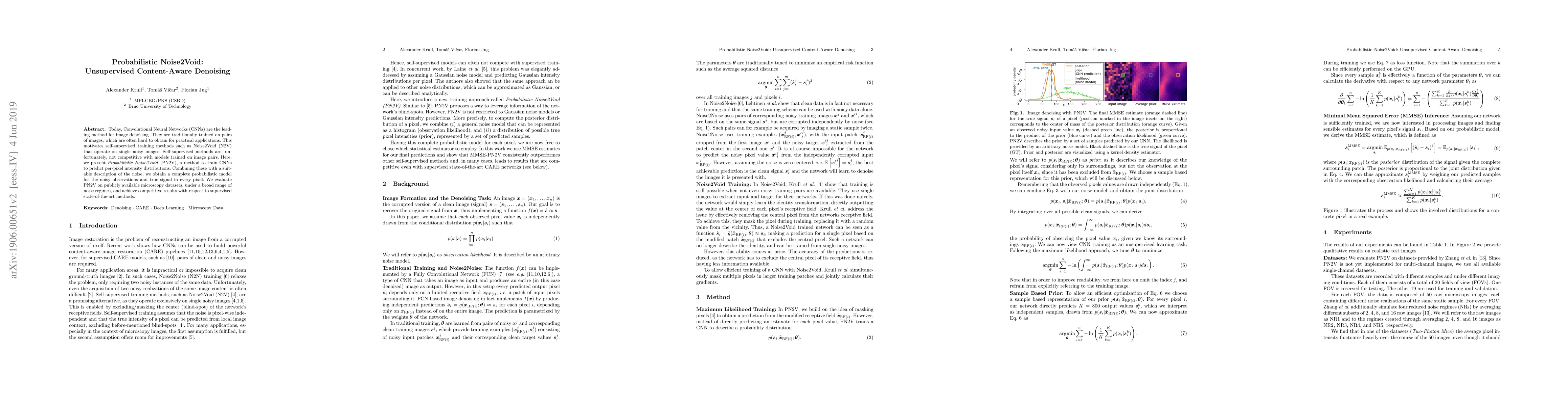
Today, Convolutional Neural Networks (CNNs) are the leading method for image denoising. They are traditionally trained on pairs of images, which are often hard to obtain for practical applications. ...
Microscopy is routinely used to image biological structures of interest. Due to imaging constraints, acquired images are typically low-SNR and contain noise. Over the last few years, regression-based ...
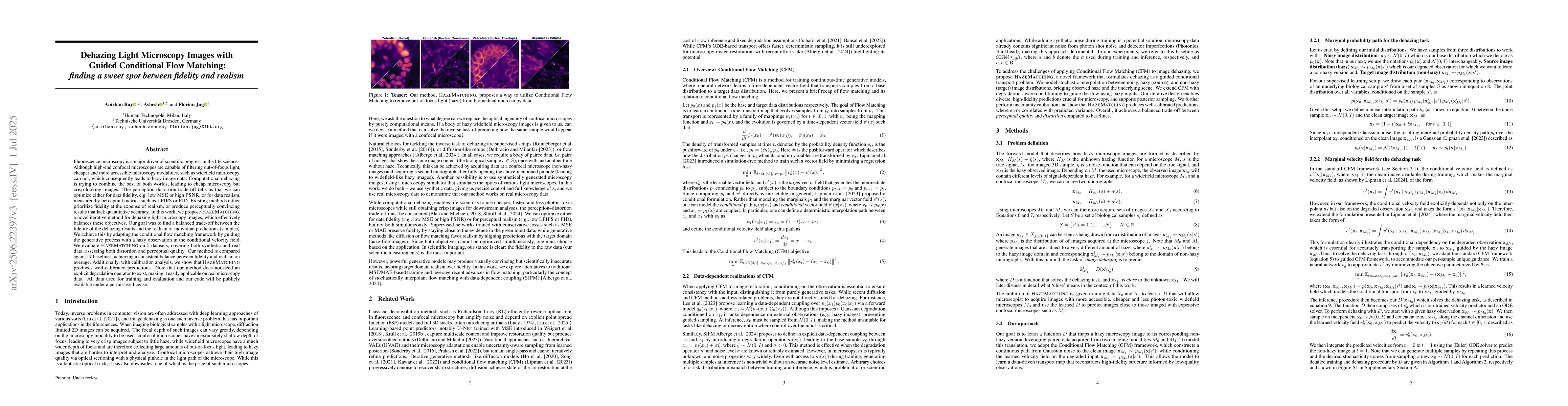
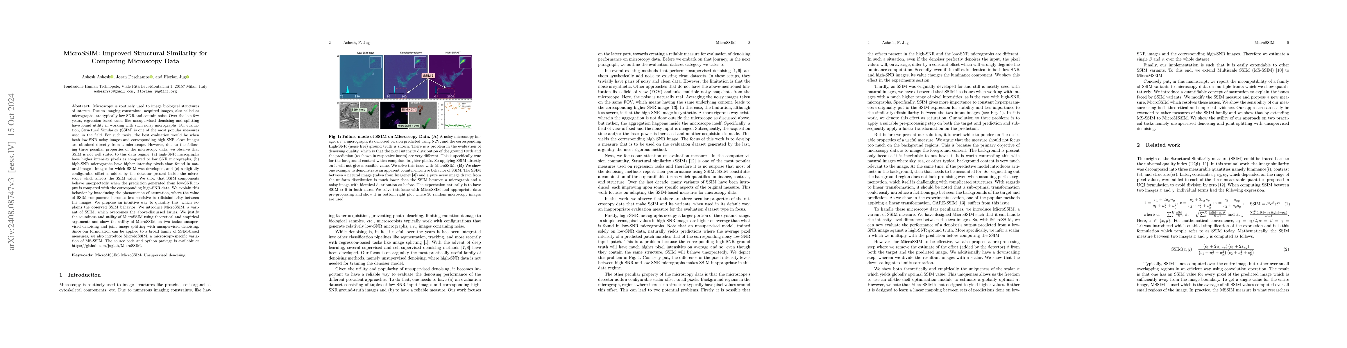
Fluorescence microscopy is a major driver of scientific progress in the life sciences. Although high-end confocal microscopes are capable of filtering out-of-focus light, cheaper and more accessible m...
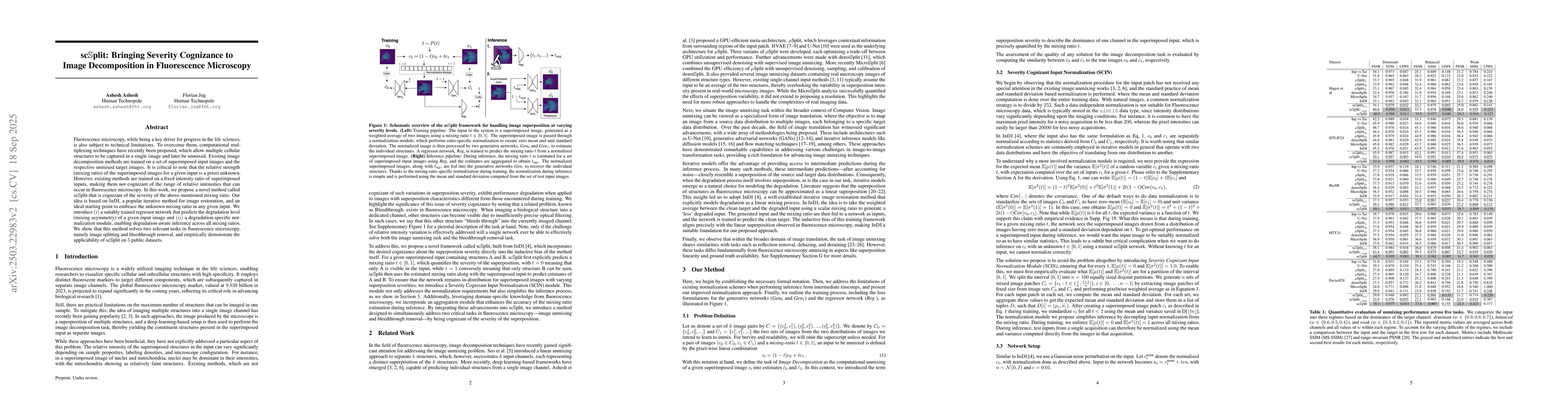
Fluorescence microscopy, while being a key driver for progress in the life sciences, is also subject to technical limitations. To overcome them, computational multiplexing techniques have recently bee...
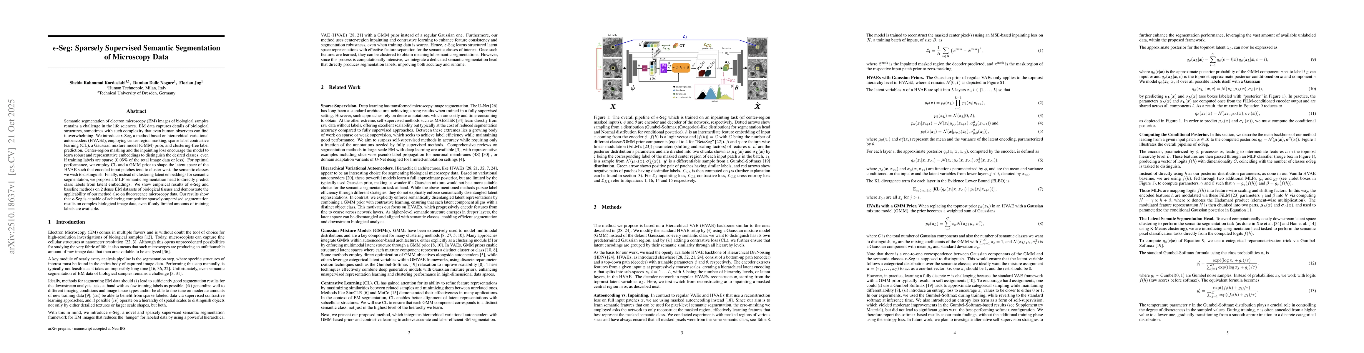
Semantic segmentation of electron microscopy (EM) images of biological samples remains a challenge in the life sciences. EM data captures details of biological structures, sometimes with such complexi...
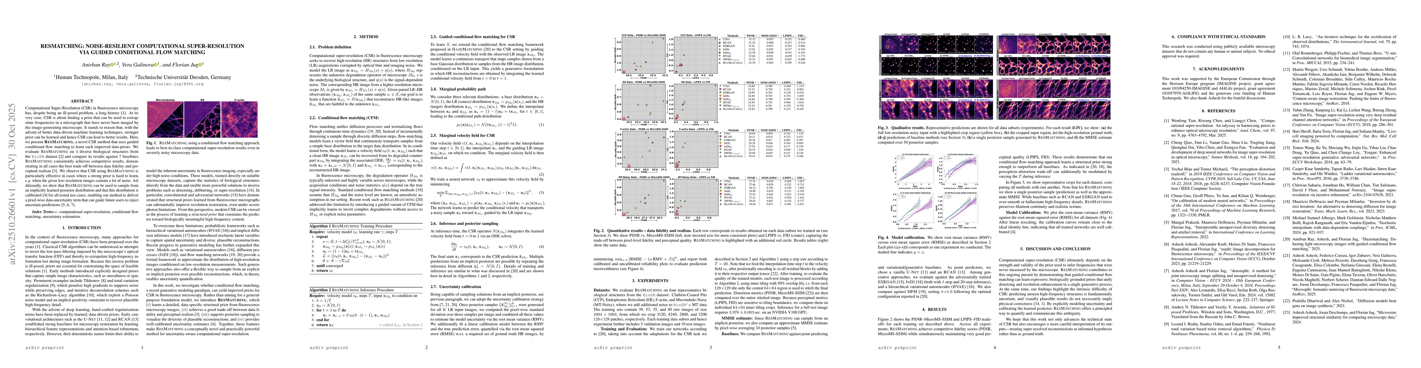
Computational Super-Resolution (CSR) in fluorescence microscopy has, despite being an ill-posed problem, a long history. At its very core, CSR is about finding a prior that can be used to extrapolate ...
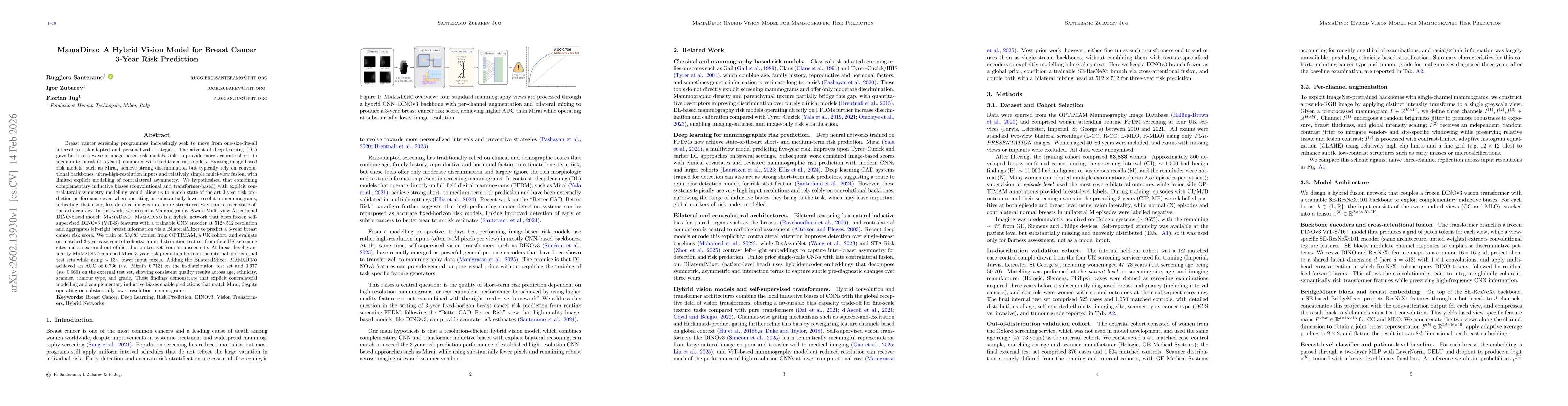
Breast cancer screening programmes increasingly seek to move from one-size-fits-all interval to risk-adapted and personalized strategies. Deep learning (DL) has enabled image-based risk models with st...
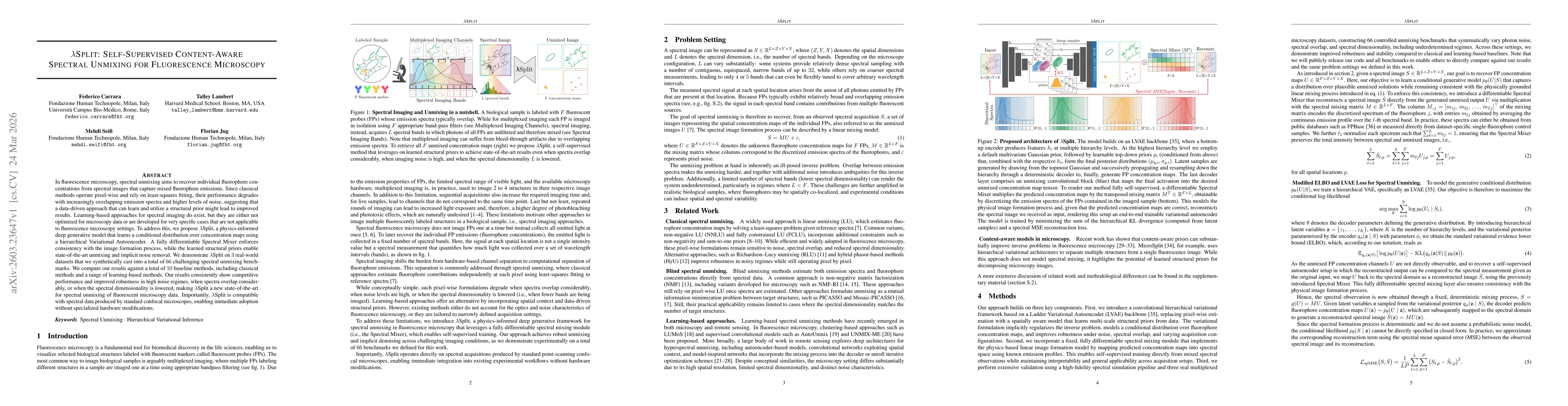
In fluorescence microscopy, spectral unmixing aims to recover individual fluorophore concentrations from spectral images that capture mixed fluorophore emissions. Since classical methods operate pixel...