Quick Summary:

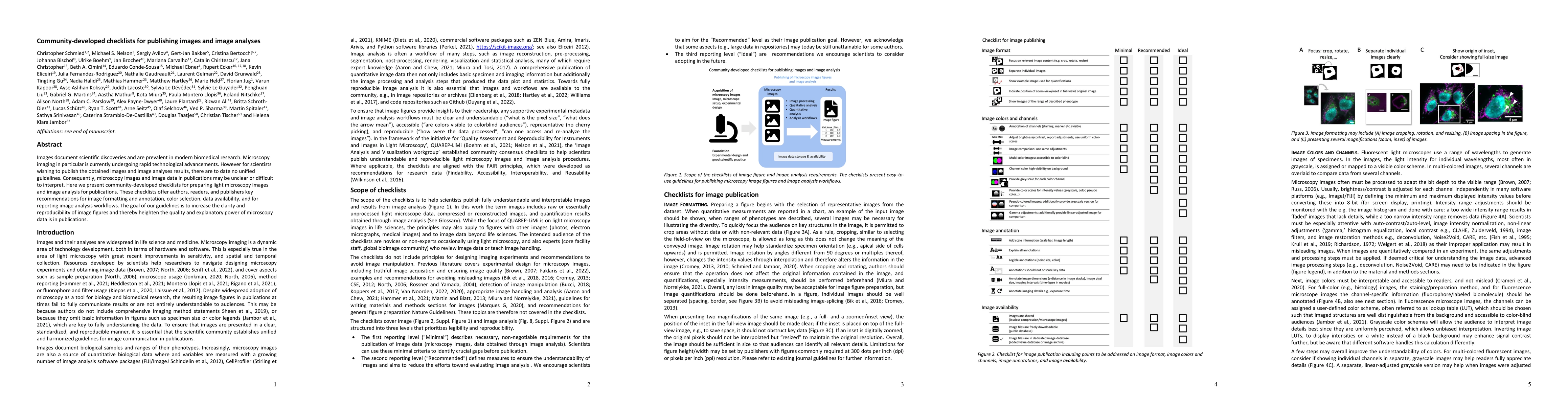
This paper introduces community-developed checklists aimed at improving the clarity and reproducibility of light microscopy images and image analyses in publications. The guidelines provide recommendations on image formatting, annotation, color selection, data availability, and reporting workflows to enhance the quality of microscopy data.

Summary

Images document scientific discoveries and are prevalent in modern biomedical research. Microscopy imaging in particular is currently undergoing rapid technological advancements. However for scientists wishing to publish the obtained images and image analyses results, there are to date no unified guidelines. Consequently, microscopy images and image data in publications may be unclear or difficult to interpret. Here we present community-developed checklists for preparing light microscopy images and image analysis for publications. These checklists offer authors, readers, and publishers key recommendations for image formatting and annotation, color selection, data availability, and for reporting image analysis workflows. The goal of our guidelines is to increase the clarity and reproducibility of image figures and thereby heighten the quality of microscopy data is in publications.

AI Key Findings

Get AI-generated insights about this paper's methodology, results, and significance.

Paper Details

PDF Preview

Download PDF

Key Terms

microscopy (0.399)
microscopy (0.375)
image (0.323)
image (0.316)
images (0.286)
images (0.275)
recommendations (0.189)
advancements (0.181)
reporting (0.181)
document (0.181)
document (0.180)
unclear (0.178)
recommendations (0.176)
advancements (0.174)
technological (0.172)
interpret (0.165)
availability (0.165)
date (0.160)
interpret (0.158)
scientific (0.158)

Citation Network

Current paper (gray), citations (green), references (blue)

Current Paper
Citations
References
Click to view

Similar Papers

No similar papers found for this research.

Title Authors Year Actions
Evaluation of image analysis tools for the measurement of cellular morphology
Matthew D. Bourn, Lauren F. Daly, Jim F. Huggett, Julian Braybrook, Jeanne F. Rivera
2025
View Paper
Colony context and size-dependent compensation mechanisms give rise to variations in nuclear growth trajectories.
Julie C. Dixon, Christopher L. Frick, Chantelle L. Leveille, Philip Garrison, Peyton A. Lee, Saurabh S. Mogre, Benjamin Morris, Nivedita Nivedita, R. Vasan, Jianxu Chen, Cameron L. Fraser, Clare R. Gamlin, Leigh K. Harris, Melissa C. Hendershott, Graham T. Johnson, Kyle N. Klein, Sandra A. Oluoch, Derek J. Thirstrup, M. F. Sluzewski, Lyndsay Wilhelm, Ruian Yang, Daniel M. Toloudis, Matheus P. Viana, J. Theriot, S. Rafelski
2025
View Paper
ReScale4DL: Balancing Pixel and Contextual Information for Enhanced Bioimage Segmentation
Mariana G. Ferreira, Bruno M. Saraiva, António D. Brito, M. G. Pinho, R. Henriques, Estibaliz Gómez-de-Mariscal
2025
View Paper
Oxidative stress and inflammation combine to exacerbate cochlear damage and sensorineural hearing loss in C57BL/6 mice
Zhongwu Su, Yuyan Chen, Yu Liu, Jinyuan Cao, Jie Cui, Haitong Chen, Qi Li
2025
View Paper
Bridging imaging-based in vitro methods from biomedical research to regulatory toxicology
M. Piergiovanni, Milena Mennecozzi, Erio Barale-Thomas, Davide Danovi, S. Dunst, David Egan, Aurora Fassi, Matthew Hartley, Philipp Kainz, Katharina Koch, Sylvia E. Le Dévédec, Iris Mangas, Elena Miranda, Jo Nyffeler, Enrico Pesenti, Fernanda Ricci, Christopher Schmied, Alexander Schreiner, Nadine Stokar-Regenscheit, Jason R Swedlow, Virginie Uhlmann, F. C. Wieland, Amy Wilson, Maurice Whelan
2025
View Paper
Addressing persistent challenges in digital image analysis of cancer tissue: resources developed from a hackathon.
Sandhya Prabhakaran, Clarence Yapp, Gregory J. Baker, Johanna Beyer, Young Hwan Chang, A. Creason, Robert Krueger, J. Muhlich, N. Patterson, Kevin Sidak, D. Sudar, Adam J Taylor, Luke Ternes, J. Troidl, Xie Yubin, Artem Sokolov, Darren R Tyson
2025
View Paper
Evaluation of Three Different Operators for Object Highlighting in Medical RGB Images: Canny, Roberts, and LoG in Independent Color Spaces
Jafar Ababneh, A. Abu-Jassar, Suhaila Abuowaida, Valentyn Liubchenko, Vyacheslav V. Lyashenko
2024
View Paper
Simple quantitation and spatial characterization of label free cellular images
V.C.J. de Boer, Xiang Zhang
2024
View Paper
The crucial role of bioimage analysts in scientific research and publication
Beth A. Cimini, Peter Bankhead, Rocco D’Antuono, Elnaz Fazeli, J. Fernandez-Rodriguez, Caterina Fuster-Barceló, Robert Haase, H. Jambor, Martin L. Jones, Florian Jug, Anna H Klemm, A. Kreshuk, S. Marcotti, Gabriel G Martins, Sara McArdle, Kota Miura, A. Muñoz-Barrutia, Laura C Murphy, Michael S Nelson, S. Norrelykke, P. Paul-Gilloteaux, T. Pengo, Joanna W. Pylvänäinen, Lior Pytowski, Arianna Ravera, Annika Reinke, Yousr Rekik, C. Strambio‐De‐Castillia, Daniel Thédié, V. Uhlmann, Oliver Umney, Laura Wiggins, Kevin W. Eliceiri
2024
View Paper
What Are the Challenges Ahead for Training the Present and Next-Generation Microscopists in the Life Sciences (a.k.a. Structural Cell Biologists)?
Filip Braet, D. Taatjes
2024
View Paper
Morphological Profiling Dataset of EU-OPENSCREEN Bioactive Compounds Over Multiple Imaging Sites and Cell Lines
Christopher Wolff, Martin Neuenschwander, C. Beese, Divya Sitani, María C. Ramos, Alžběta Srovnalova, M. J. Varela, Pavel G. Polishchuk, Katholiki E. Skopelitou, C. Škuta, Bahne Stechmann, J. Brea, Mads Hartvig Clausen, P. Džubák, Rosario Fernández-Godino, O. Genilloud, Marián Hajdúch, M. I. Loza, Martin Lehmann, J. V. von Kries, Han Sun, Christopher Schmied
2024
View Paper
A human induced pluripotent stem (hiPS) cell model for the holistic study of epithelial to mesenchymal transitions (EMTs)
C. Hookway, Antoine Borensztejn, Leigh K. Harris, Sara Carlson, Gokhan Dalgin, Suraj Mishra, Nivedita Nivedita, Ellen M. Adams, Tiffany Barszczewski, Julie C. Dixon, Jacqueline H. Edmonds, Erik A. Ehlers, Alexandra J. Ferrante, M. Fuqua, Philip Garrison, Janani Gopalan, B. Gregor, Maxwell J. Hedayati, Kyle N. Klein, Chantelle L. Leveille, Sean L. Meharry, Haley S. Morris, Gouthamrajan Nadarajan, Sandra A. Oluoch, Serge E. Parent, Amber Phan, Brock Roberts, Emmanuel E. Sanchez, M. F. Sluzewski, Lev S. Snyder, Derek J. Thirstrup, J. P. Thottam, Julia R. Torvi, Gaea Turman, Matheus P. Viana, Lyndsay Wilhelm, C. Wijesooriya, Jie Yao, J. Theriot, S. Rafelski, R. Gunawardane
2024
View Paper
From zero to figure hero. A checklist for designing scientific data visualizations
H. K. Jambor
2024
View Paper
Arterial specification precedes microvascular restitution in the peri-infarct cortex that is driven by small microvessels
Nina Hagemann, Yachao Qi, A. Mohamud Yusuf, AnRan Li, Xiaoni Zhang, Philippa Spangenberg, Anthony Squire, Thorsten R. Doeppner, Fengyan Jin, Shuo Zhao, Jianxu Chen, Axel Mosig, Matthias Gunzer, Dirk M. Hermann
2024
View Paper
Integrating bioengineering, super-resolution microscopy and mechanobiology in autophagy research: addendum to the guidelines (4th edition)
Andrea Ravasio, D. Klionsky, Cristina Bertocchi
2024
View Paper
AI analysis of super-resolution microscopy: Biological discovery in the absence of ground truth
Ivan R. Nabi, Ben Cardoen, Ismail M. Khater, Guang Gao, Timothy H. Wong, Ghassan Hamarneh
2024
View Paper
Protein Disulfide Isomerase Endoplasmic Reticulum Protein 57 (ERp57) is Protective Against ALS-Associated Mutant TDP-43 in Neuronal Cells
S. Parakh, E. Perri, M. Vidal, Zeinab Takalloo, Cyril J Jagaraj, P. Mehta, Shu Yang, Colleen J Thomas, Ian Blair, Yuning Hong, J. Atkin
2024
View Paper
A practical guide to bioimaging research data management in core facilities
Christian Schmidt, Tom Boissonnet, Julia Dohle, Karen Bernhardt, Elisa Ferrando-May, Tobias Wernet, R. Nitschke, S. Kunis, S. Weidtkamp‐Peters
2024
View Paper
A new protocol for multispecies bacterial infections in zebrafish and their monitoring through automated image analysis
Désirée A. Schmitz, Tobias Wechsler, H. Li, Bjoern H Menze, R. Kümmerli
2024
View Paper
Staying on track – Keeping things running in a high‐end scientific imaging core facility
Oliver Renaud, Nathalie Aulner, Audrey Salles, Nadia Halidi, M. Brunstein, Adeline Mallet, Karin Aumayr, S. Terjung, Daniel Levy, Saskia Lippens, J. Verbavatz, Thomas Heuser, Rachel Santarella-Mellwig, J. Tinevez, Tatiana Woller, Alexander Botzki, Christopher J Cawthorne, Sebastian Munck
2024
View Paper
Creating and troubleshooting microscopy analysis workflows: common challenges and common solutions
Beth A. Cimini
2024
View Paper
Cell Painting Gallery: an open resource for image-based profiling
Erin Weisbart, Ankur Kumar, John Arevalo, A. Carpenter, B. Cimini, Shantanu Singh
2024
View Paper
Where imaging and metrics meet.
2024
View Paper
vEMstitch: an algorithm for fully automatic image stitching of volume electron microscopy
Bintao He, Yan Zhang, Zhenbang Zhang, Yiran Cheng, Fa Zhang, Fei Sun, Renmin Han
2024
View Paper
Believing is seeing - the deceptive influence of bias in quantitative microscopy.
Rachel M. Lee, Leanna R Eisenman, S. Khuon, Jesse S. Aaron, T. Chew
2024
View Paper
A community-driven approach to enhancing the quality and interpretability of microscopy images.
H. Jambor
2023
View Paper
What we can learn from deep space communication for reproducible bioimaging and data analysis
Tatiana Woller, Christopher J Cawthorne, Romain Raymond Agnes Slootmaekers, Ingrid Barcena Roig, Alexander Botzki, Sebastian Munck
2023
View Paper
Defining the boundaries: challenges and advances in identifying cells in microscopy images
Nodar Gogoberidze, Beth A. Cimini
2023
View Paper
Editorial: Histochemistry and Cell Biology implements new submission guidelines for image presentation and image analysis
D. Taatjes, Takehiko Koji, Michael Schrader, Jürgen Roth
2023
View Paper
Bridging imaging users to imaging analysis – A community survey
Suganya Sivagurunathan, Stefania Marcotti, Carl J Nelson, Martin L. Jones, David J. Barry, T. Slater, K. Eliceiri, Beth A. Cimini
2023
View Paper
OrgaMapper: a robust and easy-to-use workflow for analyzing organelle positioning
Christopher Schmied, Michael Ebner, Paula Samsó Ferré, V. Haucke, Martin Lehmann
2023
View Paper
The Twenty Questions of bioimage object analysis
B. Cimini, K. Eliceiri
2023
View Paper
Building a FAIR image data ecosystem for microscopy communities
Isabel Kemmer, Antje Keppler, Beatriz Serrano-Solano, A. Rybina, Buğra Özdemir, Johanna Bischof, Ayoub El Ghadraoui, J. Eriksson, Aastha Mathur
2023
View Paper
Bridging Imaging Users to Imaging Analysis - A community survey
Suganya Sivagurunathan, Stefania Marcotti, Carl J. Nelson, Martin L. Jones, David J. Barry, T. Slater, K. Eliceiri, B. Cimini
2023
View Paper
Made to measure: An introduction to quantifying microscopy data in the life sciences
Siân Culley, Alicia Cuber Caballero, J. Burden, V. Uhlmann
2023
View Paper
A biologist’s guide to planning and performing quantitative bioimaging experiments
Rebecca A. Senft, Barbara B. Diaz-Rohrer, P. Colarusso, Lucy Swift, Nasim Jamali, H. Jambor, T. Pengo, C. Brideau, P. Llopis, V. Uhlmann, Jason Kirk, Kevin Andrew Gonzales, P. Bankhead, Edward L. Evans, K. Eliceiri, B. Cimini
2023
View Paper
AI-based analysis of super-resolution microscopy: Biological discovery in the absence of ground truth
I. Nabi, Ben Cardoen, Ismail M. Khater, Guangpu Gao, Timothy H. Wong, Ghassan Hamarneh
2023
View Paper
Arteriosclerosis, Thrombosis, and Vascular Biology
Arterioscler Thromb, Vasc Biol, MD Dirk M. Hermann, Nina Hagemann, Yachao Qi, Ayan Mohamud Yusuf, Anthony Squire, T. Tertel, Bernd Giebel, Peter Ludewig, Philippa Spangenberg, Jianxu Chen, Axel Mosig, Matthias Gunzer
View Paper

Comments (0)

This website uses cookies to ensure you get the best experience. Learn more