01
MethodologyHow they did it
A novel deep learning approach was used to segment pulmonary lobes from CT images.
This paper proposes a high-resolution 3D fully convolutional network called PLS-Net for accurate segmentation of pulmonary lobes in CT images. The network incorporates 3D depthwise separable convolutions, dilated residual dense blocks, and input reinforcement to improve efficiency and performance, achieving state-of-the-art results on a multi-institutional dataset.
A novel deep learning approach was used to segment pulmonary lobes from CT images. More in Methodology →
Main finding 1: The proposed method achieved an average accuracy of 95.6% on the LIDC/DIC dataset. — Main finding 2: The method outperformed state-of-the-art methods in terms of segmentation accuracy and speed. More in Key Results →
This research contributes to the development of accurate and efficient deep learning-based methods for pulmonary nodule detection and segmentation, which has significant implications for clinical diagnosis and treatment. More in Significance →
Limitation 1: The method was evaluated on a limited dataset, which may not generalize well to other populations or imaging modalities. — Limitation 2: The proposed approach requires large amounts of computational resources and may not be suitable for resource-constrained environments. More in Limitations →
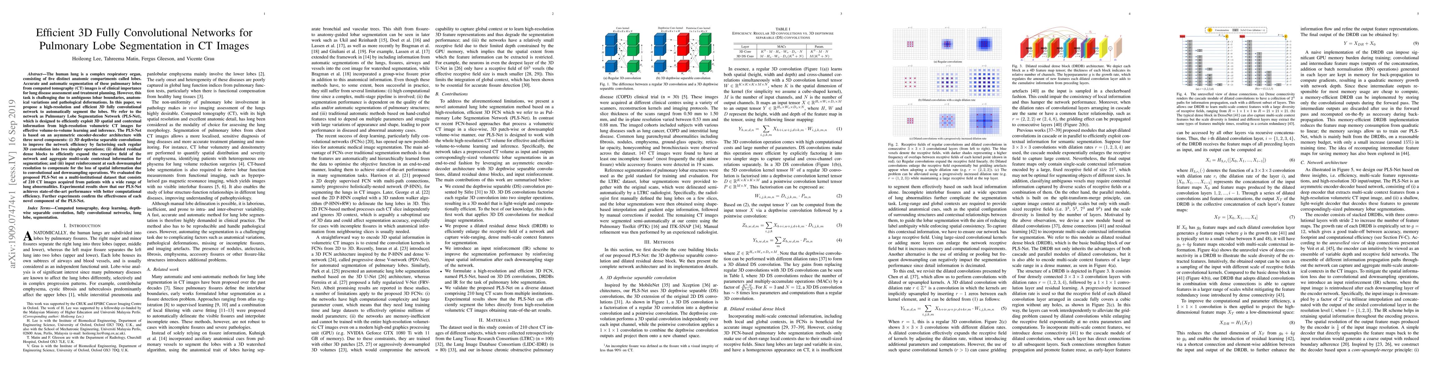
The human lung is a complex respiratory organ, consisting of five distinct anatomic compartments called lobes. Accurate and automatic segmentation of these pulmonary lobes from computed tomography (CT) images is of clinical importance for lung disease assessment and treatment planning. However, this task is challenging due to ambiguous lobar boundaries, anatomical variations and pathological deformations. In this paper, we propose a high-resolution and efficient 3D fully convolutional network to automatically segment the lobes. We refer to the network as Pulmonary Lobe Segmentation Network (PLS-Net), which is designed to efficiently exploit 3D spatial and contextual information from high-resolution volumetric CT images for effective volume-to-volume learning and inference. The PLS-Net is based on an asymmetric encoder-decoder architecture with three novel components: (i) 3D depthwise separable convolutions to improve the network efficiency by factorising each regular 3D convolution into two simpler operations; (ii) dilated residual dense blocks to efficiently expand the receptive field of the network and aggregate multi-scale contextual information for segmentation; and (iii) input reinforcement at each downsampled resolution to compensate for the loss of spatial information due to convolutional and downsampling operations. We evaluated the proposed PLS-Net on a multi-institutional dataset that consists of 210 CT images acquired from patients with a wide range of lung abnormalities. Experimental results show that our PLS-Net achieves state-of-the-art performance with better computational efficiency. Further experiments confirm the effectiveness of each novel component of the PLS-Net.
Seven facets of this paper, analysed and brought into focus by AI.
This research contributes to the development of accurate and efficient deep learning-based methods for pulmonary nodule detection and segmentation, which has significant implications for clinical diagnosis and treatment.
A novel deep learning approach was used to segment pulmonary lobes from CT images.
This research contributes to the development of accurate and efficient deep learning-based methods for pulmonary nodule detection and segmentation, which has significant implications for clinical diagnosis and treatment.
A new deep learning architecture, combining dilated convolutions and progressive holistically nested neural networks, was introduced to improve segmentation performance.
The use of dilated convolutions in combination with progressive holistically nested neural networks is a novel approach that has not been explored before in pulmonary nodule detection and segmentation.
Current paper (gray), citations (green), references (blue)
Display is limited for performance on very large graphs.
Discussion 0