Efficient, gigapixel-scale, aberration-free whole slide scanner using angular ptychographic imaging with closed-form solution
Publication
Metrics
AI Quick Summary
This paper presents WSI-APIC, a high-efficiency, aberration-free, gigapixel-scale whole slide imaging system using angular ptychographic imaging. It features GPU-accelerated phase image reconstruction and an auto-stitching technique for seamless, high-resolution pathology analysis.
Paper Preview
Abstract
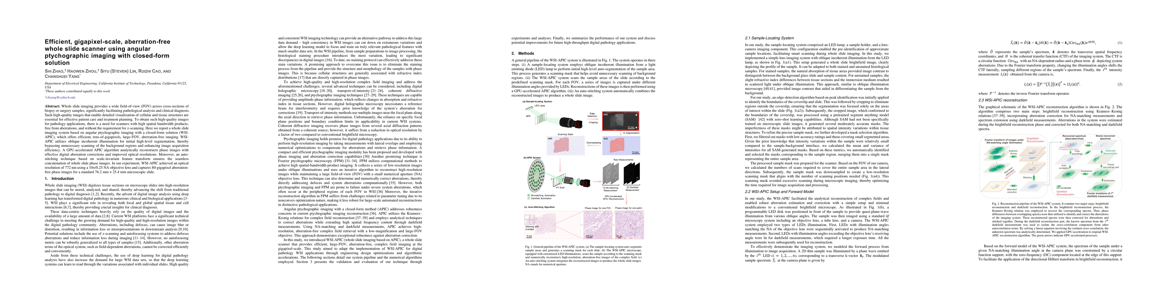
Whole slide imaging provides a wide field-of-view (FOV) across cross-sections of biopsy or surgery samples, significantly facilitating pathological analysis and clinical diagnosis. Such high-quality images that enable detailed visualization of cellular and tissue structures are essential for effective patient care and treatment planning. To obtain such high-quality images for pathology applications, there is a need for scanners with high spatial bandwidth products, free from aberrations, and without the requirement for z-scanning. Here we report a whole slide imaging system based on angular ptychographic imaging with a closed-form solution (WSI-APIC), which offers efficient, tens-of-gigapixels, large-FOV, aberration-free imaging. WSI-APIC utilizes oblique incoherent illumination for initial high-level segmentation, thereby bypassing unnecessary scanning of the background regions and enhancing image acquisition efficiency. A GPU-accelerated APIC algorithm analytically reconstructs phase images with effective digital aberration corrections and improved optical resolutions. Moreover, an auto-stitching technique based on scale-invariant feature transform ensures the seamless concatenation of whole slide phase images. In our experiment, WSI-APIC achieved an optical resolution of 772 nm using a 10x/0.25 NA objective lens and captures 80-gigapixel aberration-free phase images for a standard 76.2 mm x 25.4 mm microscopic slide.
AI Key Findings
Get AI-generated insights about this paper's methodology, results, significance, and more — seven facets brought into focus.
Impact
Authors
PDF Preview
Citation Network
Current paper (gray), citations (green), references (blue)
Display is limited for performance on very large graphs.
Discussion 0