Efficient liver segmentation with 3D CNN using computed tomography scans
Publication
Metrics
AI Quick Summary
This paper proposes an efficient liver segmentation framework using a 3D CNN DeepMedic model to segment liver regions from CT scans, improving detection of liver tumors. The model, with dual input pathways, achieved high accuracy metrics, demonstrating its effectiveness for medical treatment.
Paper Preview
Abstract
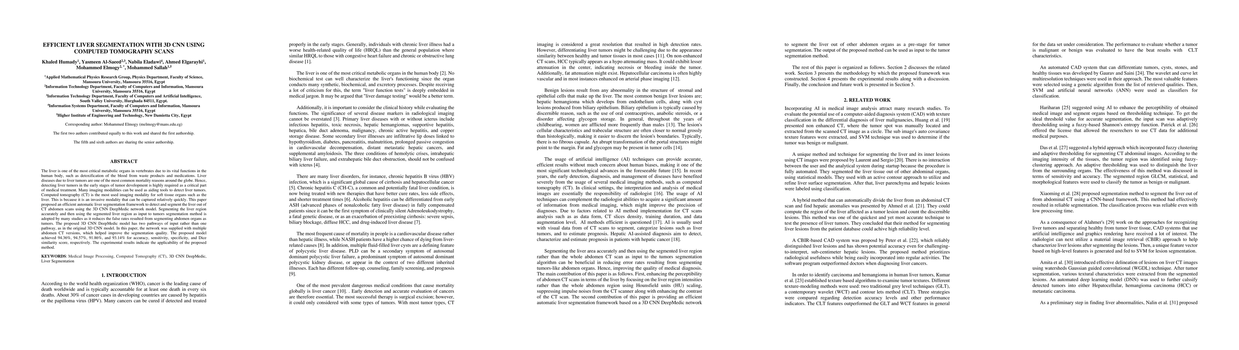
The liver is one of the most critical metabolic organs in vertebrates due to its vital functions in the human body, such as detoxification of the blood from waste products and medications. Liver diseases due to liver tumors are one of the most common mortality reasons around the globe. Hence, detecting liver tumors in the early stages of tumor development is highly required as a critical part of medical treatment. Many imaging modalities can be used as aiding tools to detect liver tumors. Computed tomography (CT) is the most used imaging modality for soft tissue organs such as the liver. This is because it is an invasive modality that can be captured relatively quickly. This paper proposed an efficient automatic liver segmentation framework to detect and segment the liver out of CT abdomen scans using the 3D CNN DeepMedic network model. Segmenting the liver region accurately and then using the segmented liver region as input to tumors segmentation method is adopted by many studies as it reduces the false rates resulted from segmenting abdomen organs as tumors. The proposed 3D CNN DeepMedic model has two pathways of input rather than one pathway, as in the original 3D CNN model. In this paper, the network was supplied with multiple abdomen CT versions, which helped improve the segmentation quality. The proposed model achieved 94.36%, 94.57%, 91.86%, and 93.14% for accuracy, sensitivity, specificity, and Dice similarity score, respectively. The experimental results indicate the applicability of the proposed method.
AI Key Findings
Get AI-generated insights about this paper's methodology, results, significance, and more — seven facets brought into focus.
Impact
Paper Details
Authors
PDF Preview
Key Terms
Citation Network
Current paper (gray), citations (green), references (blue)
Display is limited for performance on very large graphs.
Discussion 0