Multi-target and multi-stage liver lesion segmentation and detection in multi-phase computed tomography scans
Publication
Metrics
AI Quick Summary
This research proposes a multi-stage neural network for liver lesion detection in multi-phase CT scans, improving segmentation performance by 1.6% and reducing variability by 8% compared to existing models. The method uses a three-stage approach combining information from single-phase and multi-phase segmentation models.
Paper Preview
Abstract
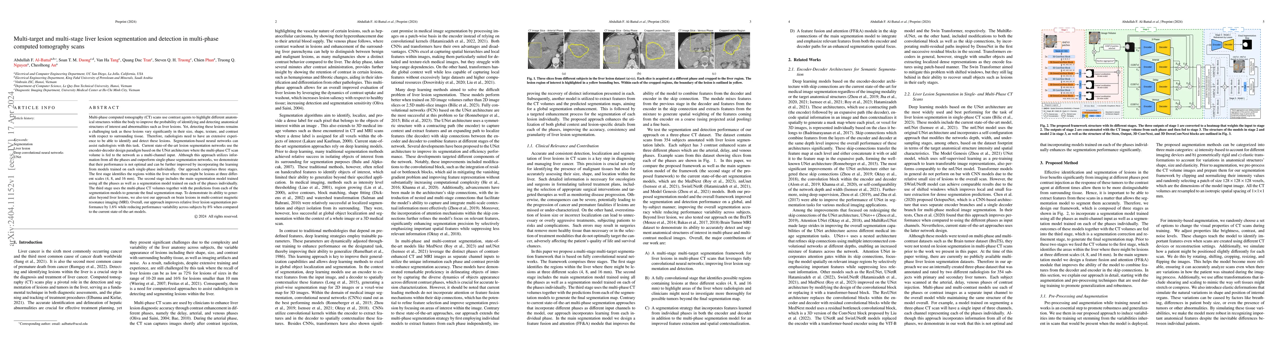
Multi-phase computed tomography (CT) scans use contrast agents to highlight different anatomical structures within the body to improve the probability of identifying and detecting anatomical structures of interest and abnormalities such as liver lesions. Yet, detecting these lesions remains a challenging task as these lesions vary significantly in their size, shape, texture, and contrast with respect to surrounding tissue. Therefore, radiologists need to have an extensive experience to be able to identify and detect these lesions. Segmentation-based neural networks can assist radiologists with this task. Current state-of-the-art lesion segmentation networks use the encoder-decoder design paradigm based on the UNet architecture where the multi-phase CT scan volume is fed to the network as a multi-channel input. Although this approach utilizes information from all the phases and outperform single-phase segmentation networks, we demonstrate that their performance is not optimal and can be further improved by incorporating the learning from models trained on each single-phase individually. Our approach comprises three stages. The first stage identifies the regions within the liver where there might be lesions at three different scales (4, 8, and 16 mm). The second stage includes the main segmentation model trained using all the phases as well as a segmentation model trained on each of the phases individually. The third stage uses the multi-phase CT volumes together with the predictions from each of the segmentation models to generate the final segmentation map. Overall, our approach improves relative liver lesion segmentation performance by 1.6% while reducing performance variability across subjects by 8% when compared to the current state-of-the-art models.
AI Key Findings
Get AI-generated insights about this paper's methodology, results, significance, and more — seven facets brought into focus.
Impact
Paper Details
Authors
PDF Preview
Key Terms
Citation Network
Current paper (gray), citations (green), references (blue)
Display is limited for performance on very large graphs.
Discussion 0