01
MethodologyHow they did it
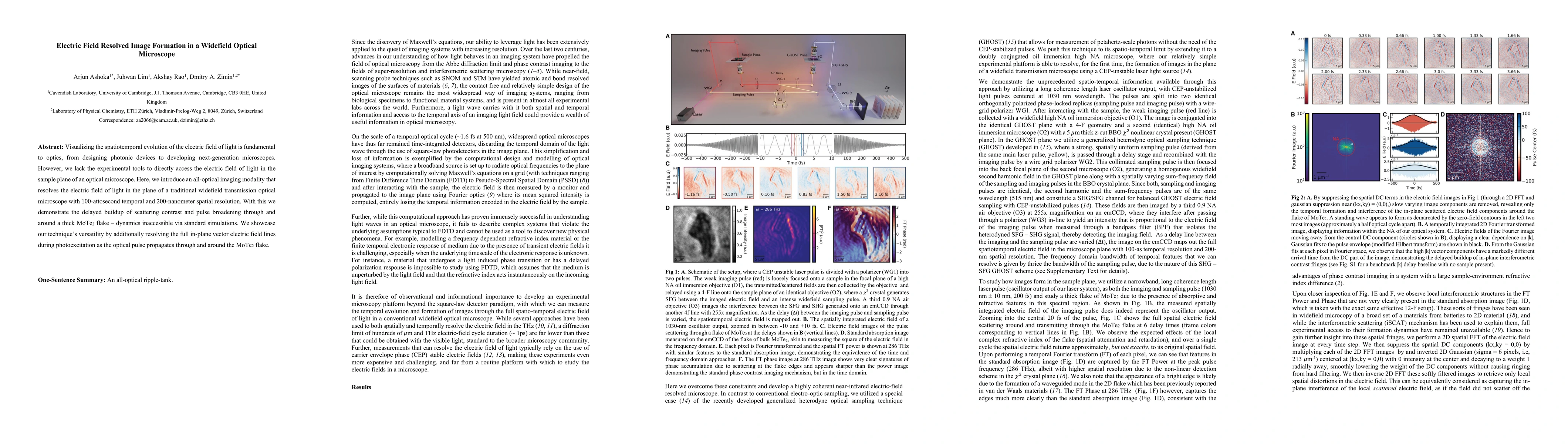
All-optical imaging using a generalized heterodyne optical sampling (GHOST) scheme with CEP‑unstable 1030 nm pulses, split into imaging and sampling replicas, a high‑NA oil immersion objective, a z‑cut BBO crystal for SHG/SFG detection, and an emCCD to capture the spatiotemporal electric field with 100‑as temporal and 200‑nm spatial resolution
Discussion 0