Enabling Collagen Quantification on HE-stained Slides Through Stain Deconvolution and Restained HE-HES
Publication
Metrics
AI Quick Summary
This paper presents a method to quantify collagen in HE-stained slides using stain deconvolution and a trained UNet model to digitally recreate HES images, thereby eliminating the need for additional Saffron staining. The model achieved a Mean Absolute Error of 0.0668, aiming to enhance clinical workflows and reduce reagent costs.
Paper Preview
Abstract
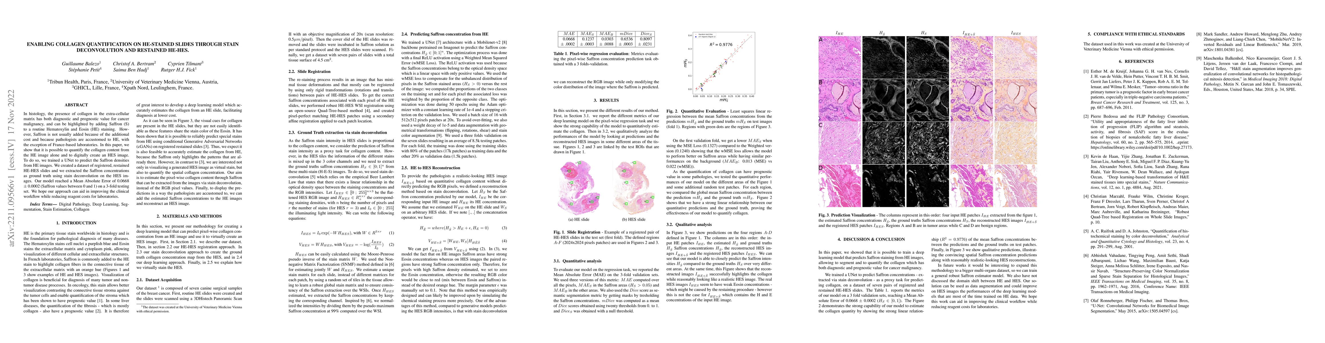
In histology, the presence of collagen in the extra-cellular matrix has both diagnostic and prognostic value for cancer malignancy, and can be highlighted by adding Saffron (S) to a routine Hematoxylin and Eosin (HE) staining. However, Saffron is not usually added because of the additional cost and because pathologists are accustomed to HE, with the exception of France-based laboratories. In this paper, we show that it is possible to quantify the collagen content from the HE image alone and to digitally create an HES image. To do so, we trained a UNet to predict the Saffron densities from HE images. We created a dataset of registered, restained HE-HES slides and we extracted the Saffron concentrations as ground truth using stain deconvolution on the HES images. Our model reached a Mean Absolute Error of 0.0668 $\pm$ 0.0002 (Saffron values between 0 and 1) on a 3-fold testing set. We hope our approach can aid in improving the clinical workflow while reducing reagent costs for laboratories.
AI Key Findings
Get AI-generated insights about this paper's methodology, results, significance, and more — seven facets brought into focus.
Impact
Paper Details
Authors
PDF Preview
Key Terms
Citation Network
Current paper (gray), citations (green), references (blue)
Display is limited for performance on very large graphs.
Discussion 0