Publication
Metrics
Quick Actions
Quick Answers
What methodology did the authors use?
The study combined 48 dimension reduction algorithms (DRAs) and 25 classification algorithms (CAs) to evaluate 1,200 learning pipelines across three multicenter datasets using rotational validation and external testing. Stability-aware scoring integrated standard deviation across cross-validation, rotations, and external tests to prioritize reproducible models. More in Methodology →
What are the key results?
MI+ETr and FEW+ETr demonstrated consistent high accuracy (0.87–0.98 externally) and stability across datasets — Rotational validation showed mean accuracies of ~0.94 in cross-validation and ~0.93 in external testing More in Key Results →
Why is this work significant?
This research addresses critical gaps in medical imaging by providing a robust framework for reproducible model selection, reducing reliance on gadolinium-based contrast agents (GBCAs) which have safety and logistical concerns. It offers a pathway for safer, more accessible imaging in clinical settings. More in Significance →
What are the main limitations?
Relied solely on T1WI-derived radiomic features — ROC-AUC variability highlights need for better threshold-independent discrimination More in Limitations →
Abstract
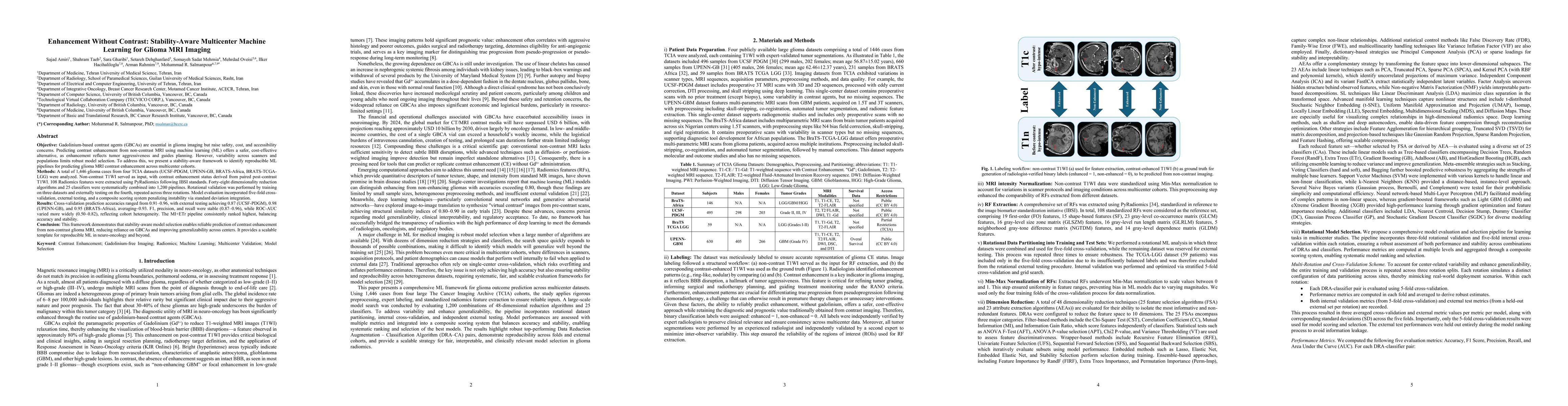
Gadolinium-based contrast agents (GBCAs) are central to glioma imaging but raise safety, cost, and accessibility concerns. Predicting contrast enhancement from non-contrast MRI using machine learning (ML) offers a safer alternative, as enhancement reflects tumor aggressiveness and informs treatment planning. Yet scanner and cohort variability hinder robust model selection. We propose a stability-aware framework to identify reproducible ML pipelines for multicenter prediction of glioma MRI contrast enhancement. We analyzed 1,446 glioma cases from four TCIA datasets (UCSF-PDGM, UPENN-GB, BRATS-Africa, BRATS-TCGA-LGG). Non-contrast T1WI served as input, with enhancement derived from paired post-contrast T1WI. Using PyRadiomics under IBSI standards, 108 features were extracted and combined with 48 dimensionality reduction methods and 25 classifiers, yielding 1,200 pipelines. Rotational validation was trained on three datasets and tested on the fourth. Cross-validation prediction accuracies ranged from 0.91 to 0.96, with external testing achieving 0.87 (UCSF-PDGM), 0.98 (UPENN-GB), and 0.95 (BRATS-Africa), with an average of 0.93. F1, precision, and recall were stable (0.87 to 0.96), while ROC-AUC varied more widely (0.50 to 0.82), reflecting cohort heterogeneity. The MI linked with ETr pipeline consistently ranked highest, balancing accuracy and stability. This framework demonstrates that stability-aware model selection enables reliable prediction of contrast enhancement from non-contrast glioma MRI, reducing reliance on GBCAs and improving generalizability across centers. It provides a scalable template for reproducible ML in neuro-oncology and beyond.
AI Key Findings
Generated Oct 02, 2025
Methodology — What approach did the authors take?
The study combined 48 dimension reduction algorithms (DRAs) and 25 classification algorithms (CAs) to evaluate 1,200 learning pipelines across three multicenter datasets using rotational validation and external testing. Stability-aware scoring integrated standard deviation across cross-validation, rotations, and external tests to prioritize reproducible models.
Key Results — What are the main findings?
- MI+ETr and FEW+ETr demonstrated consistent high accuracy (0.87–0.98 externally) and stability across datasets
- Rotational validation showed mean accuracies of ~0.94 in cross-validation and ~0.93 in external testing
- Accuracy, F1, precision, and recall remained stable across internal and external evaluations, while ROC-AUC showed greater variability
Significance — Why does this research matter?
This research addresses critical gaps in medical imaging by providing a robust framework for reproducible model selection, reducing reliance on gadolinium-based contrast agents (GBCAs) which have safety and logistical concerns. It offers a pathway for safer, more accessible imaging in clinical settings.
Technical Contribution — What is the technical contribution?
Developed a stability-aware framework that systematically evaluates DRA-CA combinations using rotational validation and integrates standard deviation as a key metric for model reproducibility
Novelty — What is new about this work?
Systematically tested 1,200 pipelines across multicenter datasets with rotational validation, emphasizing stability over peak performance, and provided a comprehensive evaluation of reproducible model selection in glioma imaging
Limitations — What are the limitations of this study?
- Relied solely on T1WI-derived radiomic features
- ROC-AUC variability highlights need for better threshold-independent discrimination
Future Work — What did the authors propose for future work?
- Incorporate multi-parametric MRI and deep learning features
- Apply explainability methods like SHAP values for interpretability
- Explore federated learning for improved generalization without data sharing
Paper Details
How to Cite This Paper
@article{rahmim2025enhancement,
title = {Enhancement Without Contrast: Stability-Aware Multicenter Machine
Learning for Glioma MRI Imaging},
author = {Rahmim, Arman and Hacihaliloglu, Ilker and Oveisi, Mehrdad and others},
year = {2025},
eprint = {2509.10767},
archivePrefix = {arXiv},
primaryClass = {cs.CV},
}
Rahmim, A., Hacihaliloglu, I., Oveisi, M., Salmanpour, M., Amiri, S., Gharibi, S., Dehghanfard, S., Taeb, S., & Mehrnia, S. (2025). Enhancement Without Contrast: Stability-Aware Multicenter Machine
Learning for Glioma MRI Imaging. arXiv. https://arxiv.org/abs/2509.10767
Rahmim, Arman, et al. "Enhancement Without Contrast: Stability-Aware Multicenter Machine
Learning for Glioma MRI Imaging." arXiv, 2025, arxiv.org/abs/2509.10767.
PDF Preview
Similar Papers
Found 4 papersAdaptive PromptNet For Auxiliary Glioma Diagnosis without Contrast-Enhanced MRI
Weijian Huang, Yusong Lin, Shanshan Wang et al.
Radiogenomics: A Machine Learning Augmentation to MRI-Based Glioma Profiling.
Dsouza, Ashel Chelsea, Dsouza, Aster Chelson
Comments (0)