Academic Profile
Statistics
Similar Authors
Papers on arXiv
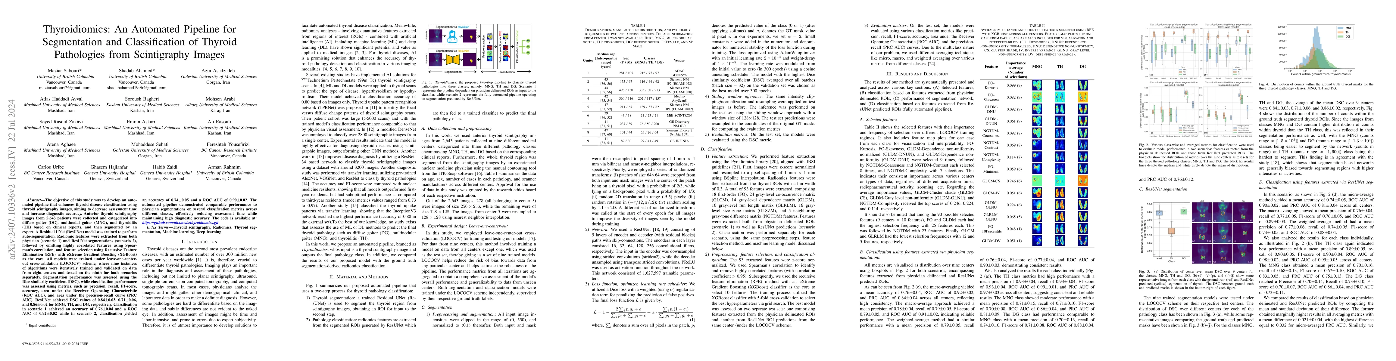
The objective of this study was to develop an automated pipeline that enhances thyroid disease classification using thyroid scintigraphy images, aiming to decrease assessment time and increase diagnos...
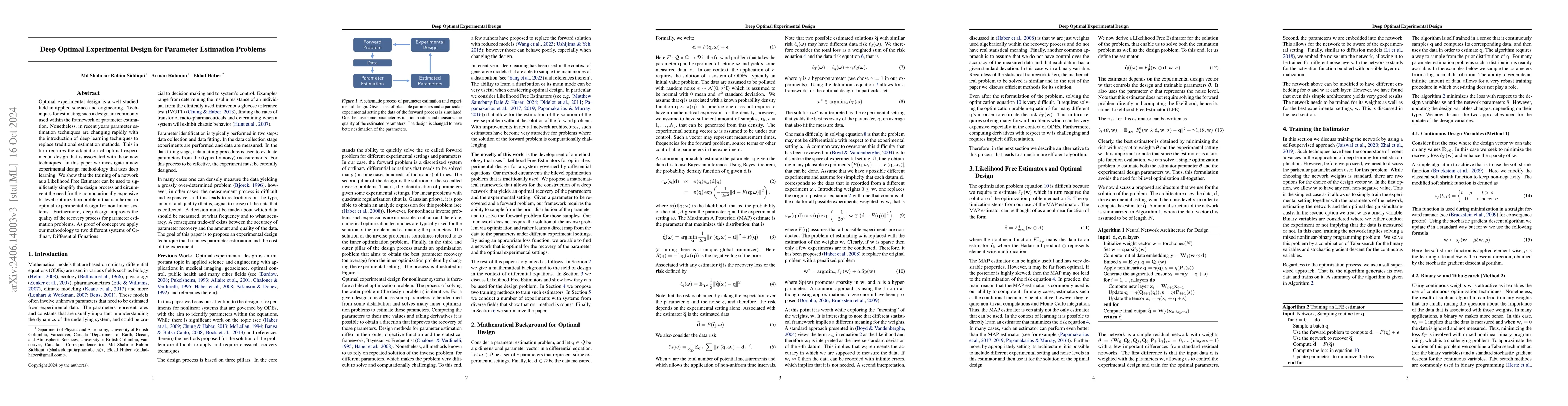
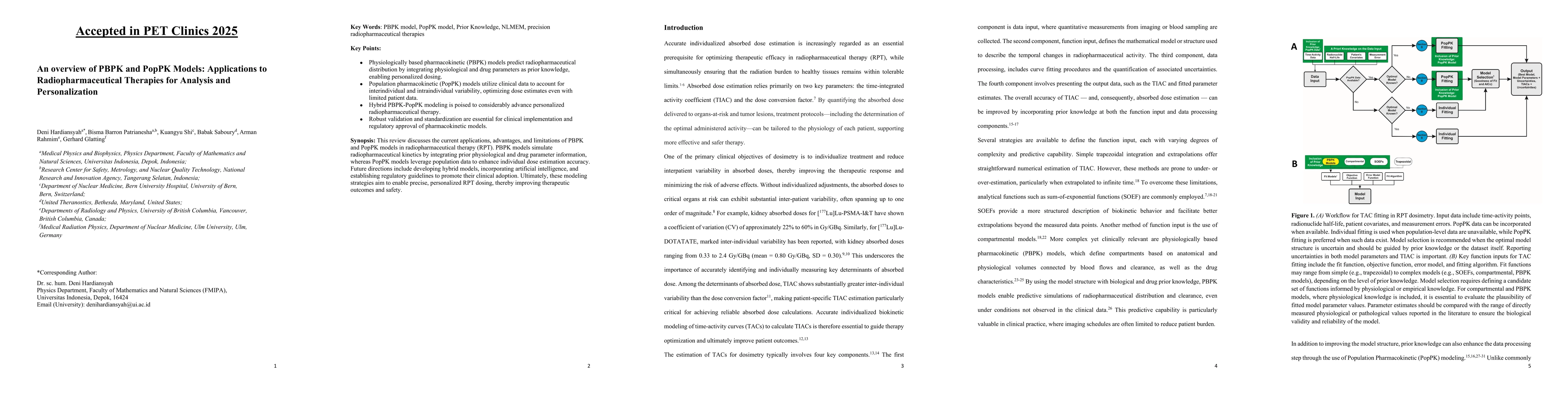
Optimal experimental design is a well studied field in applied science and engineering. Techniques for estimating such a design are commonly used within the framework of parameter estimation. Noneth...
The 2nd SNMMI Artificial Intelligence (AI) Summit, organized by the SNMMI AI Task Force, took place in Bethesda, MD, on February 29 - March 1, 2024. Bringing together various community members and s...
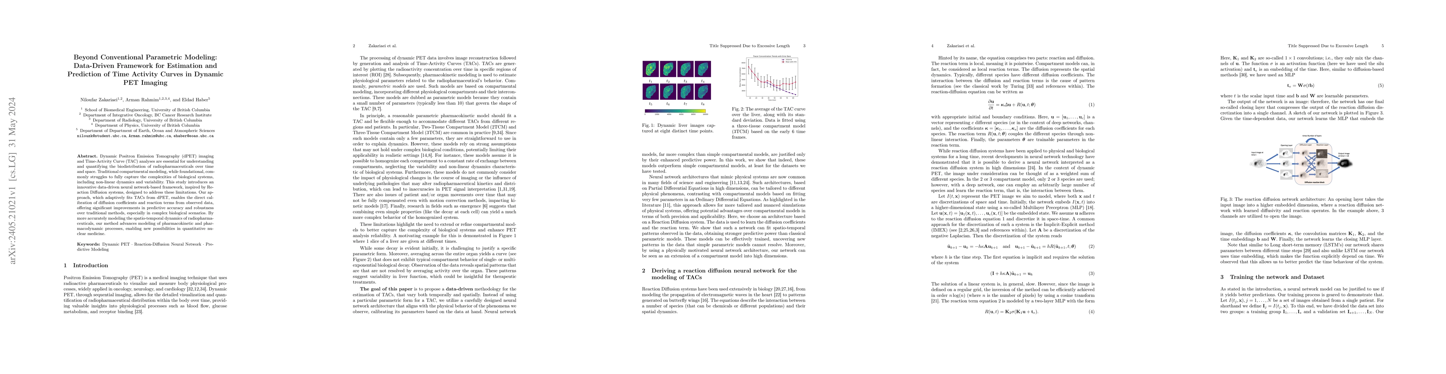
Dynamic Positron Emission Tomography (dPET) imaging and Time-Activity Curve (TAC) analyses are essential for understanding and quantifying the biodistribution of radiopharmaceuticals over time and s...
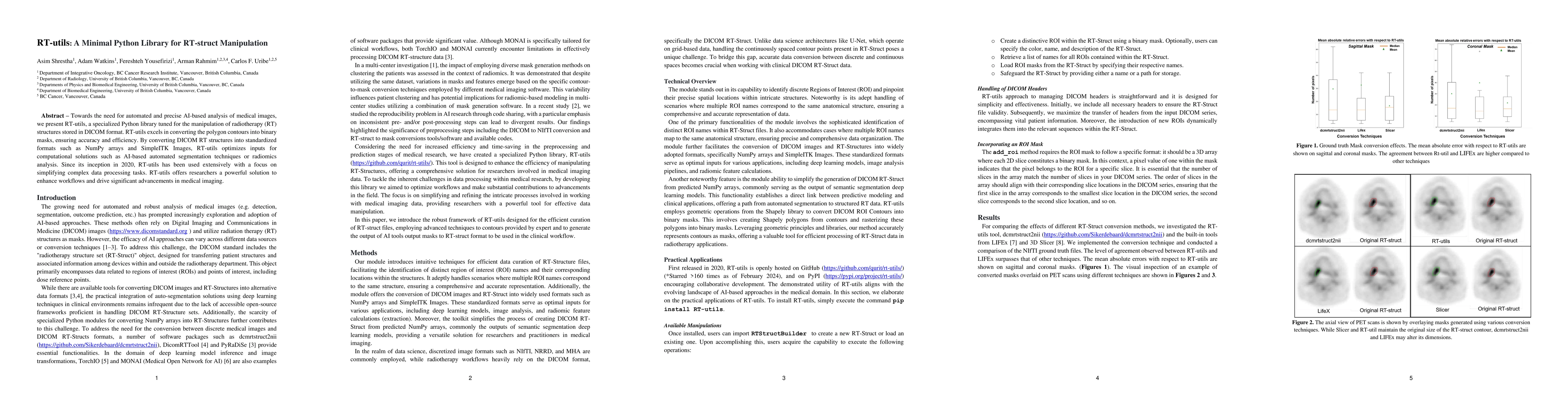
Towards the need for automated and precise AI-based analysis of medical images, we present RT-utils, a specialized Python library tuned for the manipulation of radiotherapy (RT) structures stored in...
$^{18}$F-Fluoromisonidazole ($^{18}$F-FMISO) is a highly promising positron emission tomography radiopharmaceutical for identifying hypoxic regions in solid tumors. This research employs spatiotempo...
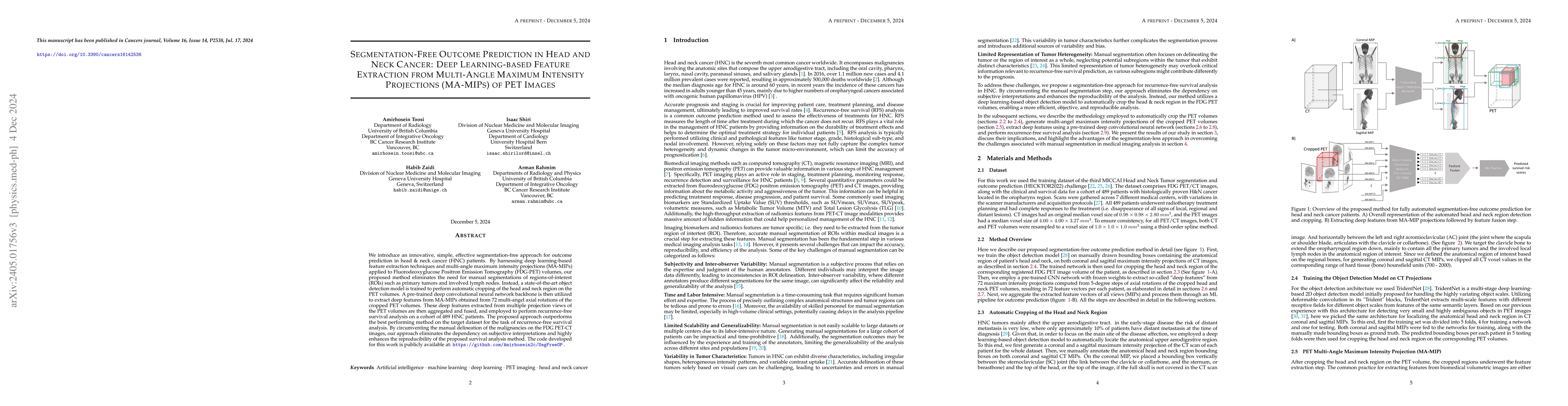
We introduce an innovative, simple, effective segmentation-free approach for outcome prediction in head \& neck cancer (HNC) patients. By harnessing deep learning-based feature extraction techniques...
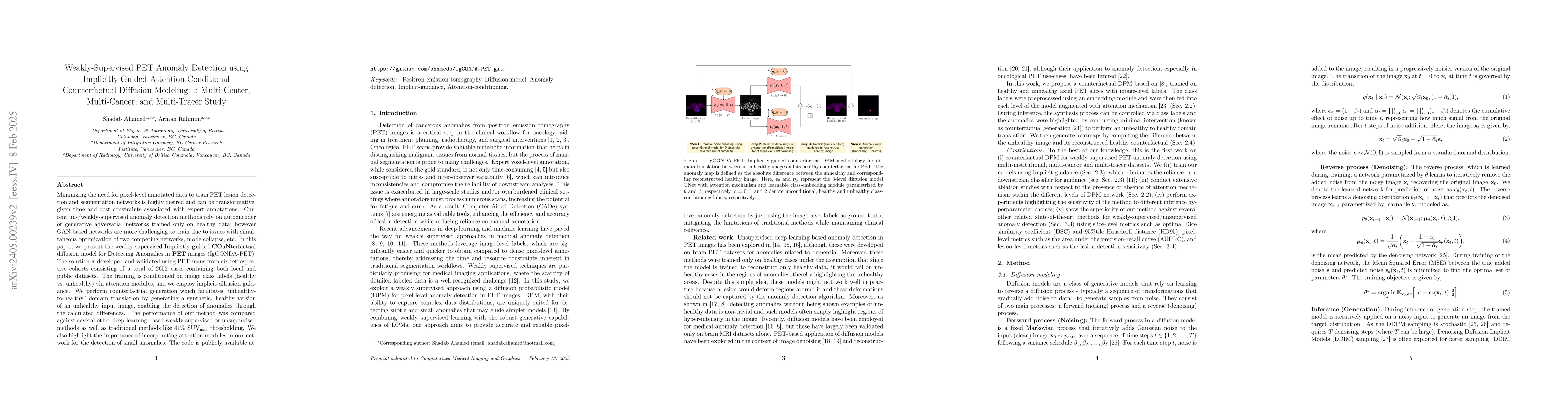
Minimizing the need for pixel-level annotated data for training PET anomaly segmentation networks is crucial, particularly due to time and cost constraints related to expert annotations. Current un-...
For whole-body (WB) kinetic modeling based on a typical PET scanner, a multi-pass multi-bed scanning protocol is necessary given the limited axial field-of-view. Such a protocol introduces loss of e...
The effectiveness of Deep Neural Networks (DNNs) heavily relies on the abundance and accuracy of available training data. However, collecting and annotating data on a large scale is often both costl...
Automated slice classification is clinically relevant since it can be incorporated into medical image segmentation workflows as a preprocessing step that would flag slices with a higher probability ...
Accurate detection and segmentation of diffuse large B-cell lymphoma (DLBCL) from PET images has important implications for estimation of total metabolic tumor volume, radiomics analysis, surgical i...
The purpose was to investigate the spatial heterogeneity of prostate-specific membrane antigen (PSMA) positron emission tomography (PET) uptake within parotid glands. We aim to quantify patterns in ...
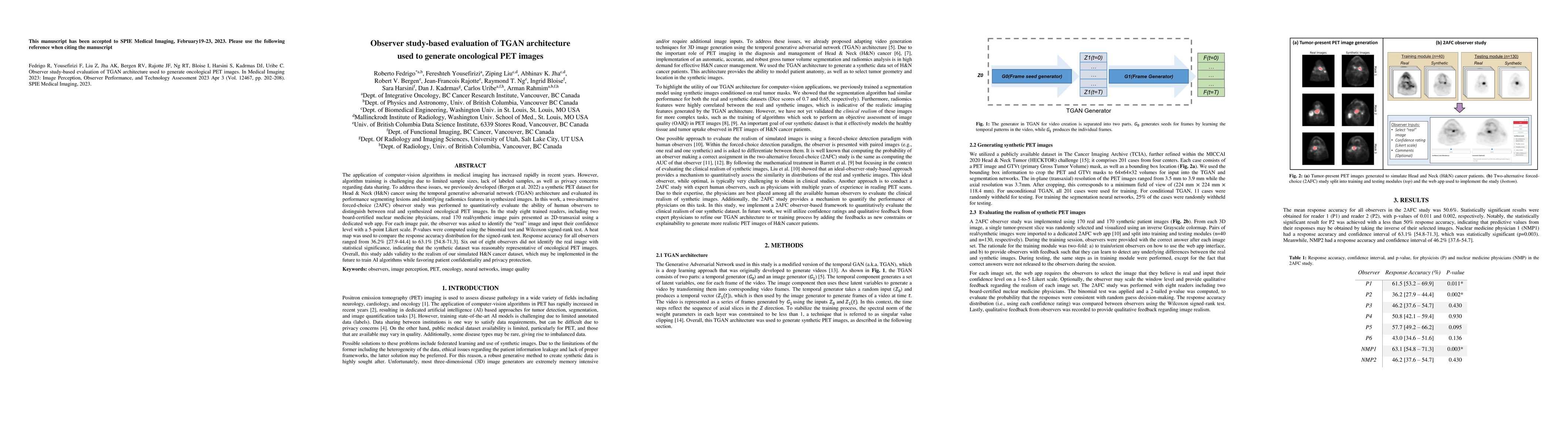
The application of computer-vision algorithms in medical imaging has increased rapidly in recent years. However, algorithm training is challenging due to limited sample sizes, lack of labeled sample...
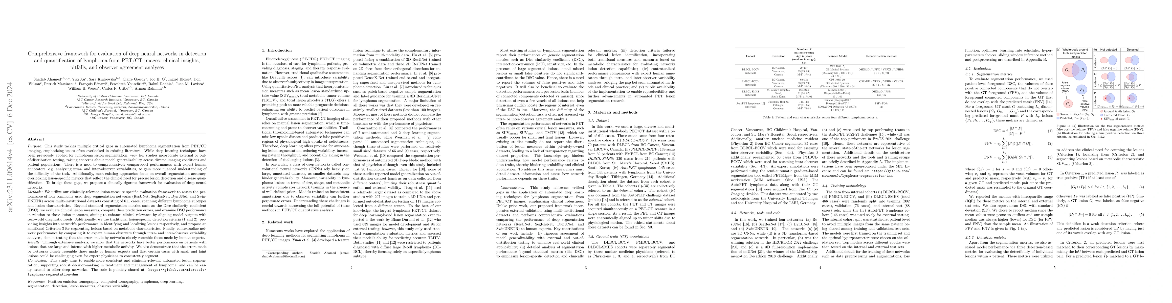
This study performs comprehensive evaluation of four neural network architectures (UNet, SegResNet, DynUNet, and SwinUNETR) for lymphoma lesion segmentation from PET/CT images. These networks were t...
Automated segmentation of cancerous lesions in PET/CT images is a vital initial task for quantitative analysis. However, it is often challenging to train deep learning-based segmentation methods to ...
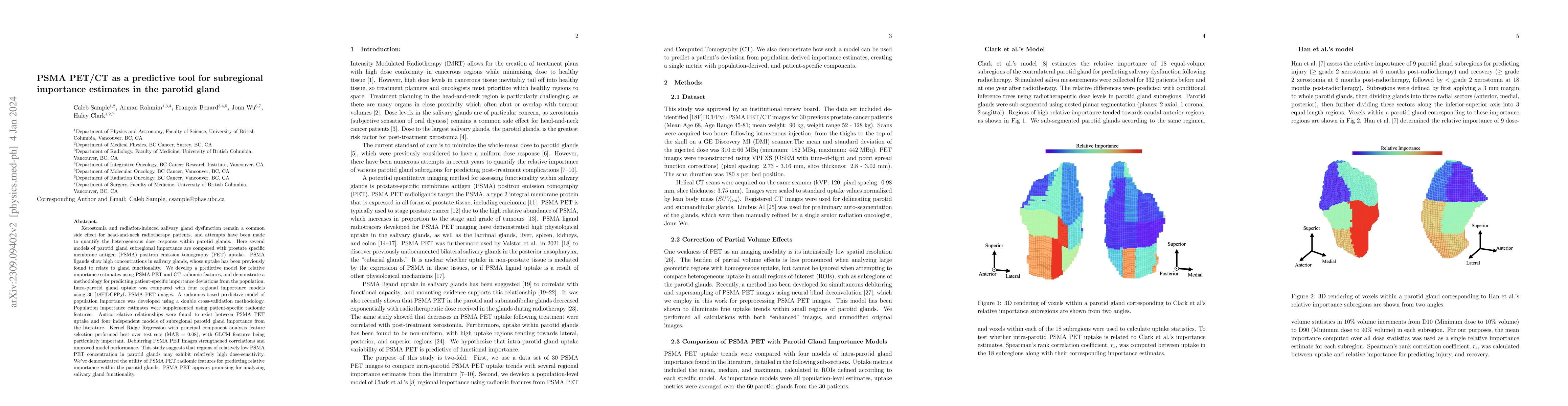
Xerostomia and radiation-induced salivary gland dysfunction remain a common side effect for head-and-neck radiotherapy patients, and attempts have been made to quantify the heterogeneous dose respon...
Background: There is an absence of open-source libraries in emission tomography that (i) use modern and popular backend code to encourage community contributions and (ii) offer support for the multi...
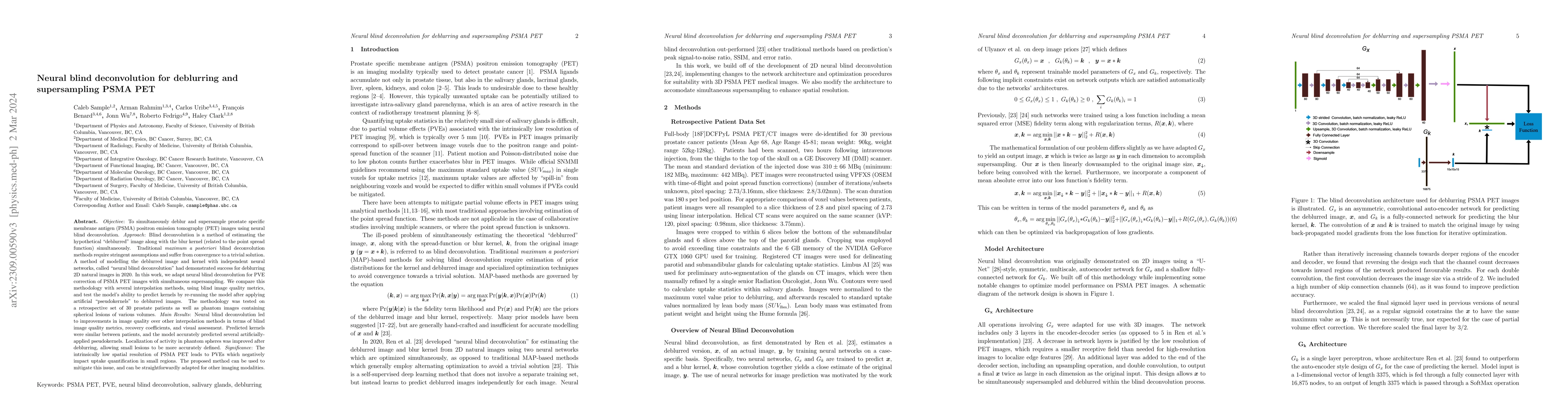
Objective: To simultaneously deblur and supersample prostate specific membrane antigen (PSMA) positron emission tomography (PET) images using neural blind deconvolution. Approach: Blind deconvolutio...
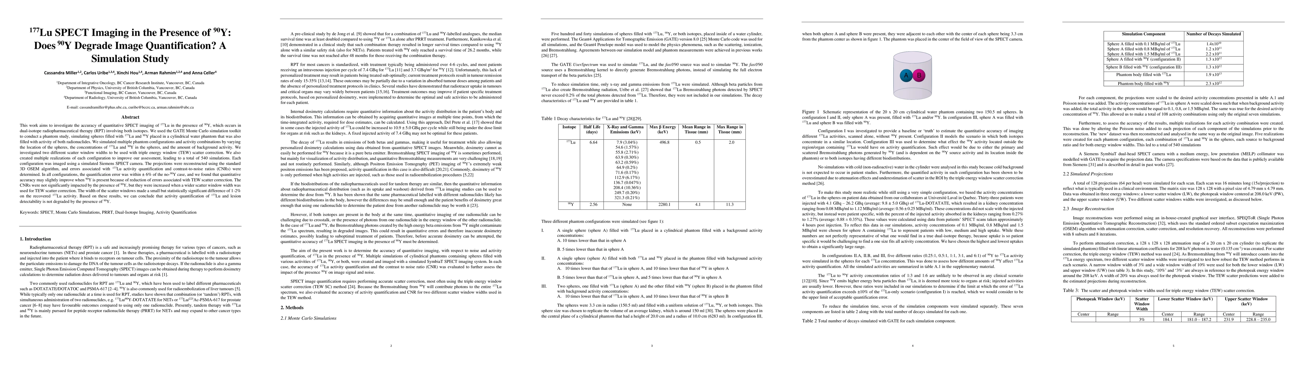
This work aims to investigate the accuracy of quantitative SPECT imaging of $^{177}$Lu in the presence of $^{90}$Y, which occurs in dual-isotope radiopharmaceutical therapy (RPT) involving both isot...
The time-consuming task of manual segmentation challenges routine systematic quantification of disease burden. Convolutional neural networks (CNNs) hold significant promise to reliably identify loca...
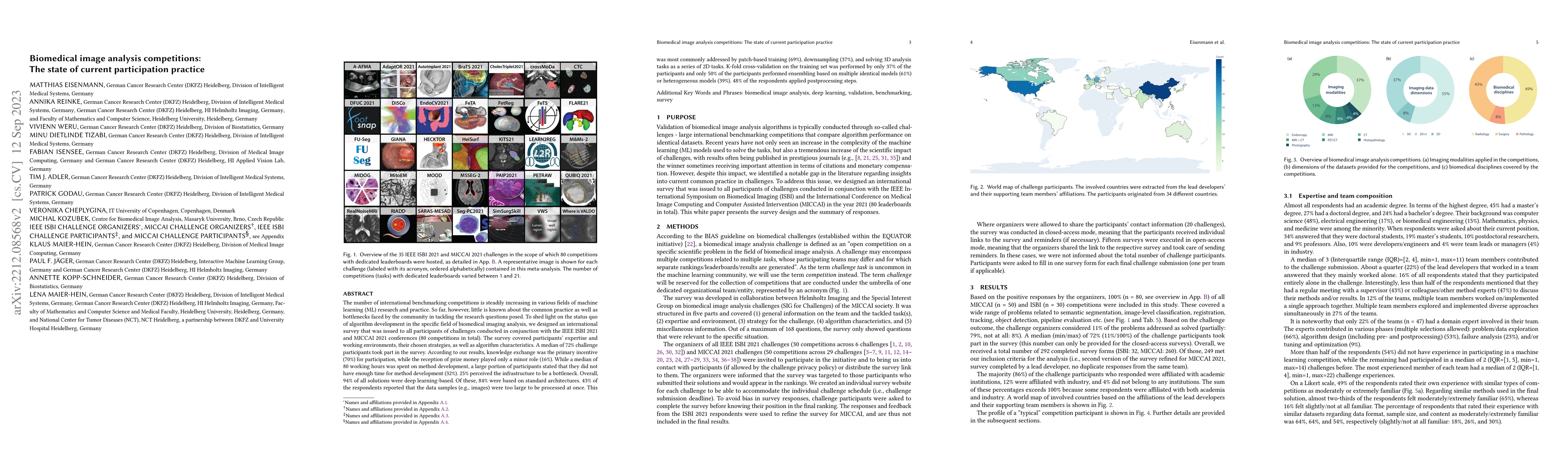
The number of international benchmarking competitions is steadily increasing in various fields of machine learning (ML) research and practice. So far, however, little is known about the common pract...
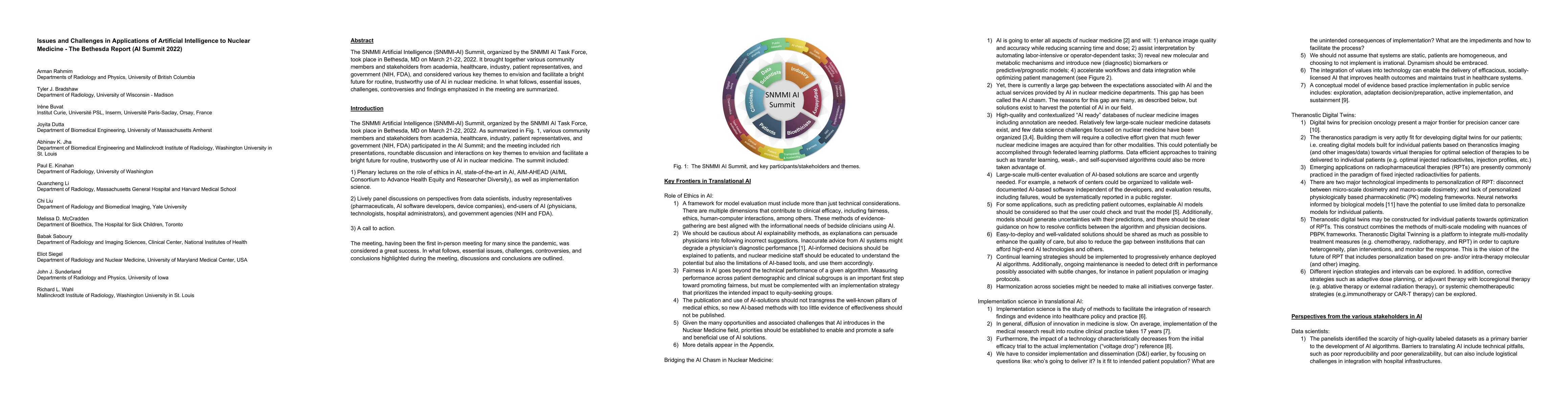
The SNMMI Artificial Intelligence (SNMMI-AI) Summit, organized by the SNMMI AI Task Force, took place in Bethesda, MD on March 21-22, 2022. It brought together various community members and stakehol...
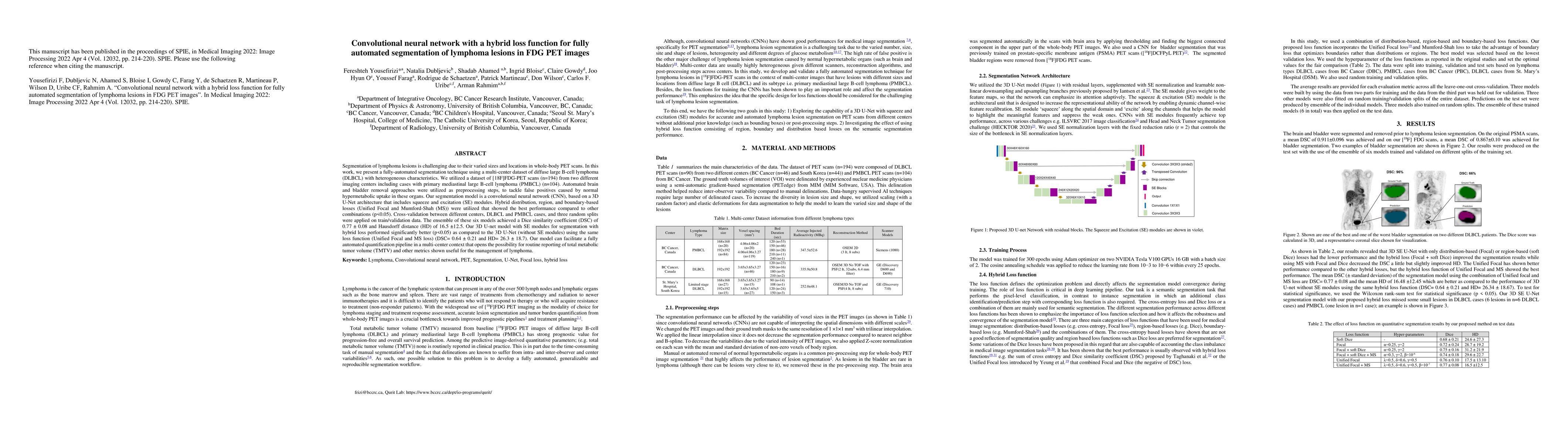
Segmentation of lymphoma lesions is challenging due to their varied sizes and locations in whole-body PET scans. This work presents a fully-automated segmentation technique using a multi-center data...
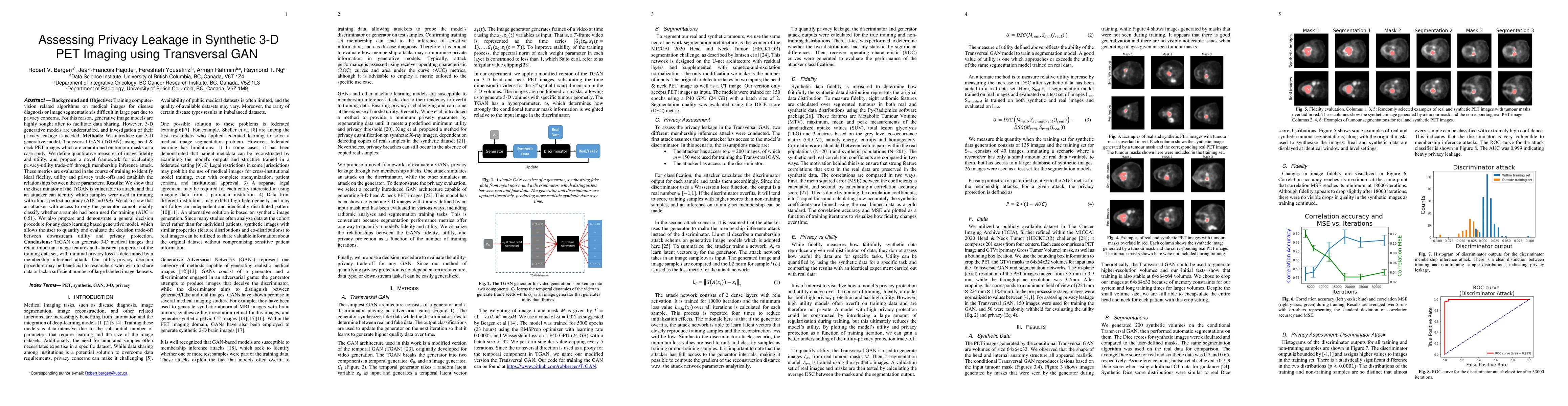
Training computer-vision related algorithms on medical images for disease diagnosis or image segmentation is difficult in large part due to privacy concerns. For this reason, generative image models...
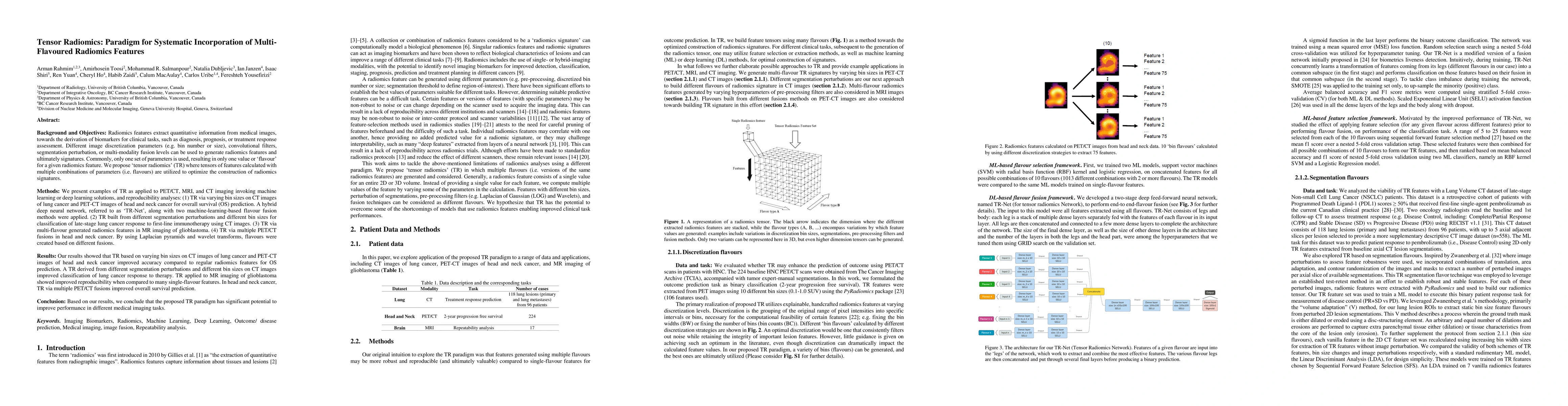
Radiomics features extract quantitative information from medical images, towards the derivation of biomarkers for clinical tasks, such as diagnosis, prognosis, or treatment response assessment. Diff...
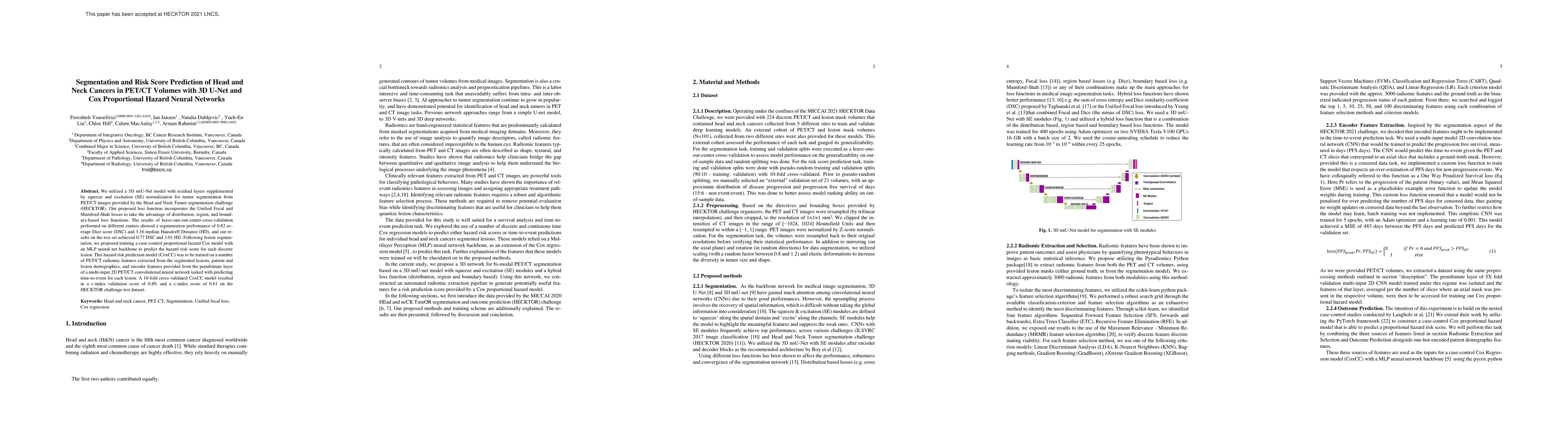
We utilized a 3D nnU-Net model with residual layers supplemented by squeeze and excitation (SE) normalization for tumor segmentation from PET/CT images provided by the Head and Neck Tumor segmentati...
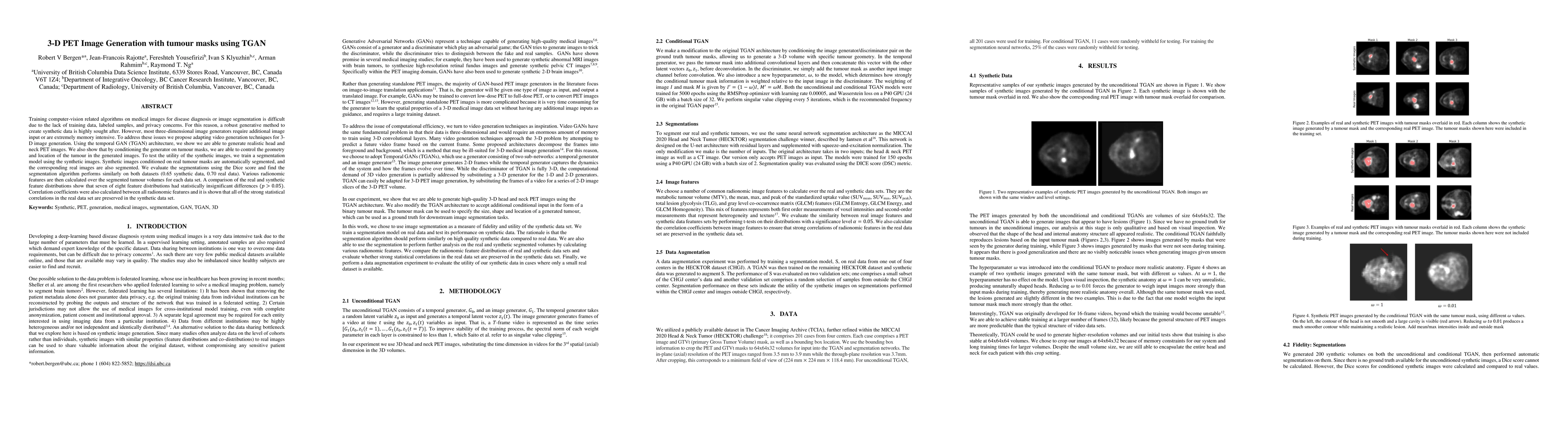
Training computer-vision related algorithms on medical images for disease diagnosis or image segmentation is difficult due to the lack of training data, labeled samples, and privacy concerns. For th...
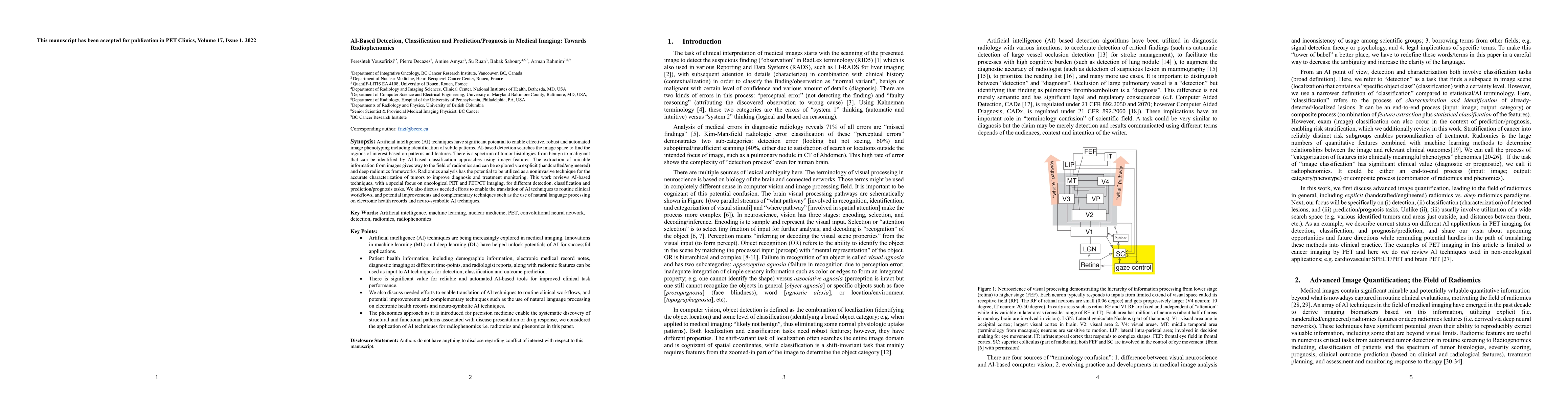
Artificial intelligence (AI) techniques have significant potential to enable effective, robust and automated image phenotyping including identification of subtle patterns. AI-based detection searche...
The field of artificial intelligence (AI), regarded as one of the most enigmatic areas of science, has witnessed exponential growth in the past decade including a remarkably wide array of applicatio...
Artificial intelligence (AI) techniques for image-based segmentation have garnered much attention in recent years. Convolutional neural networks (CNNs) have shown impressive results and potential to...
Purpose: The XCAT phantom allows for highly sophisticated multimodality imaging research. It includes a complete set of organs, muscle, bone, soft tissue, while also accounting for age, sex, and bod...
Artificial intelligence (AI) has significant potential to positively impact and advance medical imaging, including positron emission tomography (PET) imaging applications. AI has the ability to enha...
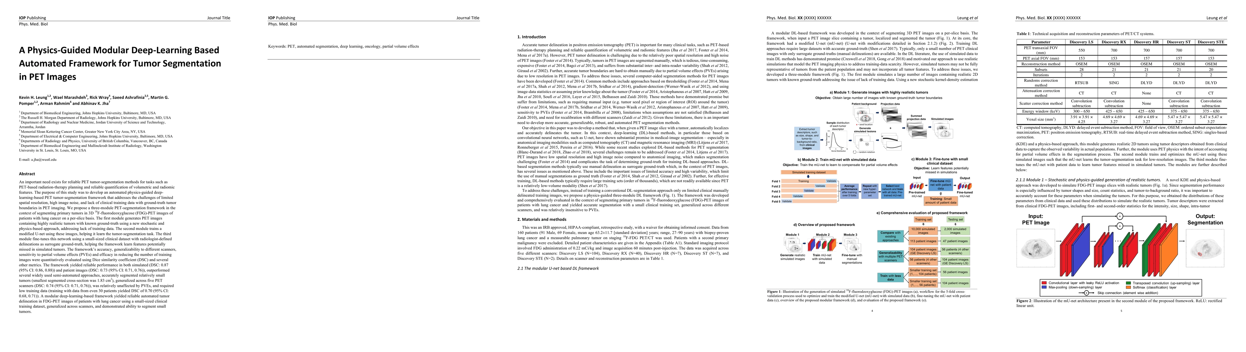
The objective of this study was to develop a PET tumor-segmentation framework that addresses the challenges of limited spatial resolution, high image noise, and lack of clinical training data with g...
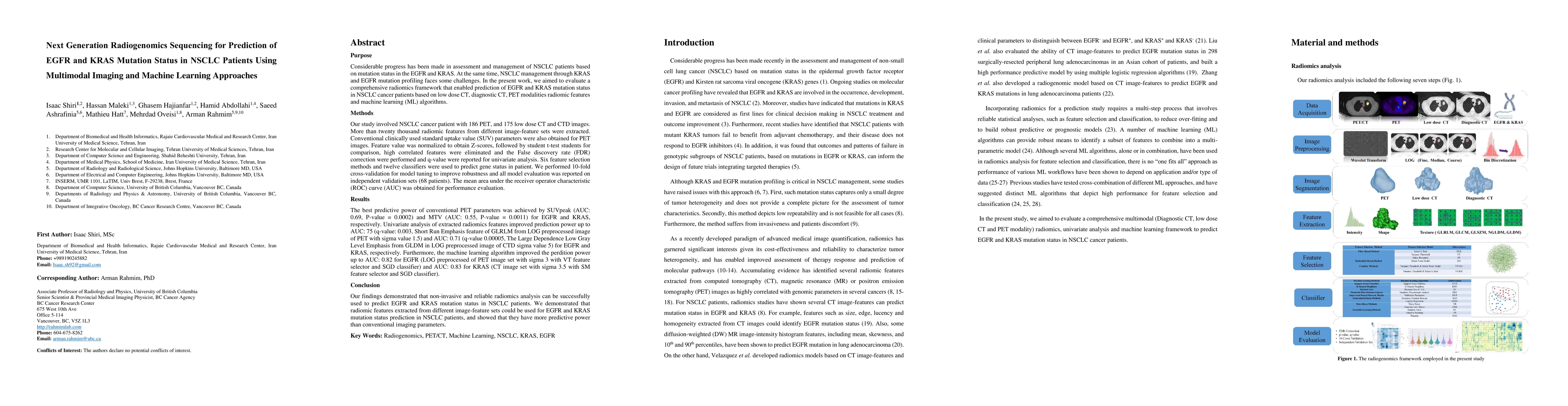
Aim: In the present work, we aimed to evaluate a comprehensive radiomics framework that enabled prediction of EGFR and KRAS mutation status in NSCLC cancer patients based on PET and CT multi-modalit...

The aim of this study was to develop radiomic models using PET/CT radiomic features with different machine learning approaches for finding best predictive epidermal growth factor receptor (EGFR) and...
Prostate specific membrane antigen (PSMA) positron emission tomography/computed tomography (PET/CT) imaging provides a tremendously exciting frontier in visualization of prostate cancer (PCa) metastat...
Purpose: Artificial intelligence (AI) techniques have been extensively utilized for diagnosing and prognosis of several diseases in recent years. This study identifies, appraises and synthesizes publi...
This study evaluates metrics for tasks such as classification, regression, clustering, correlation analysis, statistical tests, segmentation, and image-to-image (I2I) translation. Metrics were compare...
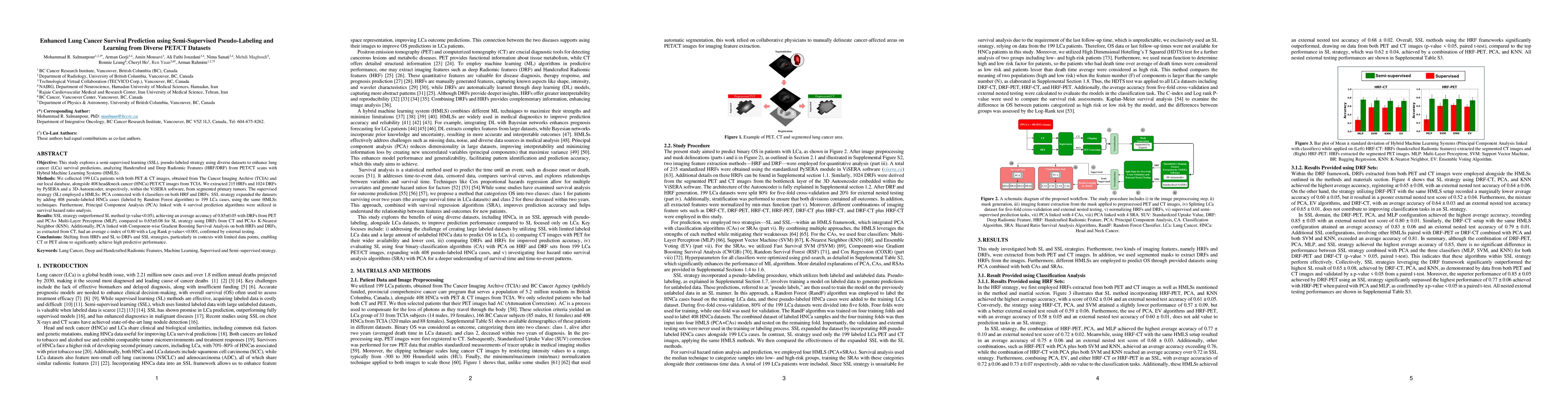
Objective: This study explores a semi-supervised learning (SSL), pseudo-labeled strategy using diverse datasets to enhance lung cancer (LCa) survival predictions, analyzing Handcrafted and Deep Radiom...
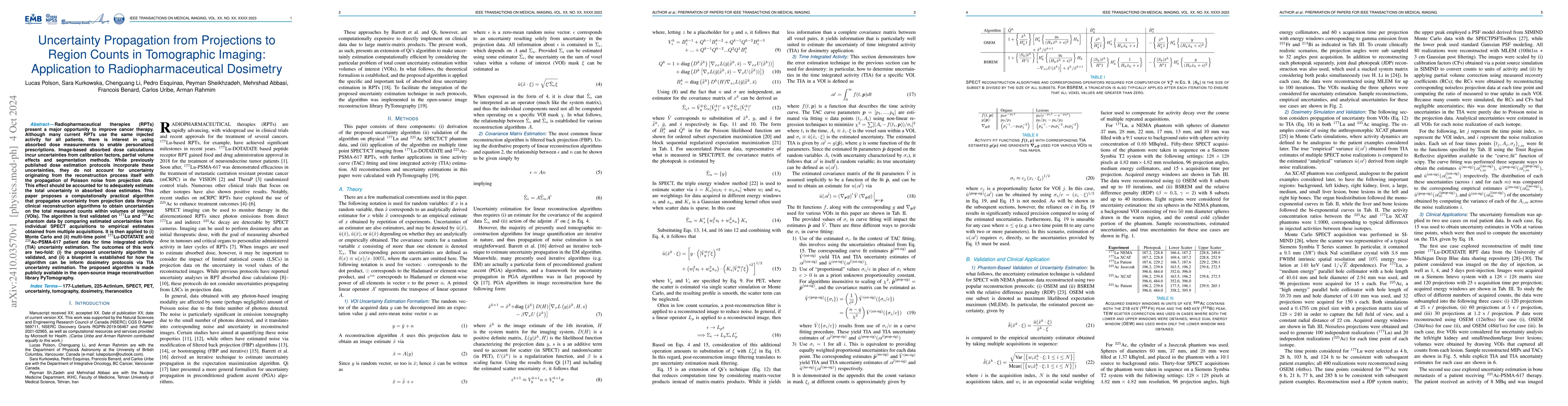
Radiopharmaceutical therapies (RPTs) present a major opportunity to improve cancer therapy. Although many current RPTs use the same injected activity for all patients, there is interest in using absor...
Modeling of the collimator-detector response (CDR) in SPECT reconstruction enables improved resolution and more accurate quantitation, especially for higher energy imaging (e.g.Lu-177 and Ac-225). Suc...
We investigate the connection between visual semantic features defined in PI-RADS and associated risk factors, moving beyond abnormal imaging findings, establishing a shared framework between medical ...
Objectives: Lung cancer poses a significant global health challenge, necessitating improved prognostic methods for personalized treatment. This study introduces a censor-aware semi-supervised learning...
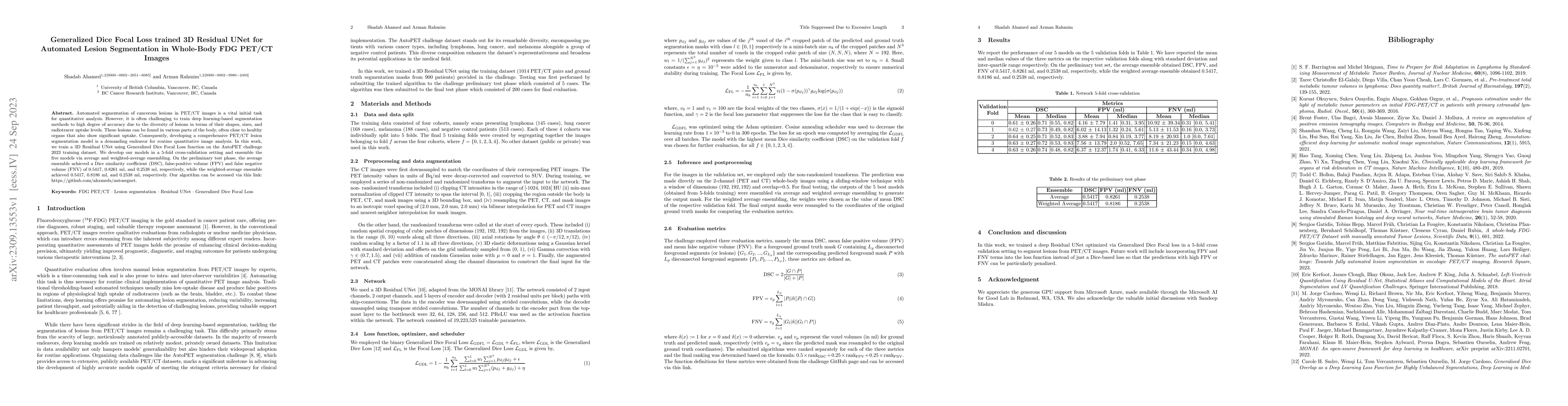
This study proposes a new loss function for deep neural networks, L1-weighted Dice Focal Loss (L1DFL), that leverages L1 norms for adaptive weighting of voxels based on their classification difficulty...
Thyroid scintigraphy is a key imaging modality for diagnosing thyroid disorders. Deep learning models for thyroid scintigraphy classification often face challenges due to limited and imbalanced datase...
This study performs a comprehensive evaluation of quantitative measurements as extracted from automated deep-learning-based segmentation methods, beyond traditional Dice Similarity Coefficient assessm...
Artificial intelligence (AI) holds strong potential for medical diagnostics, yet its clinical adoption is limited by a lack of interpretability and generalizability. This study introduces the Pathobio...
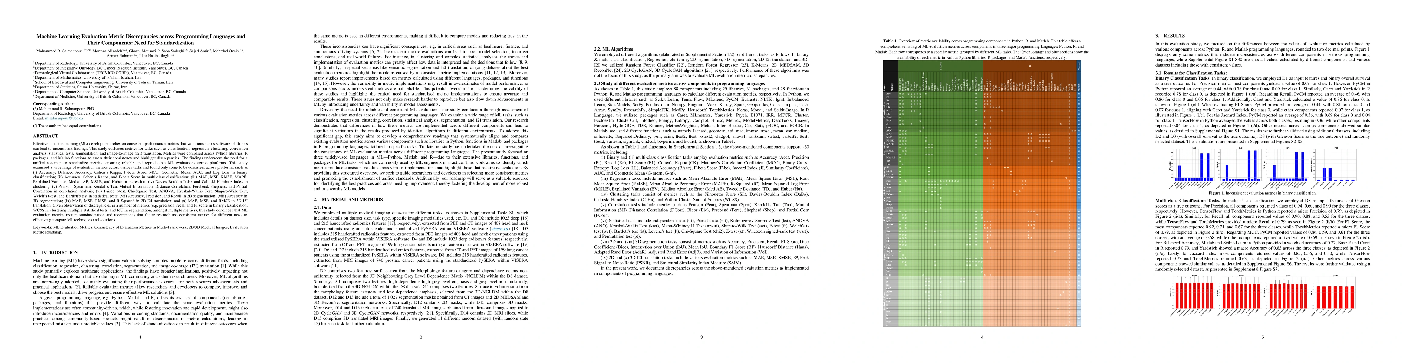
Machine learning (ML) models rely heavily on consistent and accurate performance metrics to evaluate and compare their effectiveness. However, existing libraries often suffer from fragmentation, incon...
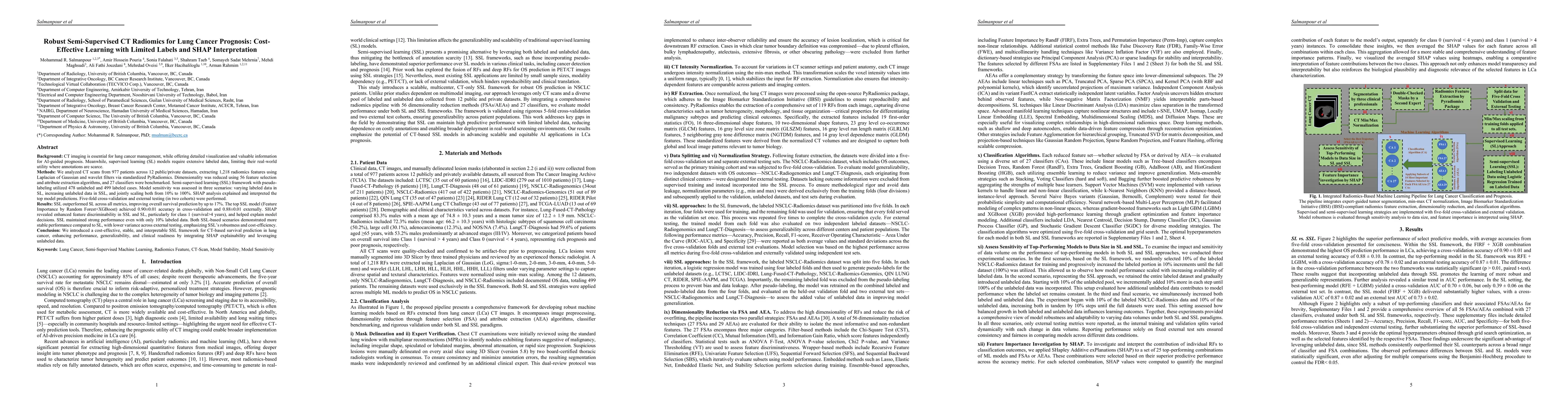
Background: CT imaging is vital for lung cancer management, offering detailed visualization for AI-based prognosis. However, supervised learning SL models require large labeled datasets, limiting thei...
Quantitative imaging (QI) is demonstrating strong promise across multiple clinical applications. For clinical translation of QI methods, objective evaluation on clinically relevant tasks is essential....
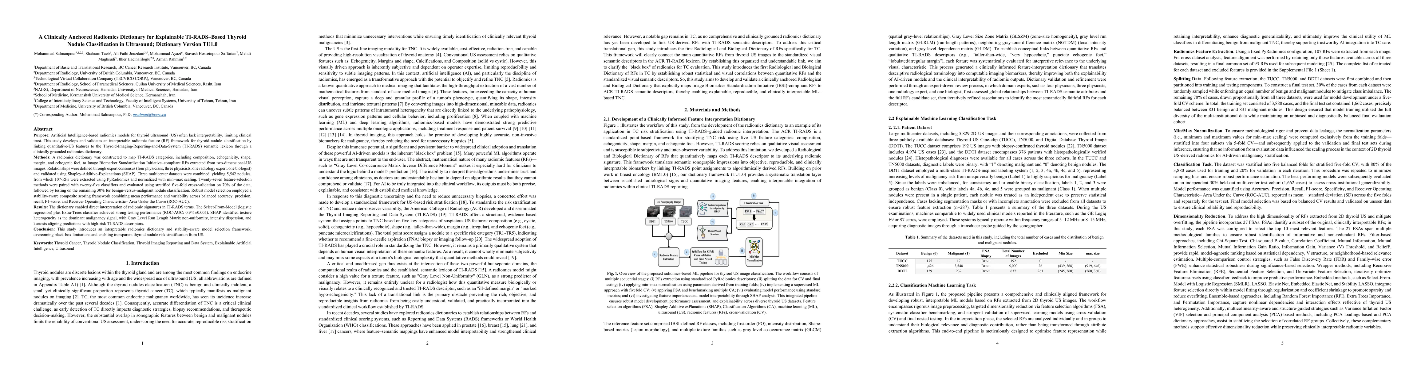
Radiomics-based AI models show promise for breast cancer diagnosis but often lack interpretability, limiting clinical adoption. This study addresses the gap between radiomic features (RF) and the stan...
Machine learning (ML), including deep learning (DL) and radiomics-based methods, is increasingly used for cancer outcome prediction with PET and SPECT imaging. However, the comparative performance of ...
LAFOV PET/CT has the potential to unlock new applications such as ultra-low dose PET/CT imaging, multiplexed imaging, for biomarker development and for faster AI-driven reconstruction, but further wor...
MTV is increasingly recognized as an accurate estimate of disease burden, which has prognostic value, but its implementation has been hindered by the time-consuming need for manual segmentation of ima...
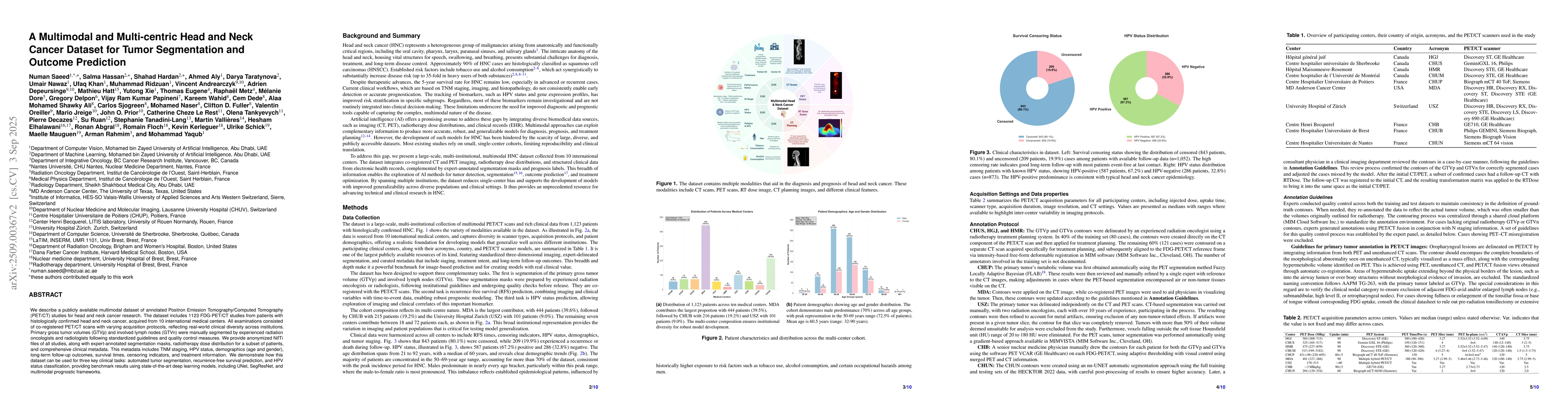
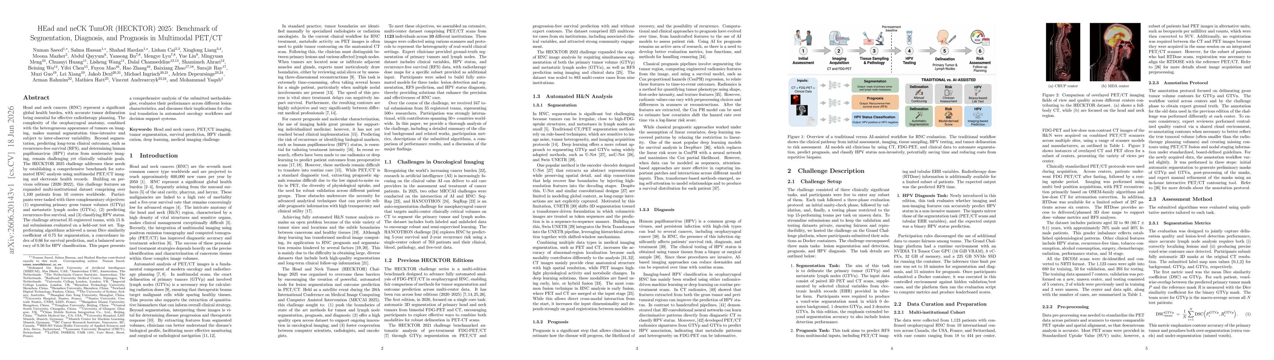
We describe a publicly available multimodal dataset of annotated Positron Emission Tomography/Computed Tomography (PET/CT) studies for head and neck cancer research. The dataset includes 1123 FDG-PET/...
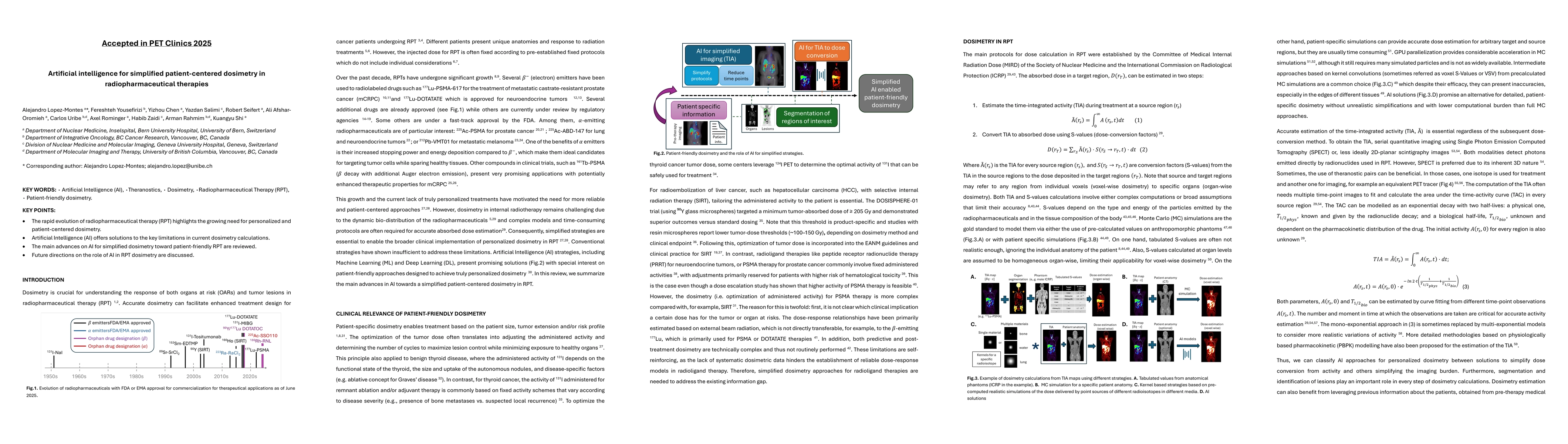
This review discusses the current applications, advantages, and limitations of PBPK and PopPK models in radiopharmaceutical therapy (RPT). PBPK models simulate radiopharmaceutical kinetics by integrat...
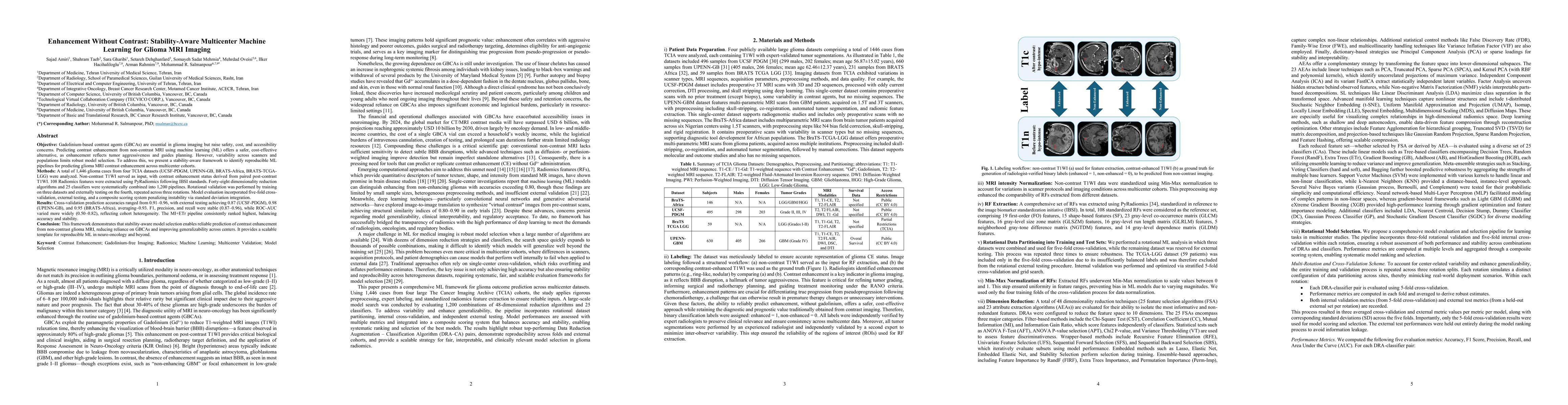
Gadolinium-based contrast agents (GBCAs) are central to glioma imaging but raise safety, cost, and accessibility concerns. Predicting contrast enhancement from non-contrast MRI using machine learning ...
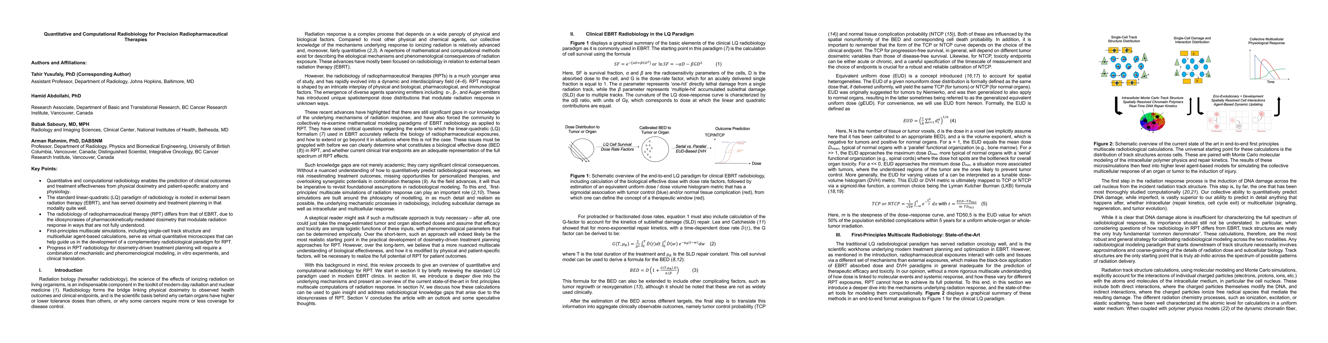
This article reviews the evolving field of radiobiology, emphasizing the need for advanced multiscale, mechanistic models to optimize radiopharmaceutical therapies (RPT). While the traditional linear-...
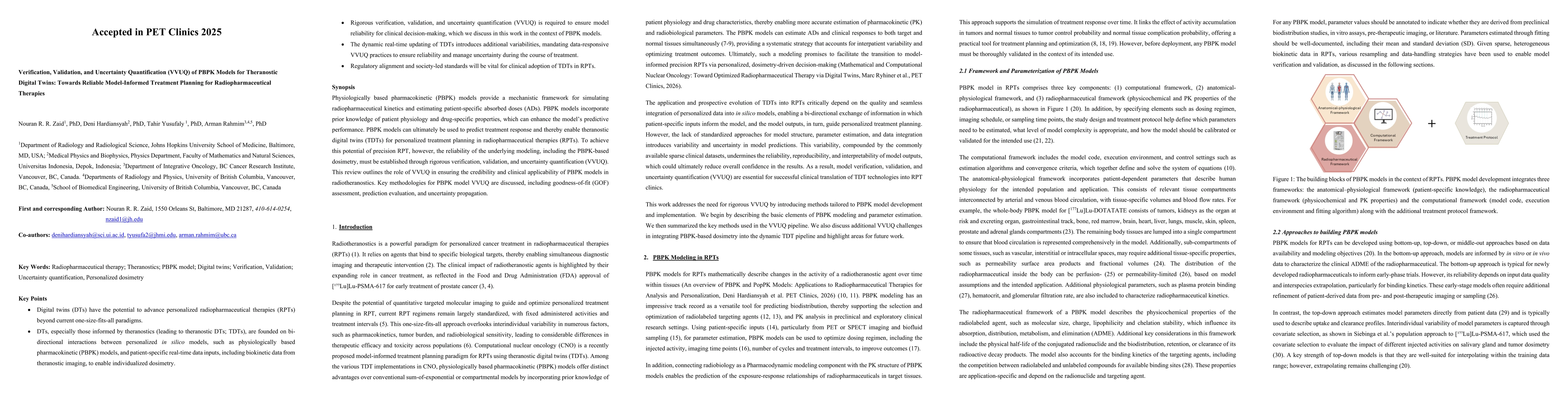
Physiologically based pharmacokinetic (PBPK) models provide a mechanistic framework for simulating radiopharmaceutical kinetics and estimating patient-specific absorbed doses (ADs). PBPK models incorp...
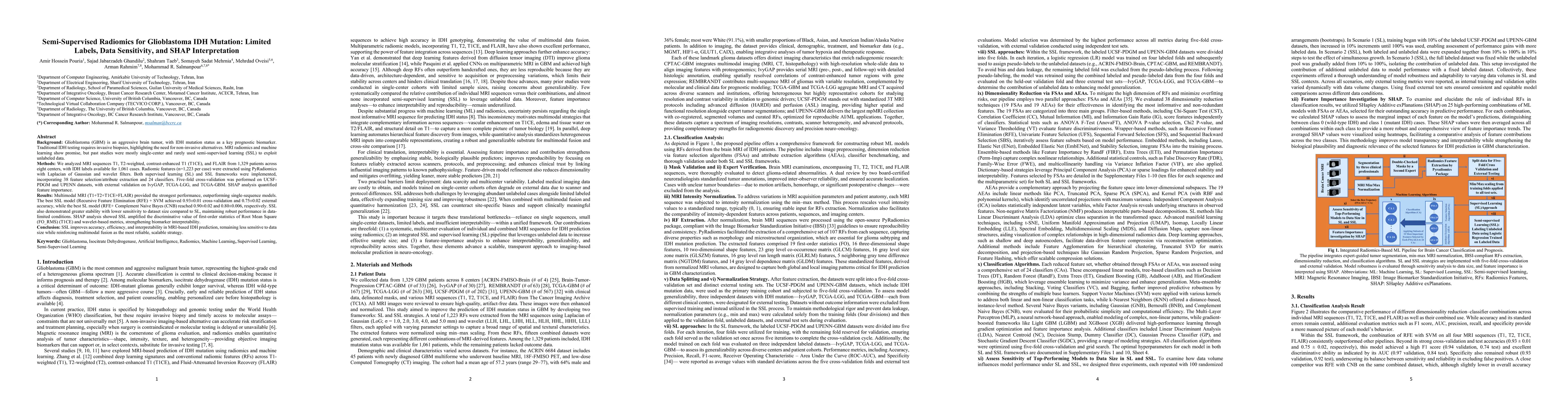
Glioblastoma (GBM) is an aggressive brain tumor in which IDH mutation status is a key prognostic biomarker, but traditional testing requires invasive biopsies, emphasizing the need for non-invasive ap...
Long-axial field-of-view (LAFOV) PET/CT has the potential to redefine the role of molecular imaging in theranostics by making multiparametric whole-body (MPWB) imaging and predictive dosimetry more cl...
KEY WORDS: Artificial Intelligence (AI), Theranostics, Dosimetry, Radiopharmaceutical Therapy (RPT), Patient-friendly dosimetry KEY POINTS - The rapid evolution of radiopharmaceutical therapy (RPT) hi...
The transformative potential of artificial intelligence (AI) in medical Imaging (MI) is well recognized. Yet despite promising reports in research settings, many AI tools fail to achieve clinical adop...
Lung cancer remains the leading cause of cancer mortality, with CT imaging central to screening, prognosis, and treatment. Manual segmentation is variable and time-intensive, while deep learning (DL) ...
Predictive dosimetry is central to enabling personalization of radiopharmaceutical therapies (RPTs) such as prostate-specific membrane antigen (PSMA) targeted RPTs. This study integrates physiological...
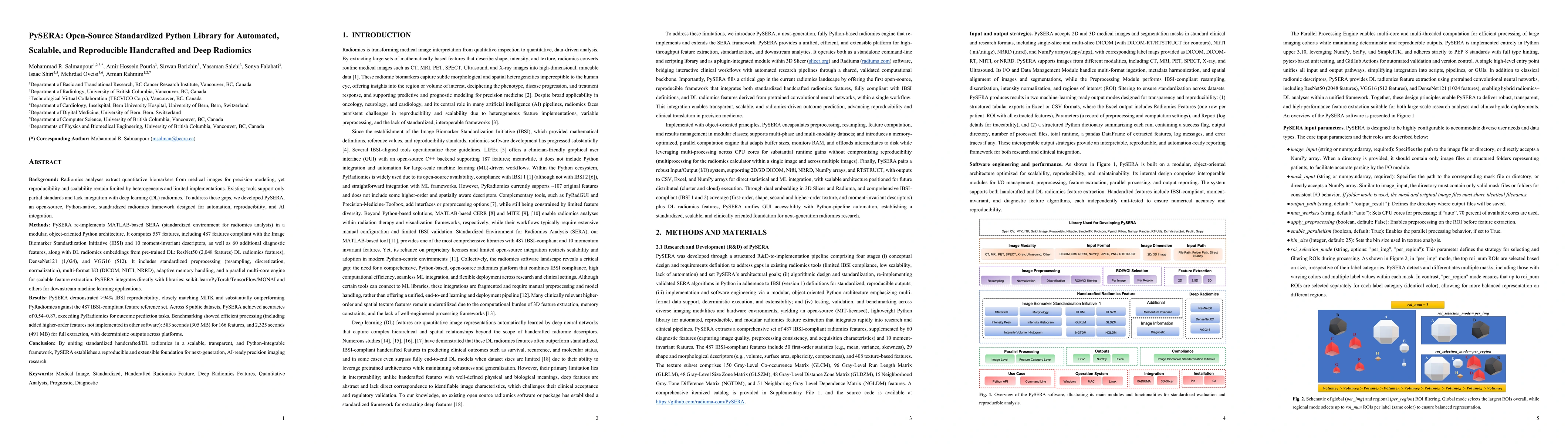
Radiomics enables the extraction of quantitative biomarkers from medical images for precision modeling, but reproducibility and scalability remain limited due to heterogeneous software implementations...
The field of Clinical-Computational Nuclear Medicine is rapidly advancing, fueled by AI, tracer kinetic modeling, radiomics, and integrated informatics. These technologies improve imaging quality, aut...
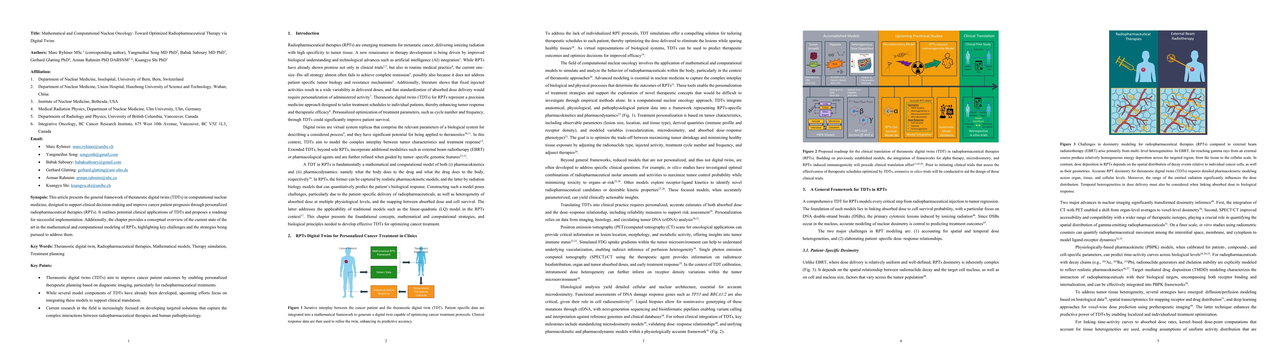
This article presents the general framework of theranostic digital twins (TDTs) in computational nuclear medicine, designed to support clinical decision-making and improve cancer patient prognosis thr...
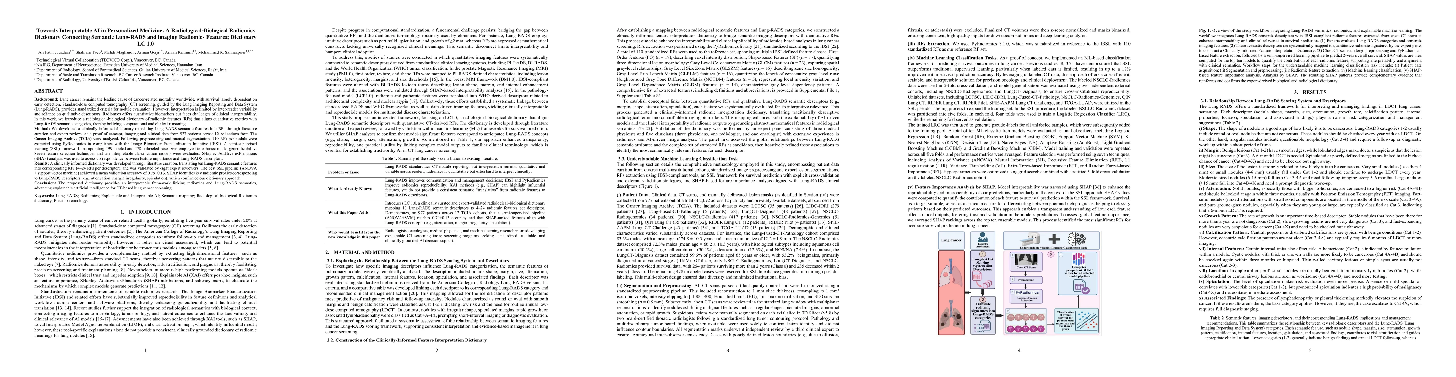
Lung cancer remains the leading cause of cancer-related mortality worldwide, with survival strongly dependent on early detection. Standard-dose computed tomography (CT) screening using the Lung Imagin...
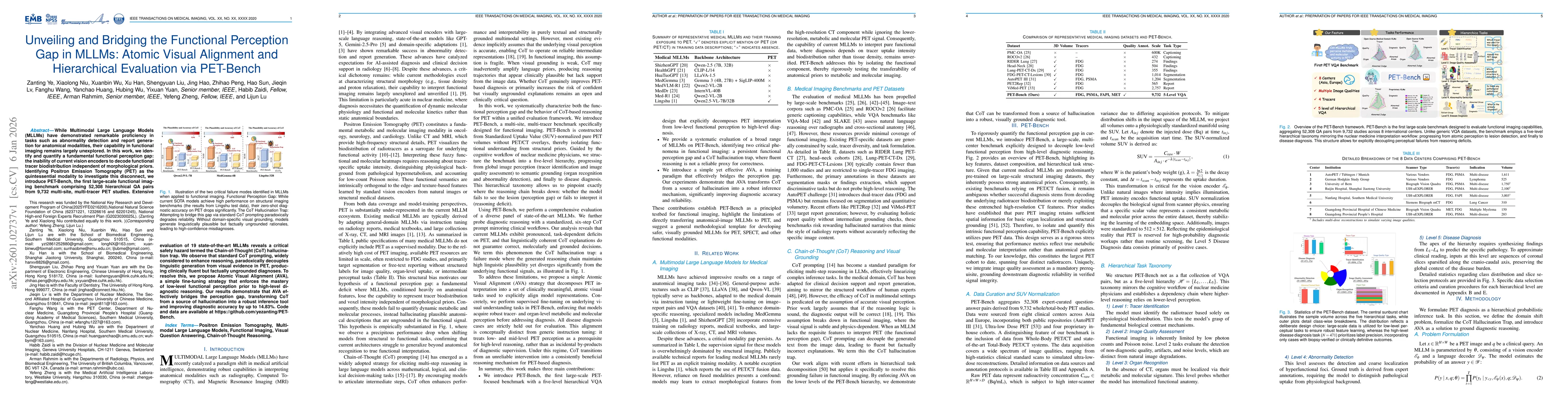
While Multimodal Large Language Models (MLLMs) have demonstrated remarkable proficiency in tasks such as abnormality detection and report generation for anatomical modalities, their capability in func...
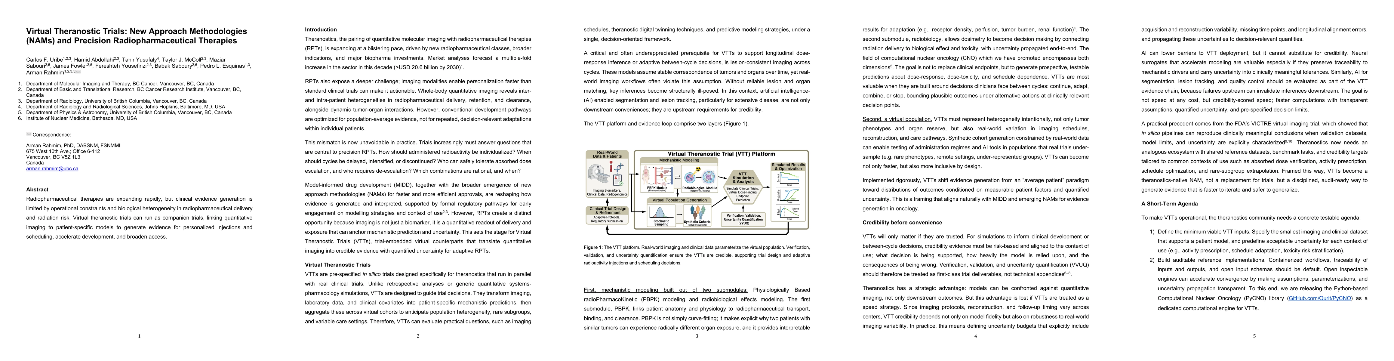
Radiopharmaceutical therapies are expanding rapidly, but clinical evidence generation is limited by operational constraints and biological heterogeneity in radiopharmaceutical delivery and radiation r...
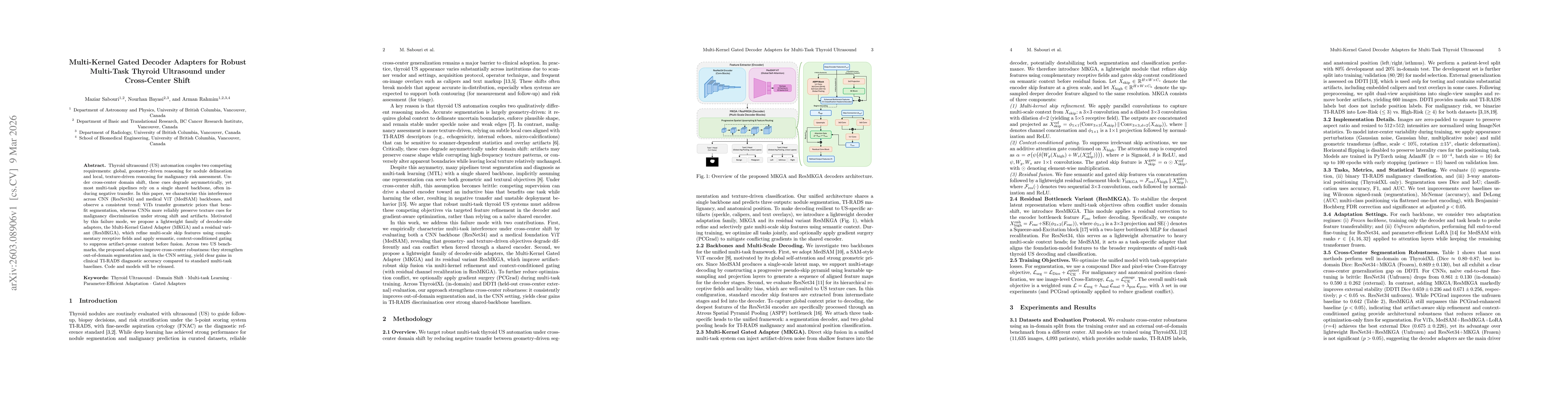
Thyroid ultrasound (US) automation couples two competing requirements: global, geometry-driven reasoning for nodule delineation and local, texture-driven reasoning for malignancy risk assessment. Unde...
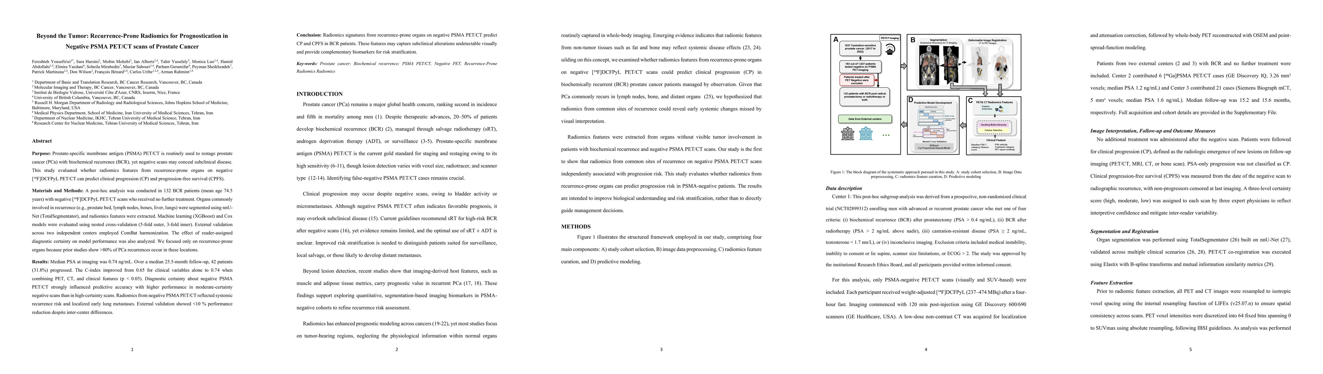
In patients with biochemical recurrence of prostate cancer and negative PSMA PET/CT, radiomics features extracted from recurrence-prone organs can predict clinical progression and progression-free sur...
Artificial intelligence based radiomics models for thyroid ultrasound (US) often achieve strong diagnostic performance but remain difficult to interpret, limiting clinical trust and adoption. We devel...
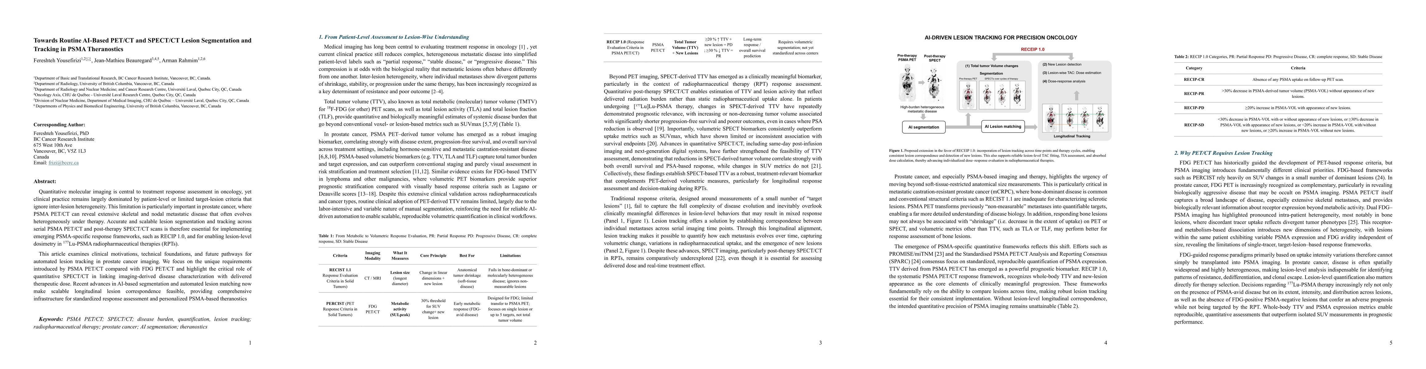
Quantitative molecular imaging is central to treatment response assessment in oncology, yet clinical practice remains largely dominated by patient-level or limited target-lesion criteria that ignore i...
Background: Renal blood flow (RBF) is an important marker of kidney health, but noninvasive assessment is not routinely used in clinical imaging. We evaluated the feasibility and physiologic validity ...
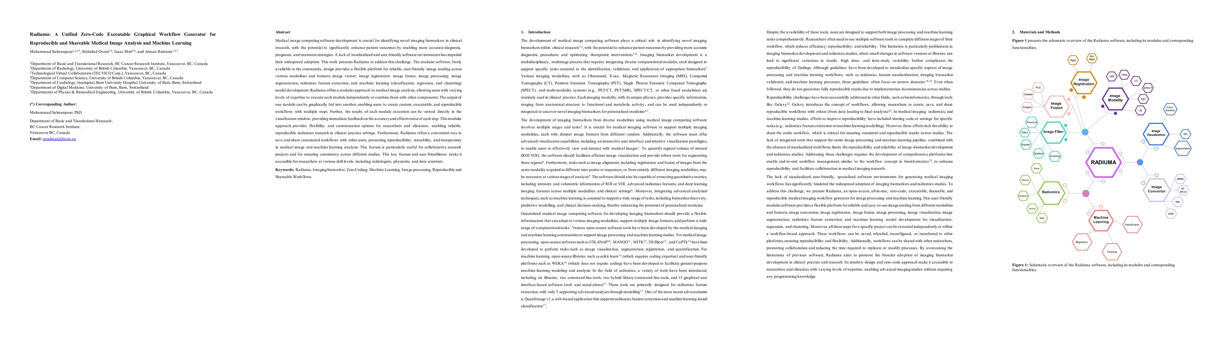
Medical image computing software is essential for identifying imaging biomarkers that can support diagnosis, prognosis, treatment planning, and clinical research. However, the lack of standardized, us...
Head and neck cancers (HNC) represent a significant global health burden, with accurate tumor delineation being essential for effective radiotherapy planning. The complexity of the oropharyngeal anato...