Summary
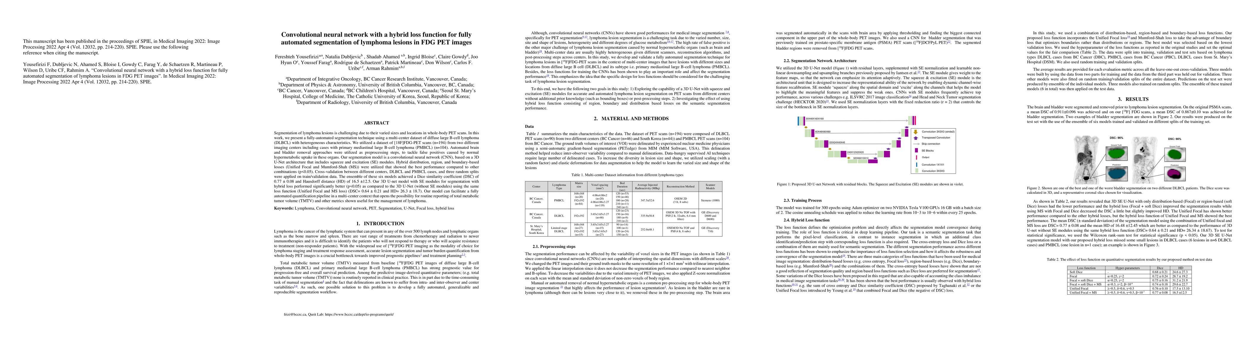
Segmentation of lymphoma lesions is challenging due to their varied sizes and locations in whole-body PET scans. This work presents a fully-automated segmentation technique using a multi-center dataset of diffuse large B-cell lymphoma (DLBCL) with heterogeneous characteristics. We utilized a dataset of [18F]FDG-PET scans (n=194) from two different imaging centers, including cases with primary mediastinal large B-cell lymphoma (PMBCL) (n=104). Automated brain and bladder removal approaches were utilized as preprocessing steps to tackle false positives caused by normal hypermetabolic uptake in these organs. Our segmentation model is a convolutional neural network (CNN) based on a 3D U-Net architecture that includes squeeze and excitation (SE) modules. Hybrid distribution, region, and boundary-based losses (Unified Focal and Mumford-Shah (MS)) were utilized that showed the best performance compared to other combinations (p<0.05). Cross-validation between different centers, DLBCL and PMBCL cases, and three random splits were applied on train/validation data. The ensemble of these six models achieved a Dice similarity coefficient (DSC) of 0.77 +- 0.08 and Hausdorff distance (HD) of 16.5 +-12.5. Our 3D U-net model with SE modules for segmentation with hybrid loss performed significantly better (p<0.05) as compared to the 3D U-Net (without SE modules) using the same loss function (Unified Focal and MS loss) (DSC= 0.64 +-0.21 and HD= 26.3 +- 18.7). Our model can facilitate a fully automated quantification pipeline in a multi-center context that opens the possibility for routine reporting of total metabolic tumor volume (TMTV) and other metrics shown useful for the management of lymphoma.
AI Key Findings
Get AI-generated insights about this paper's methodology, results, and significance.
Paper Details
PDF Preview
Key Terms
Citation Network
Current paper (gray), citations (green), references (blue)
Display is limited for performance on very large graphs.
Similar Papers
Found 4 papersSemi-supervised learning towards automated segmentation of PET images with limited annotations: Application to lymphoma patients
Arman Rahmim, François Bénard, Fereshteh Yousefirizi et al.
Adaptive Voxel-Weighted Loss Using L1 Norms in Deep Neural Networks for Detection and Segmentation of Prostate Cancer Lesions in PET/CT Images
Arman Rahmim, Shadab Ahamed, Xiaoxiao Li et al.
Whole-body tumor segmentation of 18F -FDG PET/CT using a cascaded and ensembled convolutional neural networks
Xinrui Zhan, Lei Xiang, Ludovic Sibille
| Title | Authors | Year | Actions |
|---|
Comments (0)