Academic Profile
Statistics
Similar Authors
Papers on arXiv
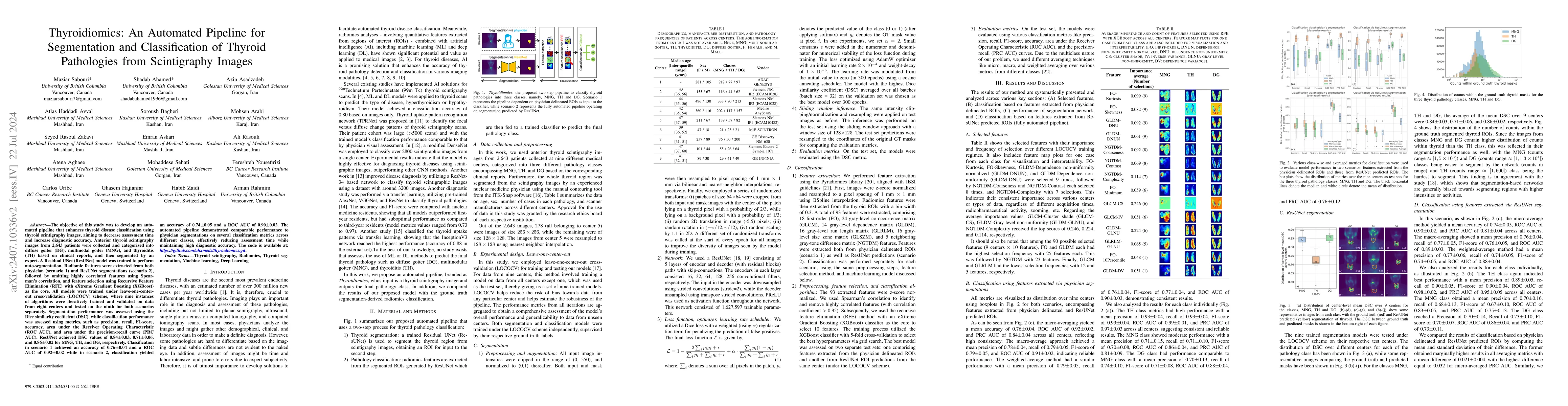
The objective of this study was to develop an automated pipeline that enhances thyroid disease classification using thyroid scintigraphy images, aiming to decrease assessment time and increase diagnos...
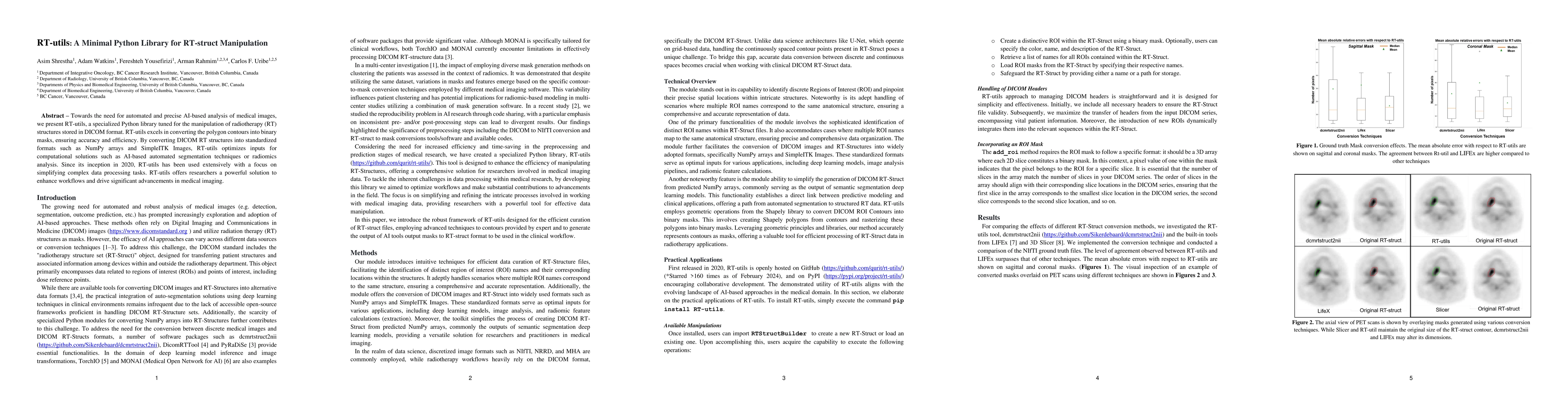
Towards the need for automated and precise AI-based analysis of medical images, we present RT-utils, a specialized Python library tuned for the manipulation of radiotherapy (RT) structures stored in...
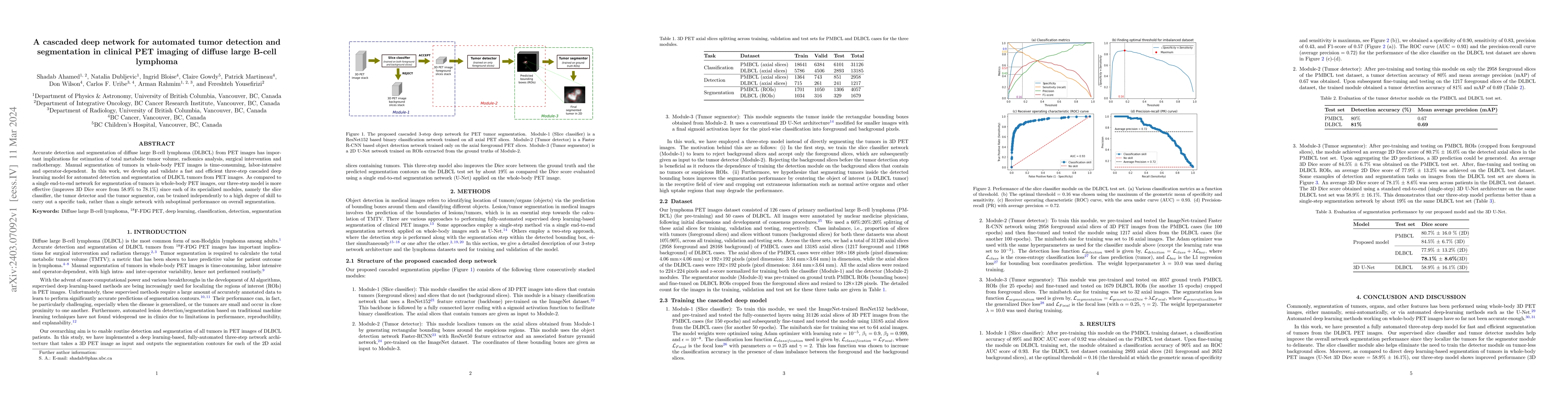
Accurate detection and segmentation of diffuse large B-cell lymphoma (DLBCL) from PET images has important implications for estimation of total metabolic tumor volume, radiomics analysis, surgical i...
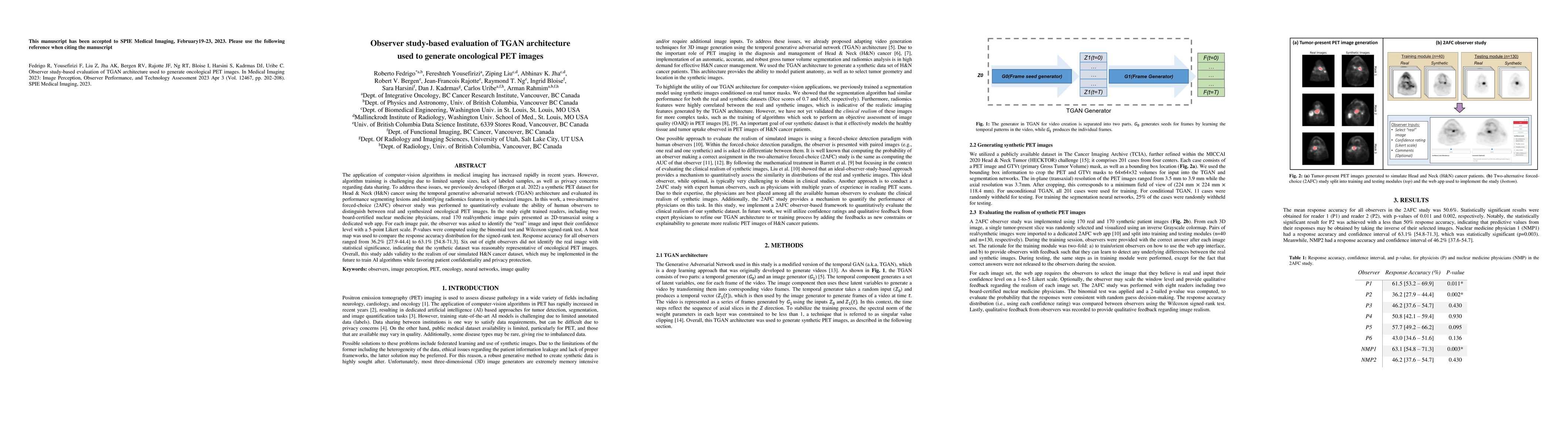
The application of computer-vision algorithms in medical imaging has increased rapidly in recent years. However, algorithm training is challenging due to limited sample sizes, lack of labeled sample...
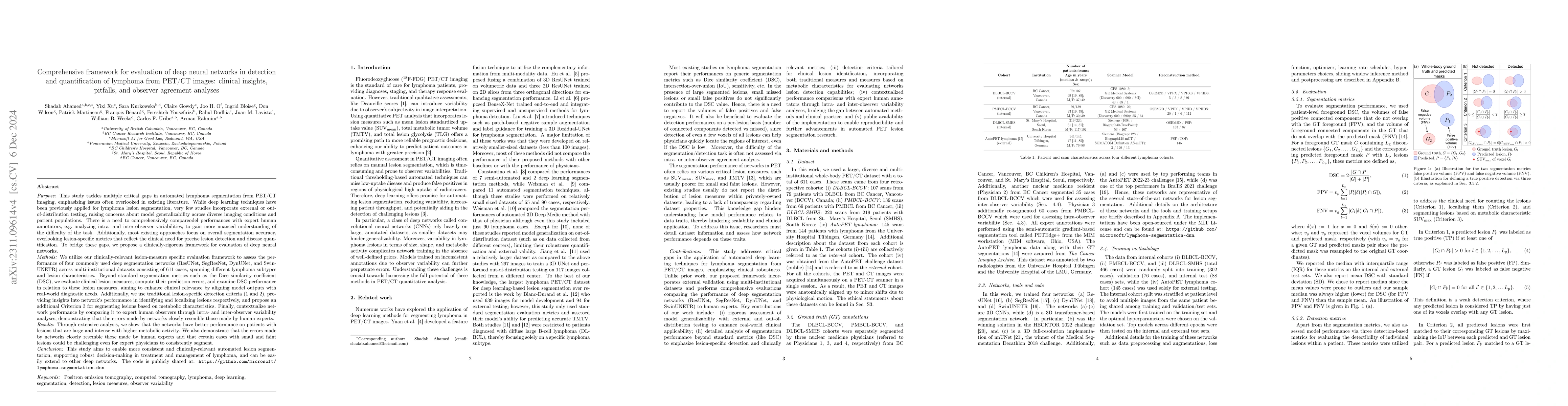
This study performs comprehensive evaluation of four neural network architectures (UNet, SegResNet, DynUNet, and SwinUNETR) for lymphoma lesion segmentation from PET/CT images. These networks were t...
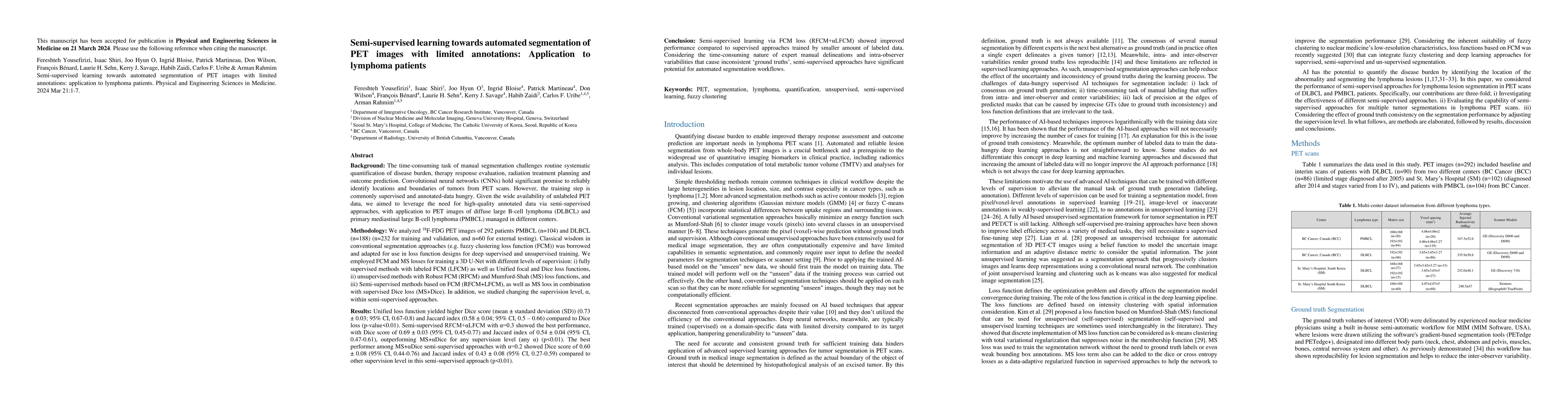
The time-consuming task of manual segmentation challenges routine systematic quantification of disease burden. Convolutional neural networks (CNNs) hold significant promise to reliably identify loca...
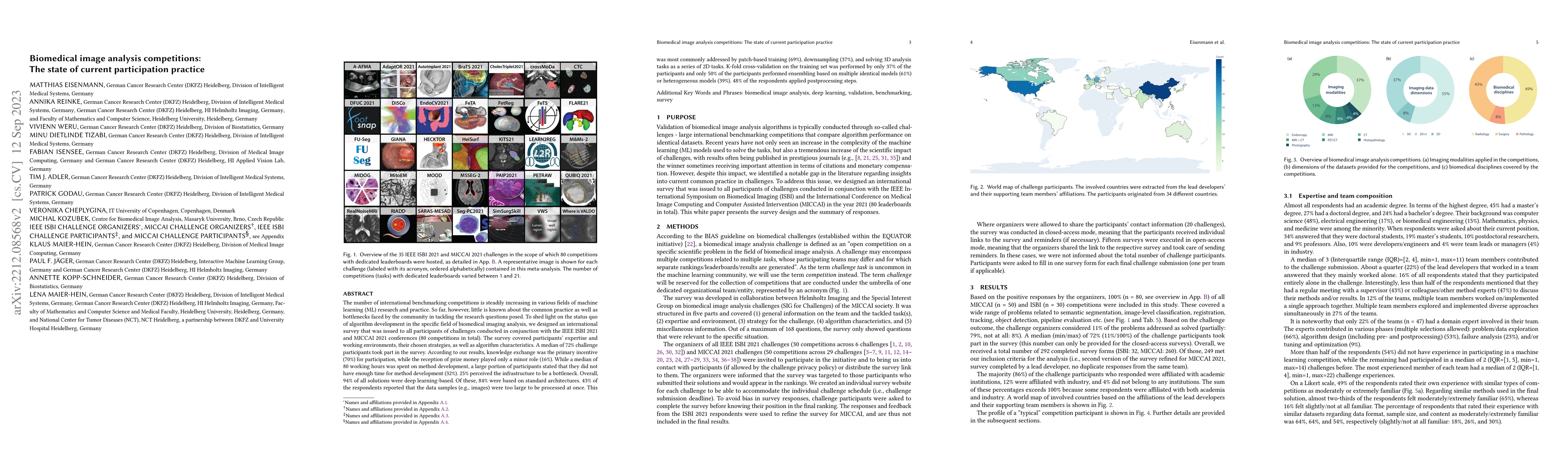
The number of international benchmarking competitions is steadily increasing in various fields of machine learning (ML) research and practice. So far, however, little is known about the common pract...
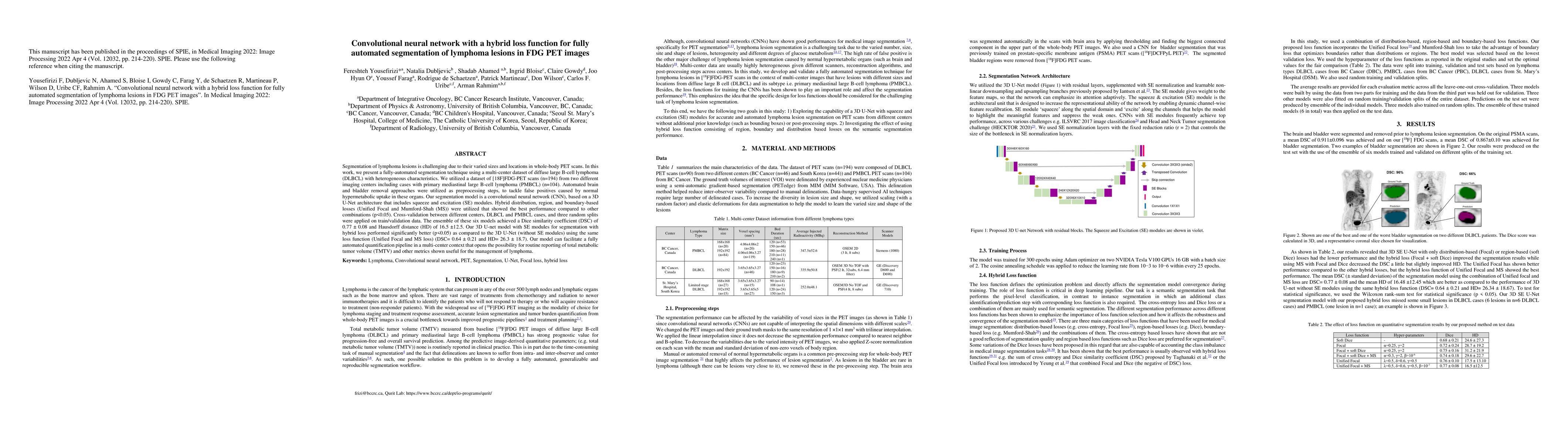
Segmentation of lymphoma lesions is challenging due to their varied sizes and locations in whole-body PET scans. This work presents a fully-automated segmentation technique using a multi-center data...
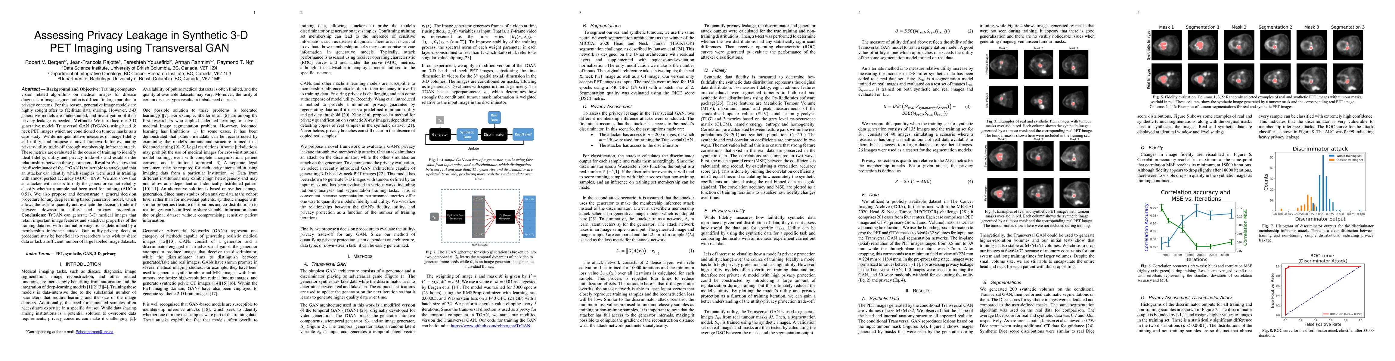
Training computer-vision related algorithms on medical images for disease diagnosis or image segmentation is difficult in large part due to privacy concerns. For this reason, generative image models...
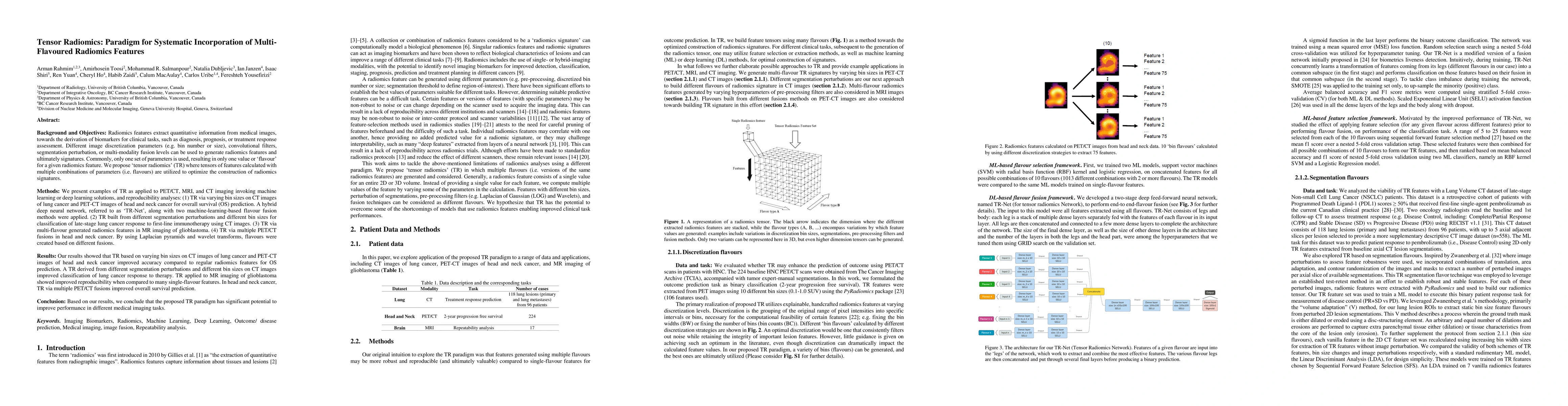
Radiomics features extract quantitative information from medical images, towards the derivation of biomarkers for clinical tasks, such as diagnosis, prognosis, or treatment response assessment. Diff...
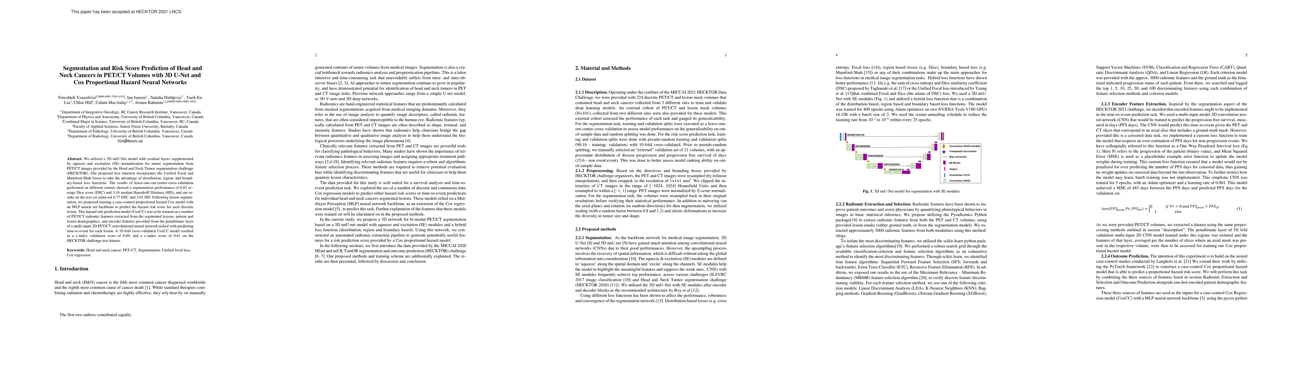
We utilized a 3D nnU-Net model with residual layers supplemented by squeeze and excitation (SE) normalization for tumor segmentation from PET/CT images provided by the Head and Neck Tumor segmentati...
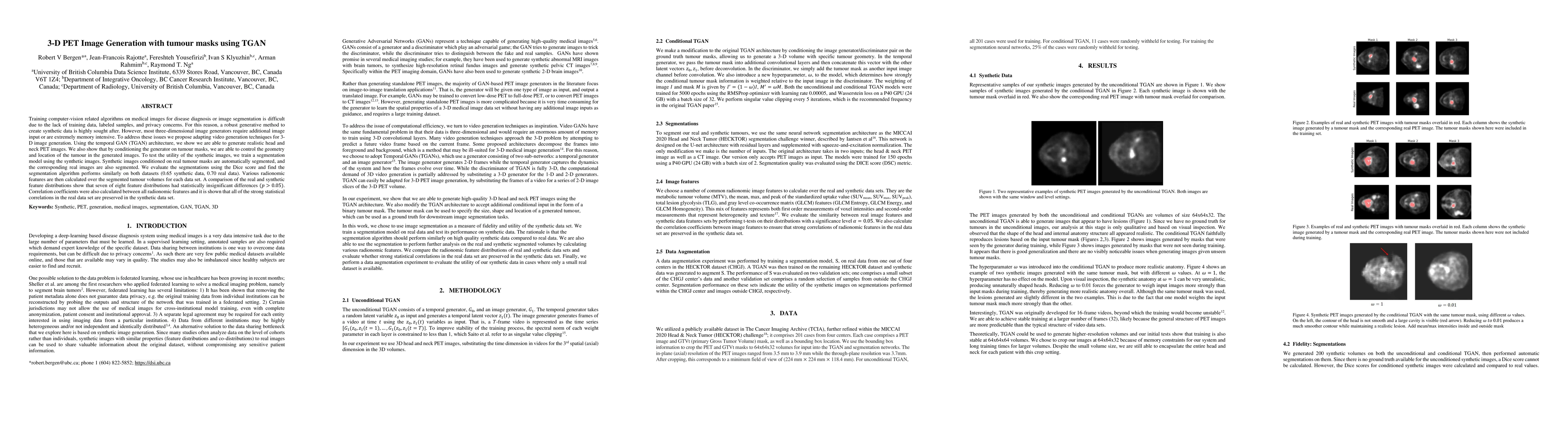
Training computer-vision related algorithms on medical images for disease diagnosis or image segmentation is difficult due to the lack of training data, labeled samples, and privacy concerns. For th...
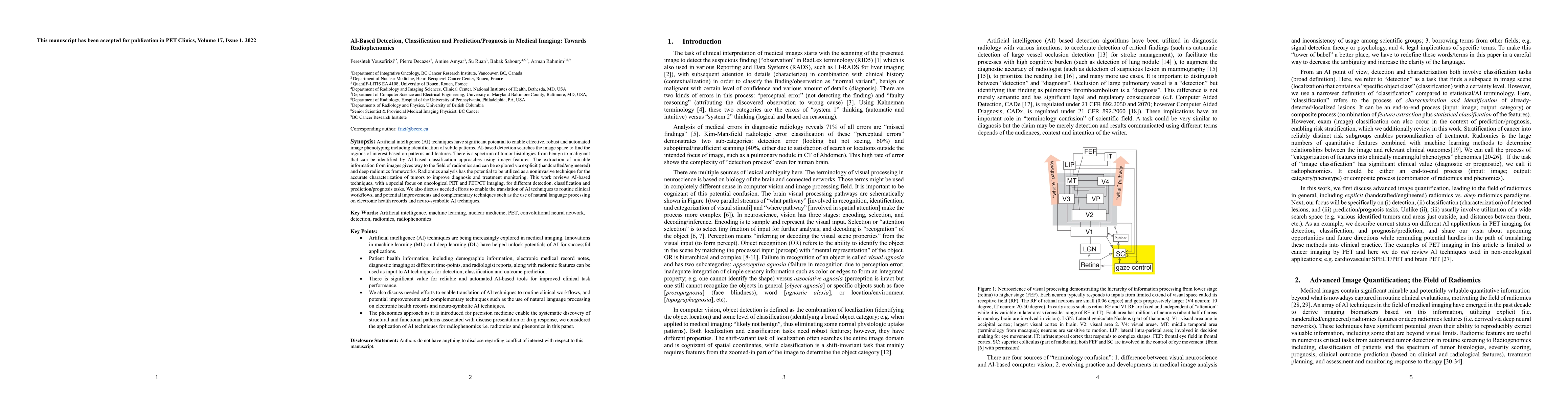
Artificial intelligence (AI) techniques have significant potential to enable effective, robust and automated image phenotyping including identification of subtle patterns. AI-based detection searche...
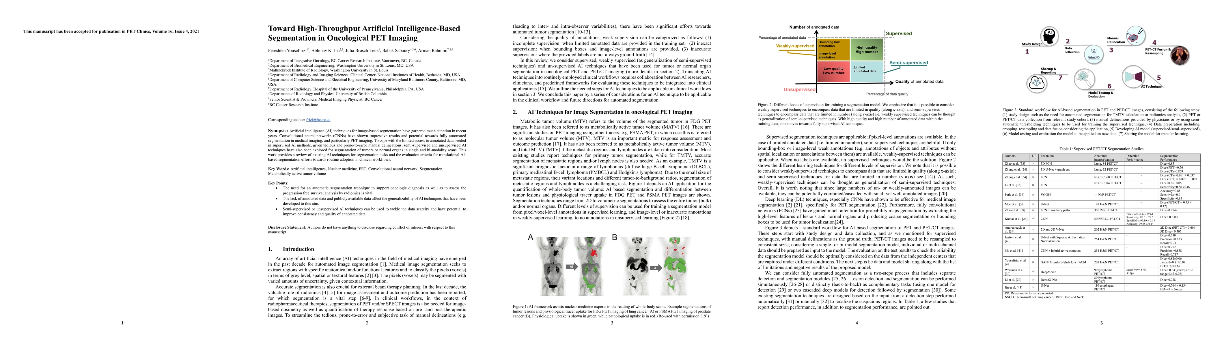
Artificial intelligence (AI) techniques for image-based segmentation have garnered much attention in recent years. Convolutional neural networks (CNNs) have shown impressive results and potential to...
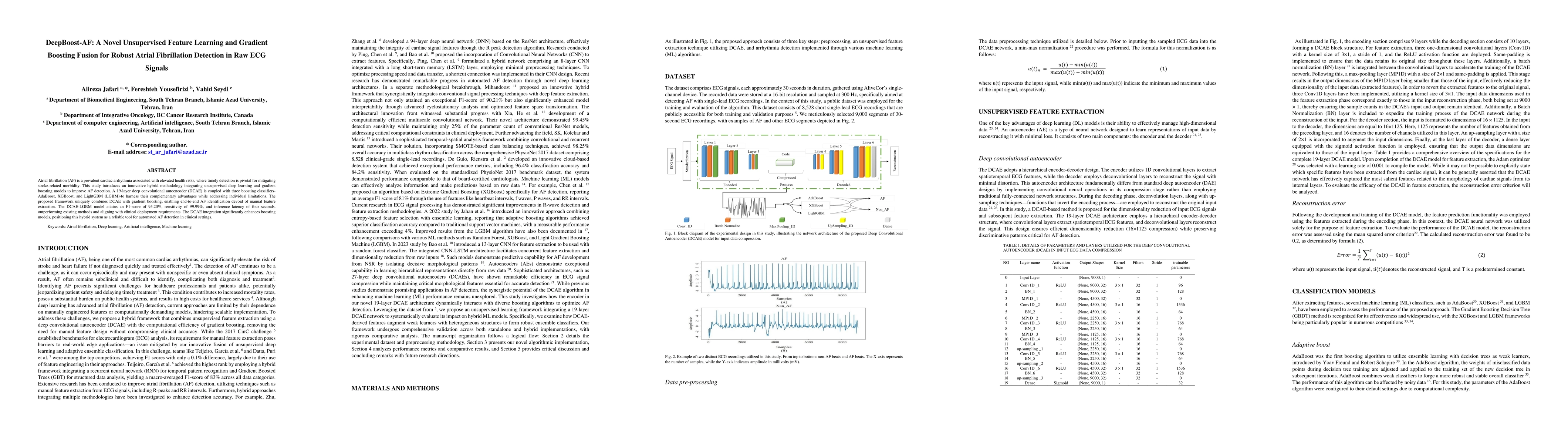
Atrial fibrillation (AF) is a prevalent cardiac arrhythmia associated with elevated health risks, where timely detection is pivotal for mitigating stroke-related morbidity. This study introduces an in...
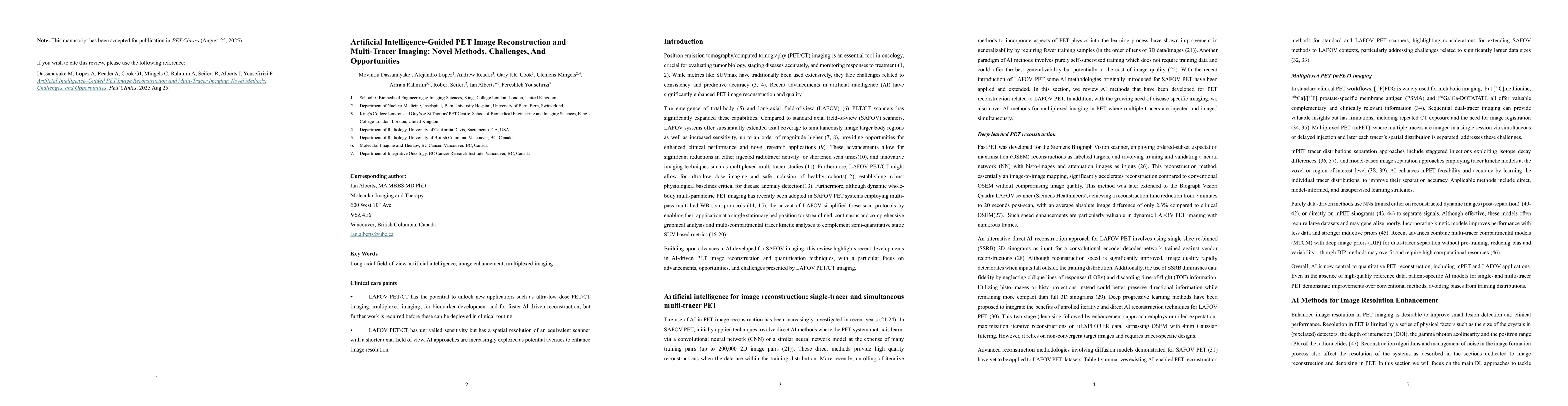
LAFOV PET/CT has the potential to unlock new applications such as ultra-low dose PET/CT imaging, multiplexed imaging, for biomarker development and for faster AI-driven reconstruction, but further wor...
MTV is increasingly recognized as an accurate estimate of disease burden, which has prognostic value, but its implementation has been hindered by the time-consuming need for manual segmentation of ima...
Long-axial field-of-view (LAFOV) PET/CT has the potential to redefine the role of molecular imaging in theranostics by making multiparametric whole-body (MPWB) imaging and predictive dosimetry more cl...
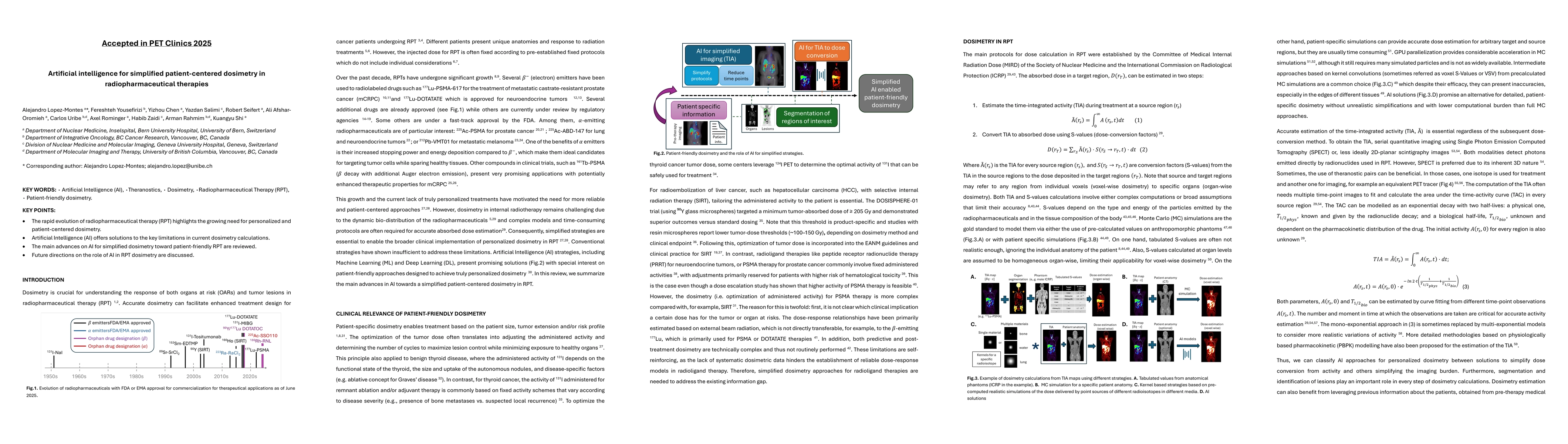
KEY WORDS: Artificial Intelligence (AI), Theranostics, Dosimetry, Radiopharmaceutical Therapy (RPT), Patient-friendly dosimetry KEY POINTS - The rapid evolution of radiopharmaceutical therapy (RPT) hi...
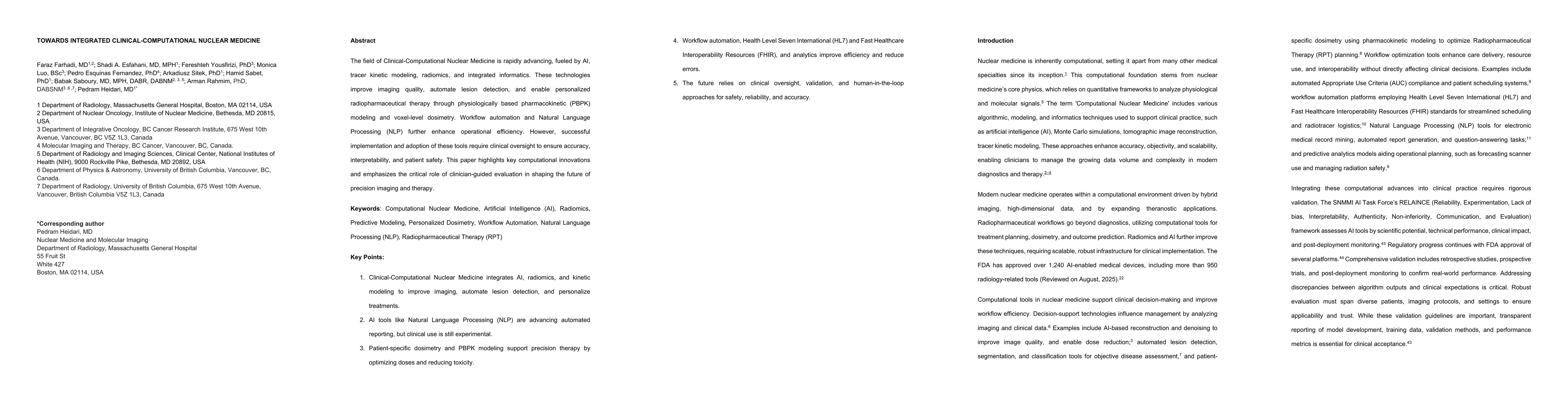
The field of Clinical-Computational Nuclear Medicine is rapidly advancing, fueled by AI, tracer kinetic modeling, radiomics, and integrated informatics. These technologies improve imaging quality, aut...
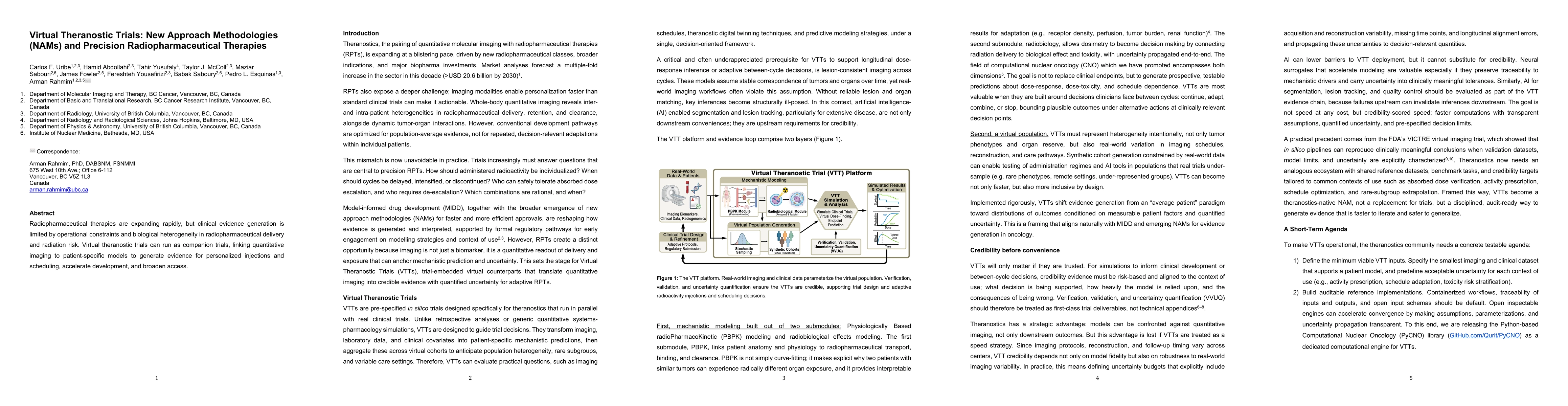
Radiopharmaceutical therapies are expanding rapidly, but clinical evidence generation is limited by operational constraints and biological heterogeneity in radiopharmaceutical delivery and radiation r...
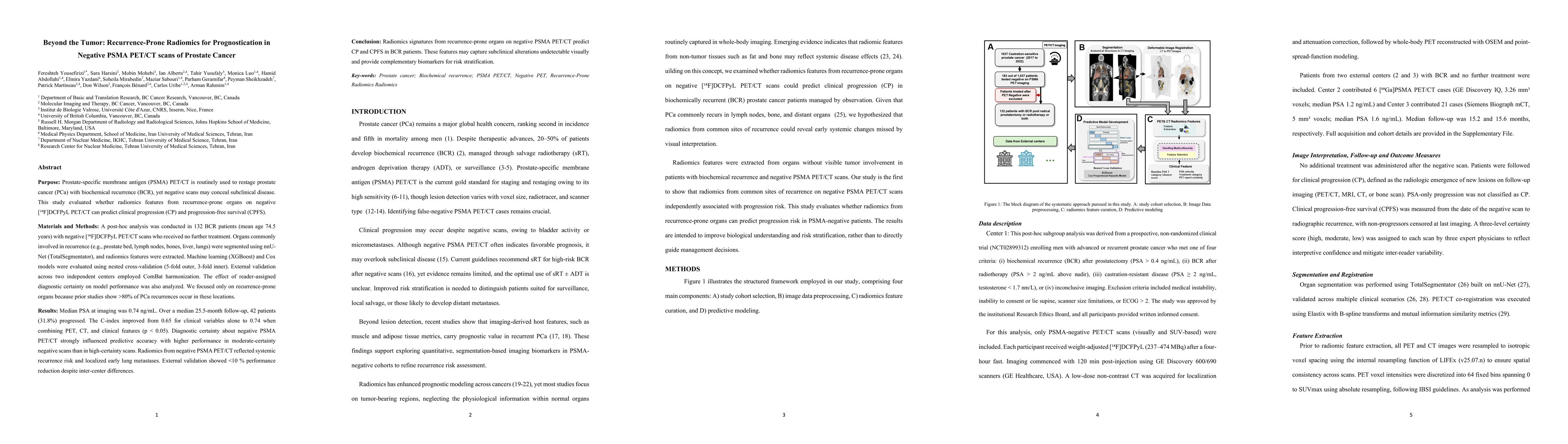
In patients with biochemical recurrence of prostate cancer and negative PSMA PET/CT, radiomics features extracted from recurrence-prone organs can predict clinical progression and progression-free sur...
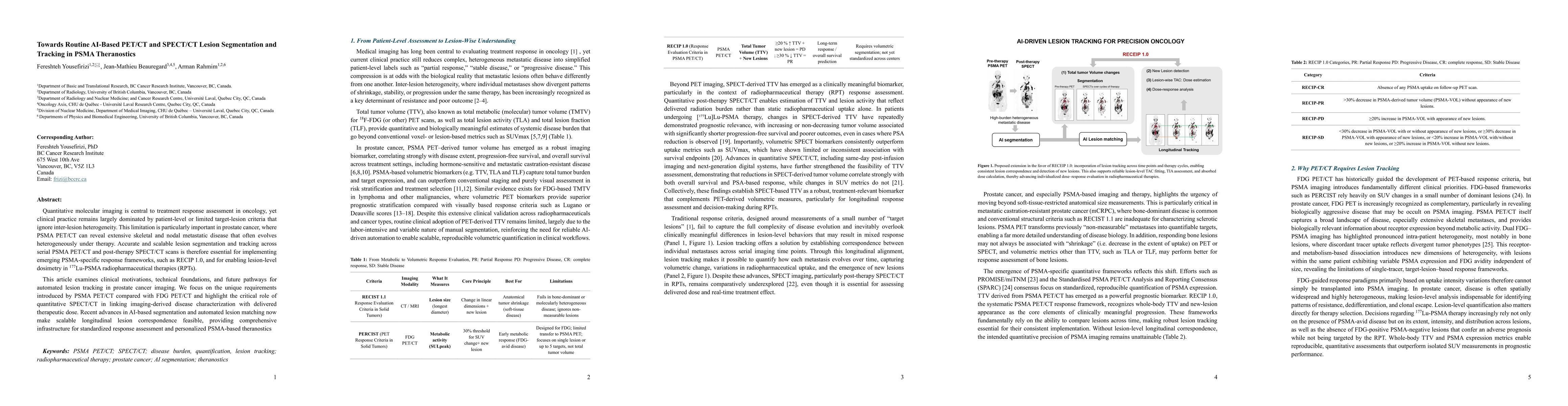
Quantitative molecular imaging is central to treatment response assessment in oncology, yet clinical practice remains largely dominated by patient-level or limited target-lesion criteria that ignore i...