Summary
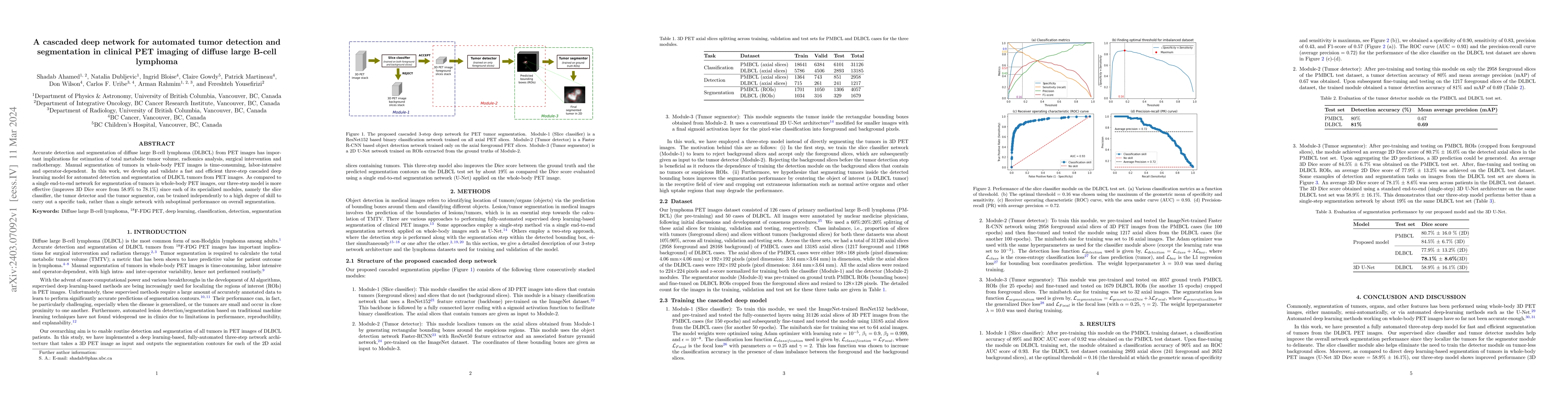
Accurate detection and segmentation of diffuse large B-cell lymphoma (DLBCL) from PET images has important implications for estimation of total metabolic tumor volume, radiomics analysis, surgical intervention and radiotherapy. Manual segmentation of tumors in whole-body PET images is time-consuming, labor-intensive and operator-dependent. In this work, we develop and validate a fast and efficient three-step cascaded deep learning model for automated detection and segmentation of DLBCL tumors from PET images. As compared to a single end-to-end network for segmentation of tumors in whole-body PET images, our three-step model is more effective (improves 3D Dice score from 58.9% to 78.1%) since each of its specialized modules, namely the slice classifier, the tumor detector and the tumor segmentor, can be trained independently to a high degree of skill to carry out a specific task, rather than a single network with suboptimal performance on overall segmentation.
AI Key Findings
Get AI-generated insights about this paper's methodology, results, and significance.
Paper Details
PDF Preview
Key Terms
Citation Network
Current paper (gray), citations (green), references (blue)
Display is limited for performance on very large graphs.
Similar Papers
Found 4 papersConvolutional neural network with a hybrid loss function for fully automated segmentation of lymphoma lesions in FDG PET images
Arman Rahmim, Shadab Ahamed, Fereshteh Yousefirizi et al.
Prognostic Value of Multiple Manual Segmentation Methods for Diffuse Large B-Cell Lymphoma with 18F-FDG PET/CT.
Doma, Andrej, Studen, Andrej, Jezeršek Novaković, Barbara
| Title | Authors | Year | Actions |
|---|
Comments (0)