Academic Profile
Statistics
Similar Authors
Papers on arXiv
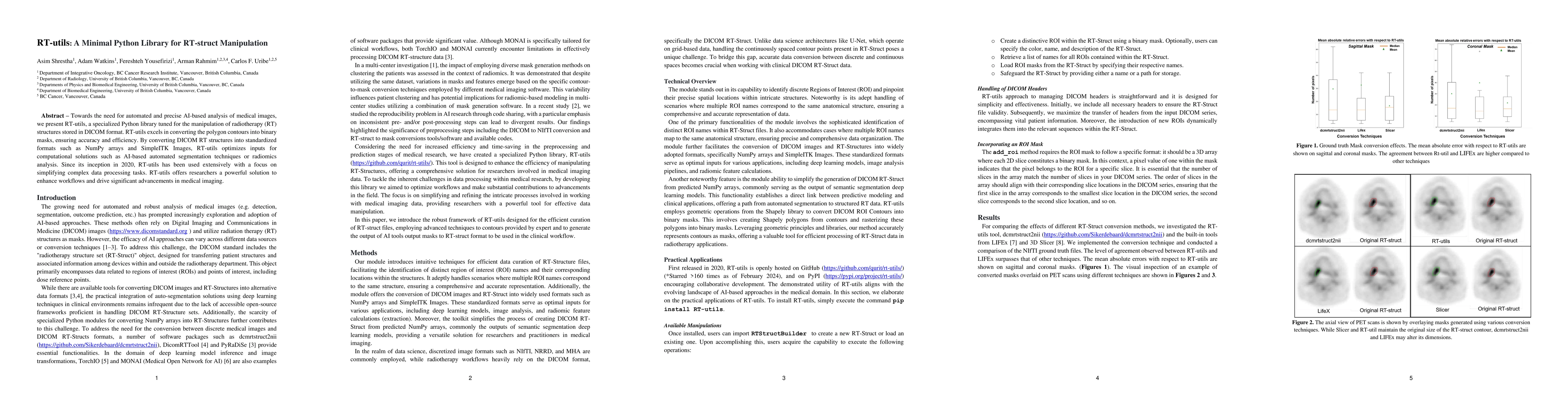
Towards the need for automated and precise AI-based analysis of medical images, we present RT-utils, a specialized Python library tuned for the manipulation of radiotherapy (RT) structures stored in...
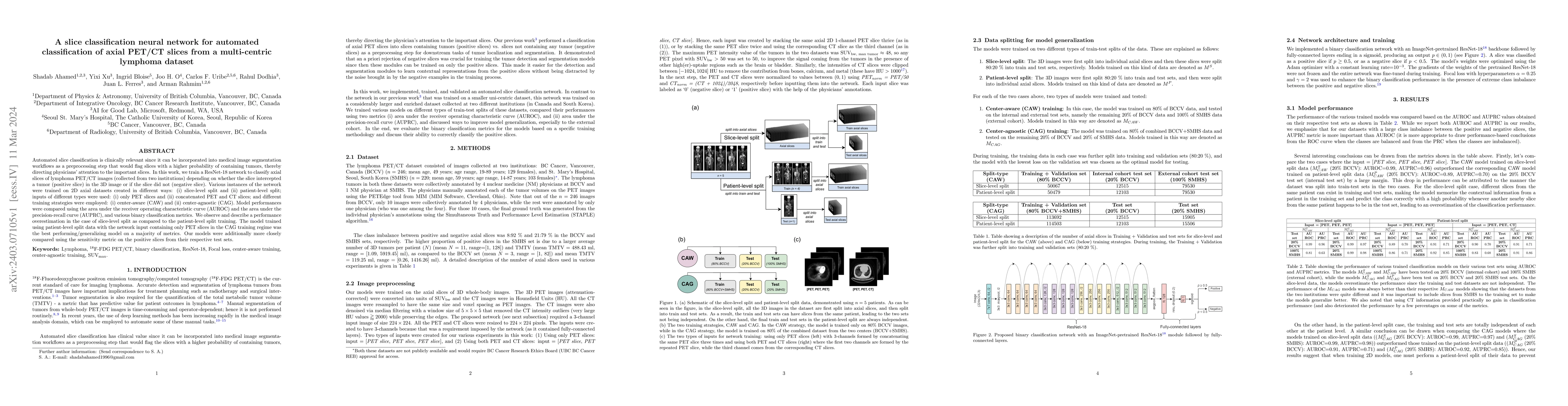
Automated slice classification is clinically relevant since it can be incorporated into medical image segmentation workflows as a preprocessing step that would flag slices with a higher probability ...
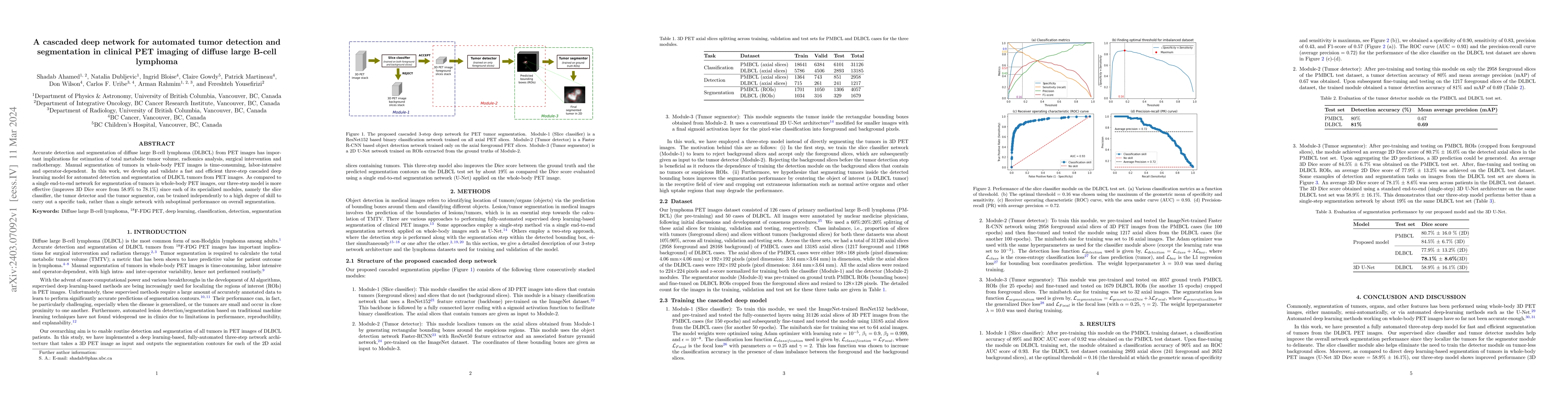
Accurate detection and segmentation of diffuse large B-cell lymphoma (DLBCL) from PET images has important implications for estimation of total metabolic tumor volume, radiomics analysis, surgical i...
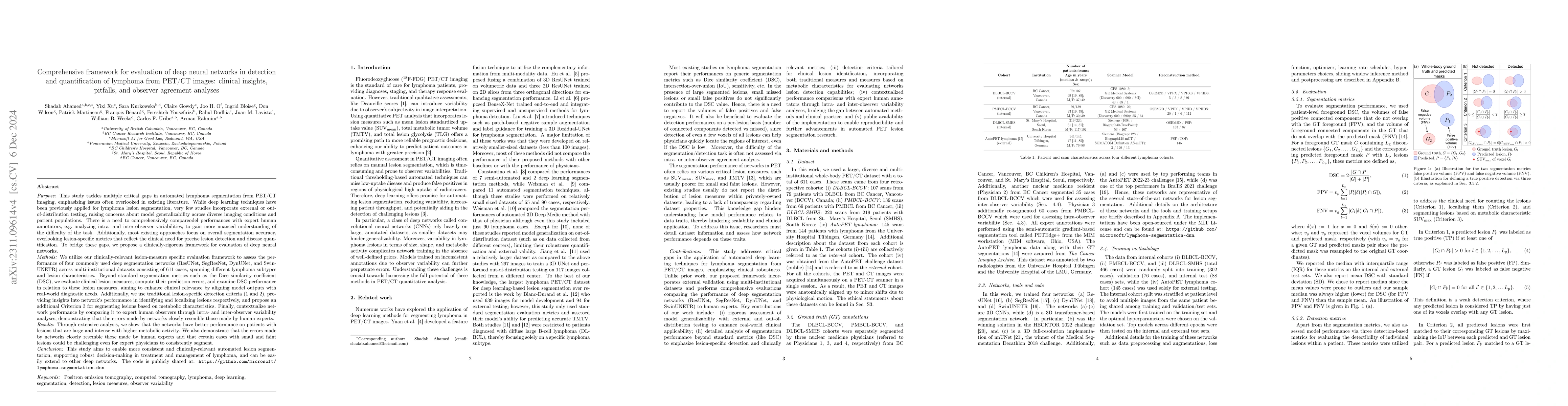
This study performs comprehensive evaluation of four neural network architectures (UNet, SegResNet, DynUNet, and SwinUNETR) for lymphoma lesion segmentation from PET/CT images. These networks were t...
The time-consuming task of manual segmentation challenges routine systematic quantification of disease burden. Convolutional neural networks (CNNs) hold significant promise to reliably identify loca...
Segmentation of lymphoma lesions is challenging due to their varied sizes and locations in whole-body PET scans. This work presents a fully-automated segmentation technique using a multi-center data...
Purpose: The XCAT phantom allows for highly sophisticated multimodality imaging research. It includes a complete set of organs, muscle, bone, soft tissue, while also accounting for age, sex, and bod...
Long-axial field-of-view (LAFOV) PET/CT has the potential to redefine the role of molecular imaging in theranostics by making multiparametric whole-body (MPWB) imaging and predictive dosimetry more cl...
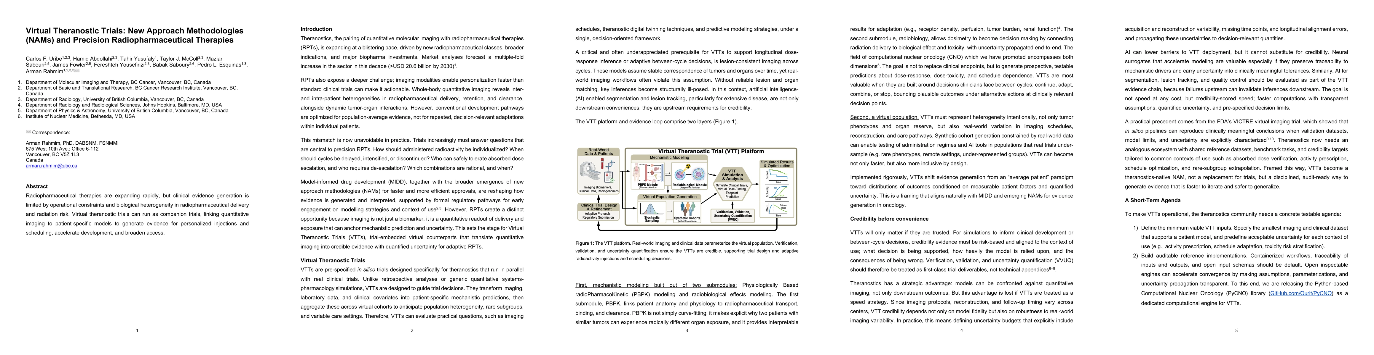
Radiopharmaceutical therapies are expanding rapidly, but clinical evidence generation is limited by operational constraints and biological heterogeneity in radiopharmaceutical delivery and radiation r...