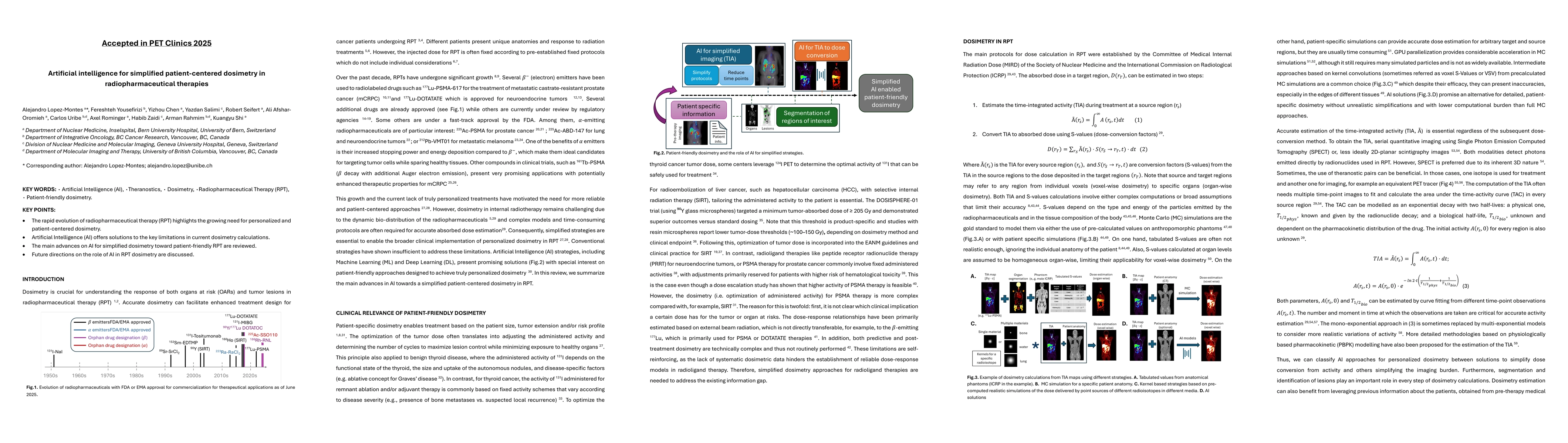
Summary

KEY WORDS: Artificial Intelligence (AI), Theranostics, Dosimetry, Radiopharmaceutical Therapy (RPT), Patient-friendly dosimetry KEY POINTS - The rapid evolution of radiopharmaceutical therapy (RPT) highlights the growing need for personalized and patient-centered dosimetry. - Artificial Intelligence (AI) offers solutions to the key limitations in current dosimetry calculations. - The main advances on AI for simplified dosimetry toward patient-friendly RPT are reviewed. - Future directions on the role of AI in RPT dosimetry are discussed.

AI Key Findings

Generated Oct 30, 2025

Methodology

The research employs a combination of deep learning techniques, including convolutional neural networks (CNNs) and information fusion strategies, to automate tumor segmentation and volume measurement in PET/CT images. It integrates multi-modal data from PET and CT scans to improve accuracy and generalizability across different patient populations and imaging centers.

Key Results

  • Development of fully automated segmentation models for various cancers including lymphoma, neuroendocrine tumors, and prostate cancer
  • Demonstration of improved accuracy and consistency in tumor volume measurements compared to manual methods
  • Successful generalizability of models across multiple centers and patient demographics

Significance

This research advances precision oncology by enabling objective, reproducible tumor volume measurements that can inform treatment decisions and monitor therapeutic response. The automated methods reduce inter-observer variability and improve efficiency in clinical workflows.

Technical Contribution

The work introduces novel information fusion frameworks and deep learning architectures specifically tailored for multi-modal PET/CT tumor segmentation, along with standardized evaluation protocols aligned with AAPM task group recommendations.

Novelty

This research stands out through its comprehensive approach combining multi-modal data integration, rigorous validation across diverse clinical settings, and the development of standardized evaluation frameworks for automated PET/CT segmentation methods.

Limitations

  • Performance may vary across different imaging protocols and scanner types
  • Requires high-quality annotated training data which may be limited in certain cancer types

Future Work

  • Integration with radiomics and biomarker analysis for personalized treatment planning
  • Development of real-time segmentation tools for intraoperative use
  • Expansion to include more cancer types and rare subtypes

Paper Details

PDF Preview

Download PDF

Comments (0)

This website uses cookies to ensure you get the best experience. Learn more