ArxivLens

Publication

Published: Apr 26, 2024
Categories: eess.IV, cs.AI, cs.CV

Metrics

Source: ArXiv

AI Quick Summary

This study proposes an AI-driven segmentation strategy utilizing convolutional neural networks (CNNs) and textural features to analyze Reflectance Confocal Microscopy (RCM) images, aiming to enhance diagnostic accuracy and confidence in dermatological practice by identifying clinically significant regions without the need for physical biopsies.

Quick Answers

What is "Enhancing Diagnosis through AI-driven Analysis of Reflectance Confocal Microscopy" about?

This study proposes an AI-driven segmentation strategy utilizing convolutional neural networks (CNNs) and textural features to analyze Reflectance Confocal Microscopy (RCM) images, aiming to enhance diagnostic accuracy and confidence in dermatological practice by identifying clinically significant regions without the need for physical biopsies.

What methodology did the authors use?

A self-supervised learning algorithm (DINO) was used to develop an ML model that can extract local textual features from image patches of Reflectance Confocal Microscopy (RCM) images. More in Methodology →

What are the key results?

The proposed approach achieved a high accuracy in downstream clinical classification tasks — Unsupervised clustering of extracted features showed promising results for RCM image segmentation More in Key Results →

Why is this work significant?

This research contributes to the development of AI-based diagnostic tools for skin diseases, improving early detection and treatment. More in Significance →

What are the main limitations?

The dataset size may not accurately represent the entire disease spectrum — Limited exploration of potential clinical applications More in Limitations →

Paper Preview

Abstract

Reflectance Confocal Microscopy (RCM) is a non-invasive imaging technique used in biomedical research and clinical dermatology. It provides virtual high-resolution images of the skin and superficial tissues, reducing the need for physical biopsies. RCM employs a laser light source to illuminate the tissue, capturing the reflected light to generate detailed images of microscopic structures at various depths. Recent studies explored AI and machine learning, particularly CNNs, for analyzing RCM images. Our study proposes a segmentation strategy based on textural features to identify clinically significant regions, empowering dermatologists in effective image interpretation and boosting diagnostic confidence. This approach promises to advance dermatological diagnosis and treatment.
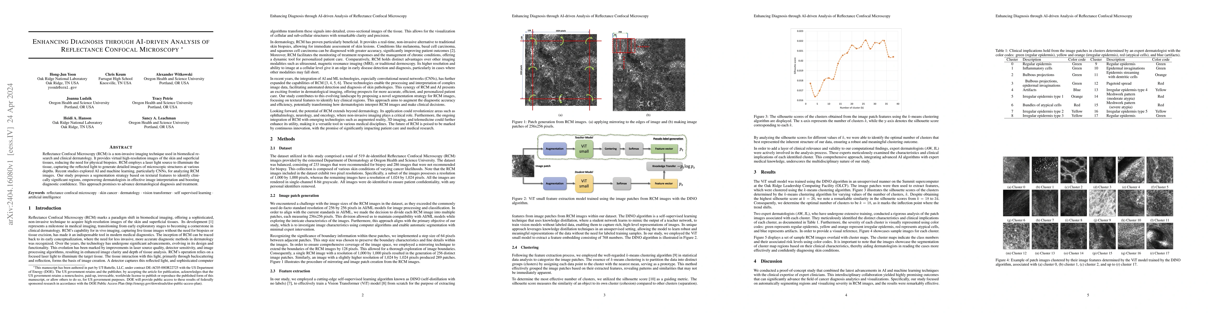
Key Findings, in focus

Seven facets of this paper, analysed and brought into focus by AI.

AI-generated · verify against source Generated Sep 05, 2025
In focus · one-line synthesis

This research contributes to the development of AI-based diagnostic tools for skin diseases, improving early detection and treatment.

01
MethodologyHow they did it

A self-supervised learning algorithm (DINO) was used to develop an ML model that can extract local textual features from image patches of Reflectance Confocal Microscopy (RCM) images.

02
ResultsWhat they found
  • The proposed approach achieved a high accuracy in downstream clinical classification tasks
  • Unsupervised clustering of extracted features showed promising results for RCM image segmentation
  • The feature map has clinical relevance and can be used as a segmentation tool for RCM images
03
SignificanceWhy it matters

This research contributes to the development of AI-based diagnostic tools for skin diseases, improving early detection and treatment.

04
Technical ContributionsWhat's new technically

The proposed self-supervised learning approach enables the extraction of local textual features from RCM images, facilitating downstream clinical classification tasks.

05
NoveltyWhat sets it apart

This work introduces a novel self-supervised learning approach for RCM image analysis, demonstrating its potential for improving diagnostic accuracy and efficiency.

06
LimitationsWhat to be cautious about
  • The dataset size may not accurately represent the entire disease spectrum
  • Limited exploration of potential clinical applications
07
Future WorkWhere it goes next
  • Investigating larger datasets to improve generalizability
  • Exploring other machine learning algorithms for RCM image analysis

Impact

18
References

Paper Details

Paper ID: 2404.16080
License: http://arxiv.org/licenses/nonexclusive-distrib/1.0/
Categories:
eess.IV cs.AI cs.CV

How to Cite This Paper

Reference manager: RIS EndNote

Key Terms

images (0.234)
skin (0.186)
reflected (0.173)
boosting (0.173)
cnns (0.173)
light (0.173)
diagnosis (0.167)
diagnostic (0.166)

Citation Network

Current paper (gray), citations (green), references (blue)

Display is limited for performance on very large graphs.

Current Paper
Citations
References
Click to view


Discussion 0

This website uses cookies to ensure you get the best experience. Learn more