01
MethodologyHow they did it
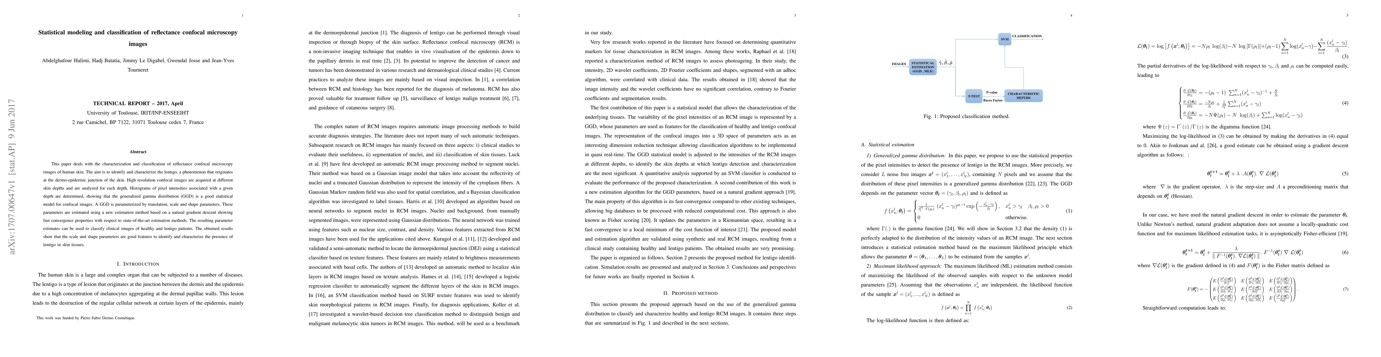
The research employs statistical modeling and classification of reflectance confocal microscopy (RCM) images of human skin to identify and characterize lentigo, a phenomenon originating at the dermo-epidermic junction. High-resolution confocal images are acquired at various skin depths, and histograms of pixel intensities are analyzed using the generalized gamma distribution (GGD) for modeling.
Discussion 0