Enhancing super-resolution ultrasound localisation through multi-frame deconvolution exploiting spatiotemporal coherence
Publication
Metrics
AI Quick Summary
This paper proposes a Multi-Frame Deconvolution (MF-Decon) framework to enhance super-resolution ultrasound imaging by improving microbubble localisation through spatiotemporal coherence exploitation. The new spatial and temporal regularisers, based on total variation and regularisation by denoising, lead to superior performance in MB localisation precision and recall compared to existing methods, resulting in clearer microvasculature maps.
Paper Preview
Abstract
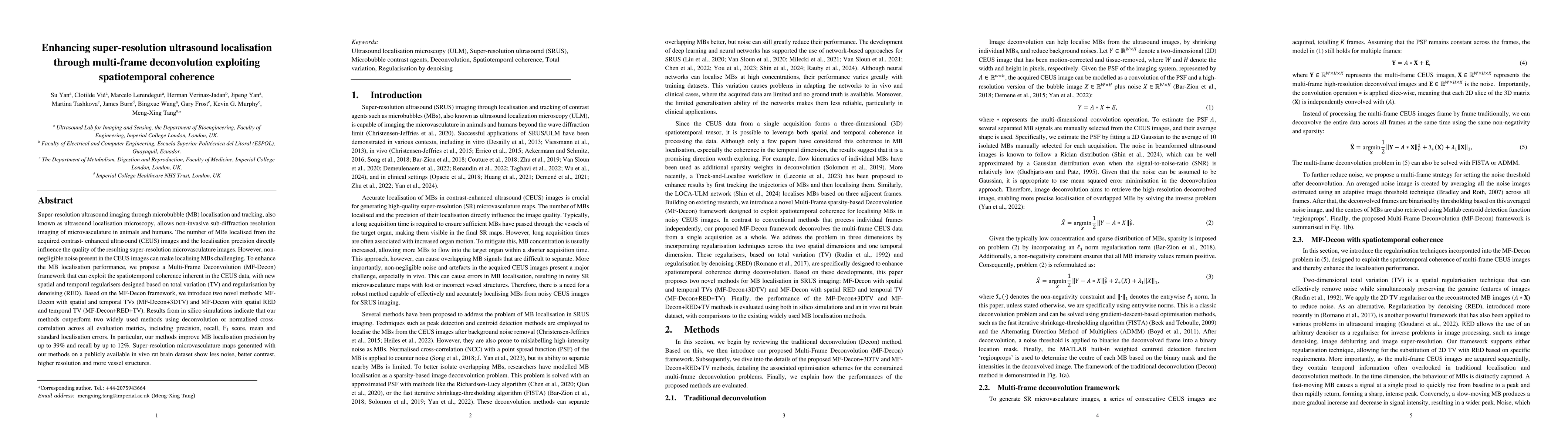
Super-resolution ultrasound imaging through microbubble (MB) localisation and tracking, also known as ultrasound localisation microscopy, allows non-invasive sub-diffraction resolution imaging of microvasculature in animals and humans. The number of MBs localised from the acquired contrast-enhanced ultrasound (CEUS) images and the localisation precision directly influence the quality of the resulting super-resolution microvasculature images. However, non-negligible noise present in the CEUS images can make localising MBs challenging. To enhance the MB localisation performance, we propose a Multi-Frame Deconvolution (MF-Decon) framework that can exploit the spatiotemporal coherence inherent in the CEUS data, with new spatial and temporal regularisers designed based on total variation (TV) and regularisation by denoising (RED). Based on the MF-Decon framework, we introduce two novel methods: MF-Decon with spatial and temporal TVs (MF-Decon+3DTV) and MF-Decon with spatial RED and temporal TV (MF-Decon+RED+TV). Results from in silico simulations indicate that our methods outperform two widely used methods using deconvolution or normalised cross-correlation across all evaluation metrics, including precision, recall, $F_1$ score, mean and standard localisation errors. In particular, our methods improve MB localisation precision by up to 39% and recall by up to 12%. Super-resolution microvasculature maps generated with our methods on a publicly available in vivo rat brain dataset show less noise, better contrast, higher resolution and more vessel structures.
AI Key Findings
Get AI-generated insights about this paper's methodology, results, significance, and more — seven facets brought into focus.
Impact
Paper Details
Authors
PDF Preview
Citation Network
Current paper (gray), citations (green), references (blue)
Display is limited for performance on very large graphs.
Discussion 0