Publication

Published: Oct 08, 2025
Categories: cs.CV

Metrics

Source: ArXiv

Quick Actions

View PDF

Quick Answers

What methodology did the authors use?

The study compared foundation models (FMs) and lightweight CNNs for diabetic macular edema (DME) detection using transfer learning. They evaluated models on IDRiD and MESSIDOR datasets with various augmentation techniques and metrics like AUC-ROC and F1-score. More in Methodology →

What are the key results?

Lightweight CNNs outperformed FMs on IDRiD with AUC-ROC of 0.98 vs 0.95 for FMs — MESSIDOR showed lower performance across all models due to domain shift and lower DME prevalence More in Key Results →

Why is this work significant?

This research highlights the importance of model architecture and data characteristics in medical image analysis, providing guidance for developing robust DME detection systems that can generalize across different clinical settings. More in Significance →

What are the main limitations?

Results may not generalize to other retinal diseases beyond DME — Dependence on specific prompt formulations for zero-shot performance More in Limitations →

Paper Preview

Abstract

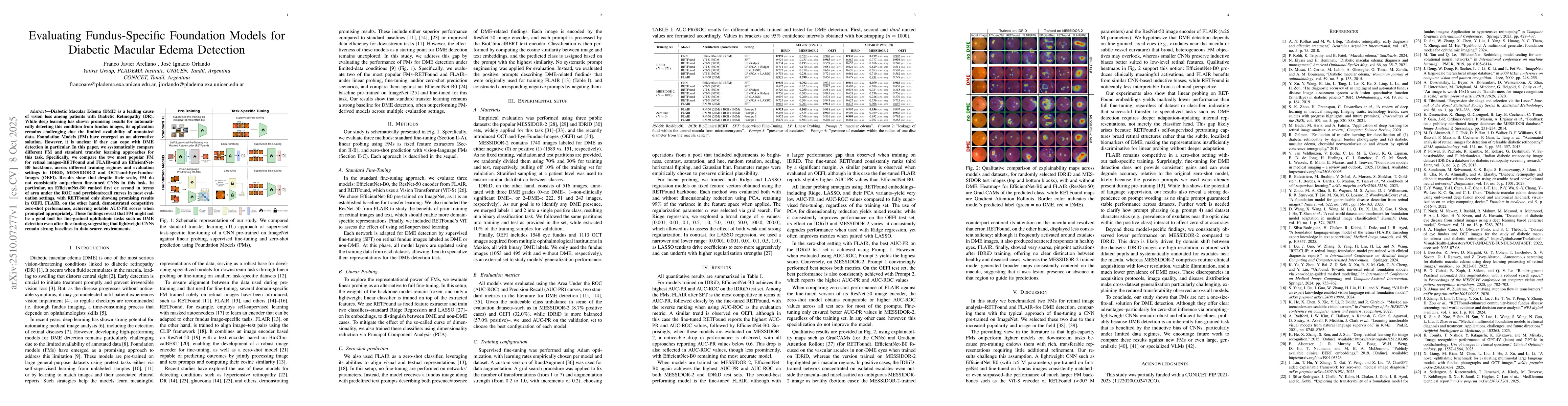
Diabetic Macular Edema (DME) is a leading cause of vision loss among patients with Diabetic Retinopathy (DR). While deep learning has shown promising results for automatically detecting this condition from fundus images, its application remains challenging due the limited availability of annotated data. Foundation Models (FM) have emerged as an alternative solution. However, it is unclear if they can cope with DME detection in particular. In this paper, we systematically compare different FM and standard transfer learning approaches for this task. Specifically, we compare the two most popular FM for retinal images--RETFound and FLAIR--and an EfficientNet-B0 backbone, across different training regimes and evaluation settings in IDRiD, MESSIDOR-2 and OCT-and-Eye-Fundus-Images (OEFI). Results show that despite their scale, FM do not consistently outperform fine-tuned CNNs in this task. In particular, an EfficientNet-B0 ranked first or second in terms of area under the ROC and precision/recall curves in most evaluation settings, with RETFound only showing promising results in OEFI. FLAIR, on the other hand, demonstrated competitive zero-shot performance, achieving notable AUC-PR scores when prompted appropriately. These findings reveal that FM might not be a good tool for fine-grained ophthalmic tasks such as DME detection even after fine-tuning, suggesting that lightweight CNNs remain strong baselines in data-scarce environments.

AI Key Findings

Generated Oct 10, 2025

Methodology — What approach did the authors take?

The study compared foundation models (FMs) and lightweight CNNs for diabetic macular edema (DME) detection using transfer learning. They evaluated models on IDRiD and MESSIDOR datasets with various augmentation techniques and metrics like AUC-ROC and F1-score.

Key Results — What are the main findings?

  • Lightweight CNNs outperformed FMs on IDRiD with AUC-ROC of 0.98 vs 0.95 for FMs
  • MESSIDOR showed lower performance across all models due to domain shift and lower DME prevalence
  • Prompt wording significantly affected zero-shot performance with no single prompt guaranteeing stable results

Significance — Why does this research matter?

This research highlights the importance of model architecture and data characteristics in medical image analysis, providing guidance for developing robust DME detection systems that can generalize across different clinical settings.

Technical Contribution — What is the technical contribution?

Proposed a systematic comparison framework between FMs and CNNs for medical image analysis, emphasizing the importance of data characteristics and model architecture selection for different clinical scenarios.

Novelty — What is new about this work?

Introduces a comprehensive analysis of zero-shot DME detection using both FMs and CNNs, revealing the critical role of prompt engineering and domain adaptation in achieving robust medical image classification.

Limitations — What are the limitations of this study?

  • Results may not generalize to other retinal diseases beyond DME
  • Dependence on specific prompt formulations for zero-shot performance
  • Limited exploration of specialized VLMs for medical imaging

Future Work — What did the authors propose for future work?

  • Investigate domain adaptation techniques to bridge IDRiD and MESSIDOR gaps
  • Develop more robust prompt engineering strategies for zero-shot scenarios
  • Explore specialized vision-language models for medical imaging tasks
  • Quantify the impact of data quality and annotation consistency on model performance

Paper Details

Paper ID: 2510.07277
Comments: Accepted for publication at SIPAIM 2025
Categories:
cs.CV

How to Cite This Paper

BibTeX
@article{arellano2025evaluating,
  title         = {Evaluating Fundus-Specific Foundation Models for Diabetic Macular Edema
  Detection},
  author        = {Arellano, Franco Javier and Orlando, José Ignacio},
  year          = {2025},
  eprint        = {2510.07277},
  archivePrefix = {arXiv},
  primaryClass  = {cs.CV},
}
APA 7
Arellano, F., & Orlando, J. (2025). Evaluating Fundus-Specific Foundation Models for Diabetic Macular Edema
  Detection. arXiv. https://arxiv.org/abs/2510.07277
MLA 9
Arellano, Franco Javier, and José Ignacio Orlando. "Evaluating Fundus-Specific Foundation Models for Diabetic Macular Edema
  Detection." arXiv, 2025, arxiv.org/abs/2510.07277.

Comments (0)

This website uses cookies to ensure you get the best experience. Learn more