Evaluation of Preprocessing Techniques for U-Net Based Automated Liver Segmentation
Publication
Metrics
AI Quick Summary
This study evaluates the impact of preprocessing techniques on U-Net based automated liver segmentation in medical images, finding that a combination of HU-windowing, median filtering, and z-score normalization achieve optimal performance. The results show promise for improving accuracy in liver analysis and quantification.
Paper Preview
Abstract
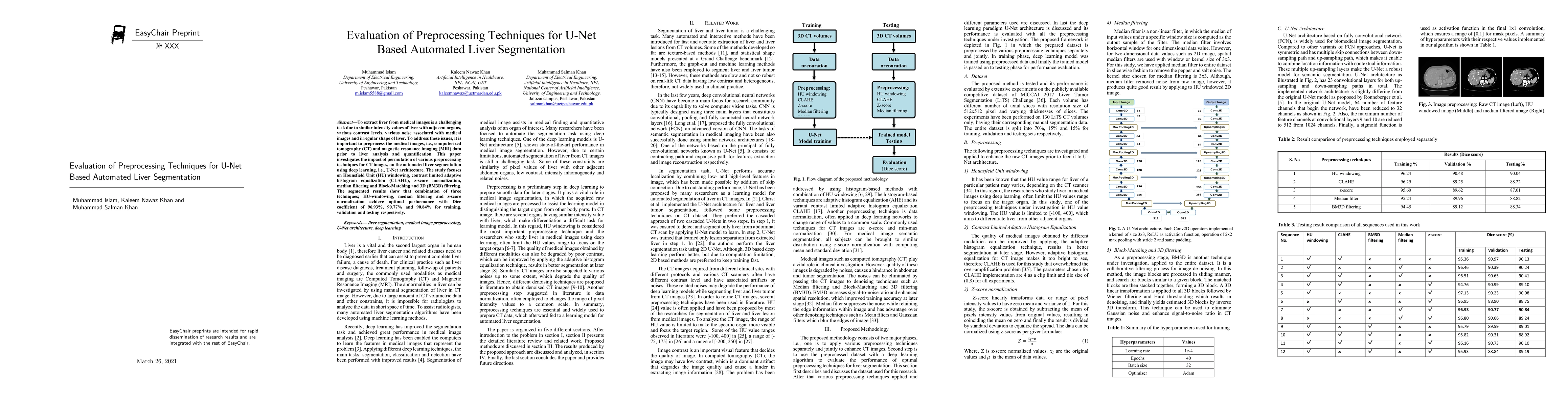
To extract liver from medical images is a challenging task due to similar intensity values of liver with adjacent organs, various contrast levels, various noise associated with medical images and irregular shape of liver. To address these issues, it is important to preprocess the medical images, i.e., computerized tomography (CT) and magnetic resonance imaging (MRI) data prior to liver analysis and quantification. This paper investigates the impact of permutation of various preprocessing techniques for CT images, on the automated liver segmentation using deep learning, i.e., U-Net architecture. The study focuses on Hounsfield Unit (HU) windowing, contrast limited adaptive histogram equalization (CLAHE), z-score normalization, median filtering and Block-Matching and 3D (BM3D) filtering. The segmented results show that combination of three techniques; HU-windowing, median filtering and z-score normalization achieve optimal performance with Dice coefficient of 96.93%, 90.77% and 90.84% for training, validation and testing respectively.
AI Key Findings
Get AI-generated insights about this paper's methodology, results, significance, and more — seven facets brought into focus.
Impact
Paper Details
Authors
PDF Preview
Key Terms
Citation Network
Current paper (gray), citations (green), references (blue)
Display is limited for performance on very large graphs.
Discussion 0