Expanding Training Data for Endoscopic Phenotyping of Eosinophilic Esophagitis

Source: ArXiv

AI Quick Summary

This study enhances the diagnostic accuracy of AI-assisted endoscopic imaging for eosinophilic esophagitis by expanding training data from 435 to 7050 images, employing a Data-efficient Image Transformer, and improving model interpretability with attention map visualizations. The results demonstrate improved classification performance and robustness.

Share: Share on X LinkedIn

Summary

Eosinophilic esophagitis (EoE) is a chronic esophageal disorder marked by eosinophil-dominated inflammation. Diagnosing EoE usually involves endoscopic inspection of the esophageal mucosa and obtaining esophageal biopsies for histologic confirmation. Recent advances have seen AI-assisted endoscopic imaging, guided by the EREFS system, emerge as a potential alternative to reduce reliance on invasive histological assessments. Despite these advancements, significant challenges persist due to the limited availability of data for training AI models - a common issue even in the development of AI for 
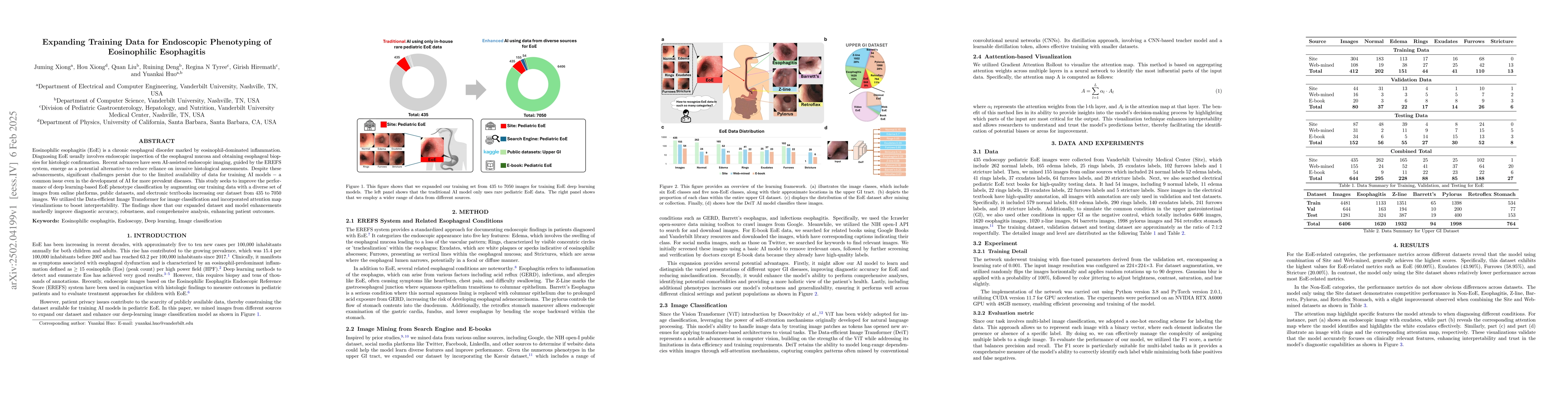
more prevalent diseases. This study seeks to improve the performance of deep learning-based EoE phenotype classification by augmenting our training data with a diverse set of images from online platforms, public datasets, and electronic textbooks increasing our dataset from 435 to 7050 images. We utilized the Data-efficient Image Transformer for image classification and incorporated attention map visualizations to boost interpretability. The findings show that our expanded dataset and model enhancements improved diagnostic accuracy, robustness, and comprehensive analysis, enhancing patient outcomes.

AI Key Findings

Generated Jun 11, 2025

Methodology

The study expanded the training dataset for deep learning models in endoscopic phenotyping of Eosinophilic Esophagitis (EoE) from 435 to 7050 images by incorporating data from diverse sources like online platforms, public datasets, and electronic textbooks. The Data-efficient Image Transformer (DeiT) was used for image classification, enhancing interpretability with attention map visualizations.

Key Results

  • Expanding the dataset improved diagnostic accuracy, robustness, and comprehensive analysis for EoE.
  • The enhanced model demonstrated better identification of EoE-related features such as Edema (60.00%), Exudates (43.90%), Furrows (58.95%), and Stricture (20.00%).
  • Attention maps highlighted clinically relevant features, validating the model's accurate focus on essential diagnostic aspects.

Significance

This research is significant as it contributes to more accurate and non-invasive diagnosis of EoE, potentially improving patient outcomes by reducing reliance on invasive histological assessments.

Technical Contribution

The study introduced an advanced endoscopy image classification model using a dual-dataset approach, combining in-house and web-mined EoE images, to improve diagnostic accuracy and robustness.

Novelty

This work stands out by emphasizing the integration of web-mined EoE images, providing a diverse and extensive dataset, which greatly enhances the model's ability to recognize various disease presentations and generalize across different patient demographics.

Limitations

  • The study did not address generalizability across different endoscopic equipment or operator variability.
  • Limited information on model performance in real-world clinical settings.

Future Work

  • Investigate model performance with diverse endoscopic equipment and operators.
  • Conduct clinical trials to validate the model's effectiveness in real-world scenarios.

Paper Details

PDF Preview

Download PDF

No citations found for this paper.

Comments (0)

This website uses cookies to ensure you get the best experience. Learn more