Academic Profile
Statistics
Similar Authors
Papers on arXiv
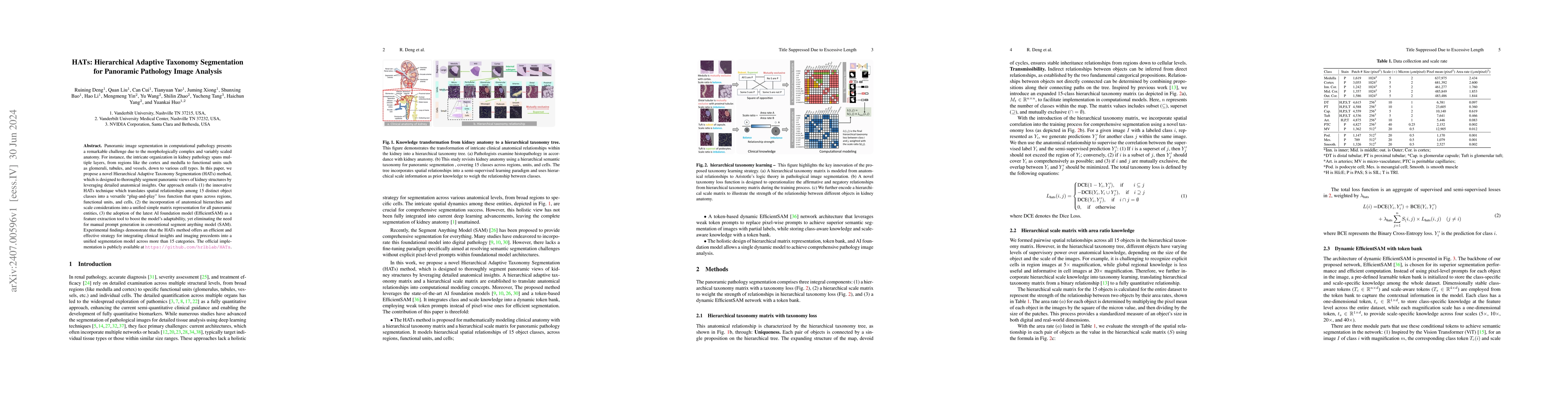
Panoramic image segmentation in computational pathology presents a remarkable challenge due to the morphologically complex and variably scaled anatomy. For instance, the intricate organization in kidn...
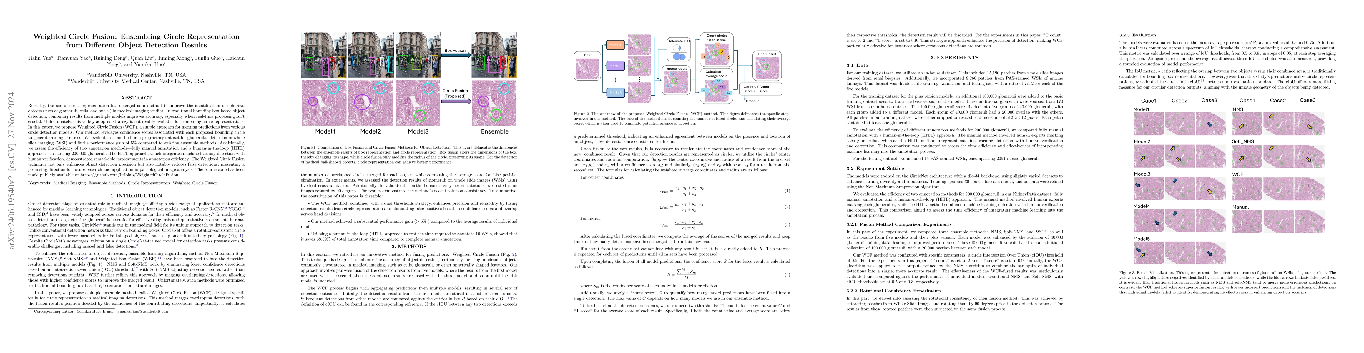
Recently, the use of circle representation has emerged as a method to improve the identification of spherical objects (such as glomeruli, cells, and nuclei) in medical imaging studies. In traditional ...
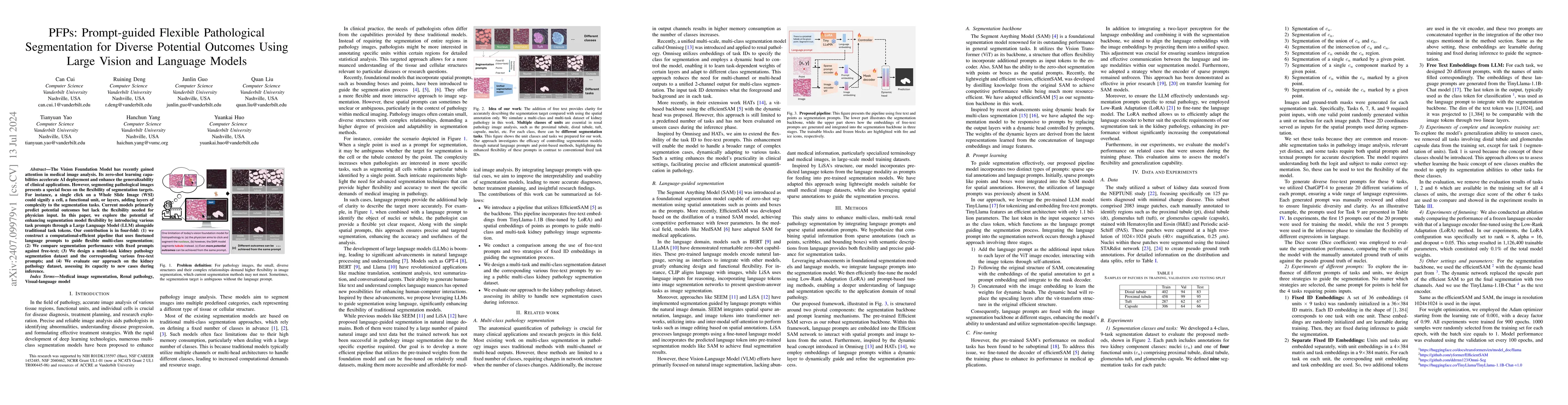
The Vision Foundation Model has recently gained attention in medical image analysis. Its zero-shot learning capabilities accelerate AI deployment and enhance the generalizability of clinical applicati...
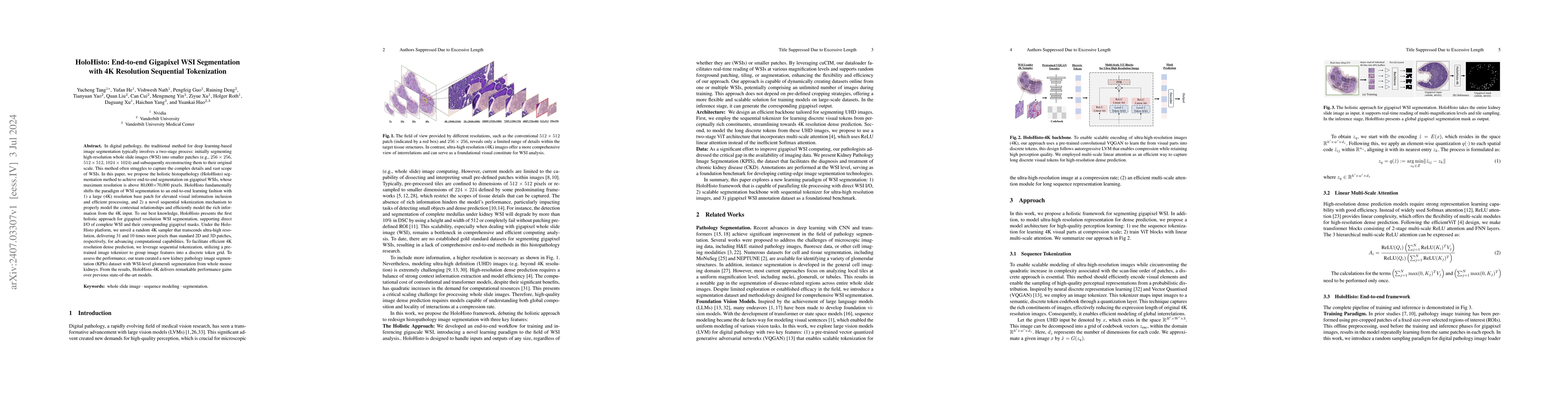
In digital pathology, the traditional method for deep learning-based image segmentation typically involves a two-stage process: initially segmenting high-resolution whole slide images (WSI) into sma...
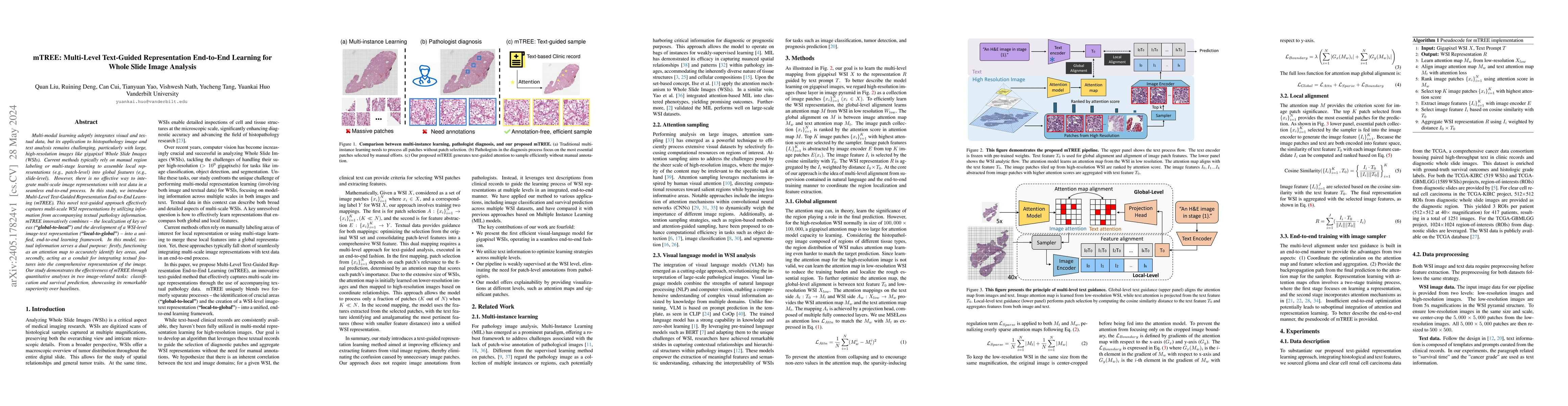
Multi-modal learning adeptly integrates visual and textual data, but its application to histopathology image and text analysis remains challenging, particularly with large, high-resolution images li...
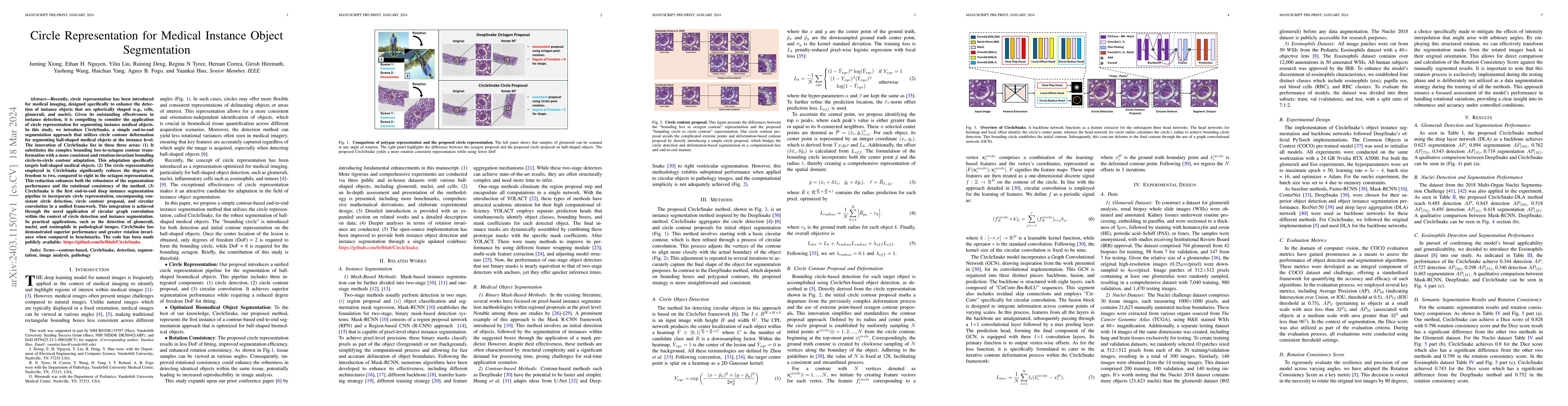
Recently, circle representation has been introduced for medical imaging, designed specifically to enhance the detection of instance objects that are spherically shaped (e.g., cells, glomeruli, and n...
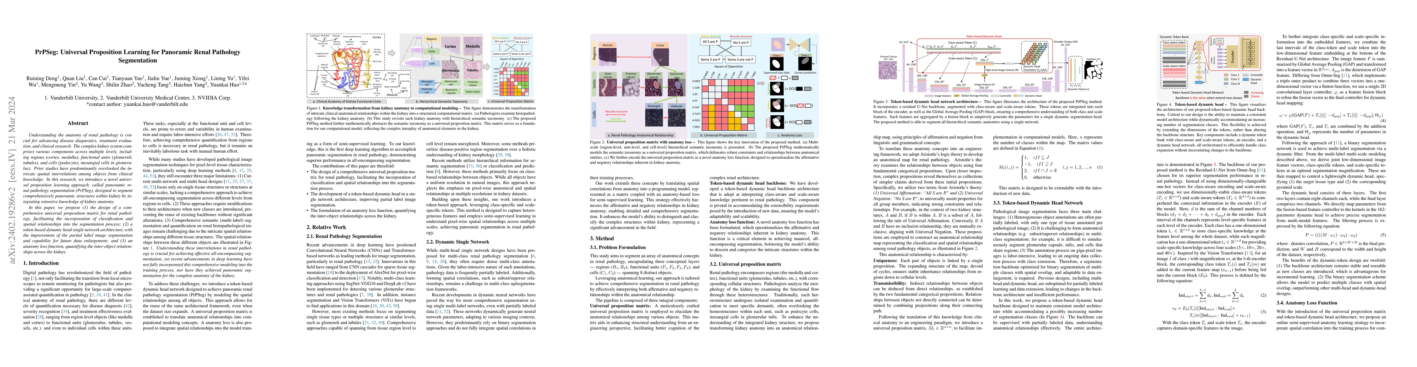
Understanding the anatomy of renal pathology is crucial for advancing disease diagnostics, treatment evaluation, and clinical research. The complex kidney system comprises various components across ...
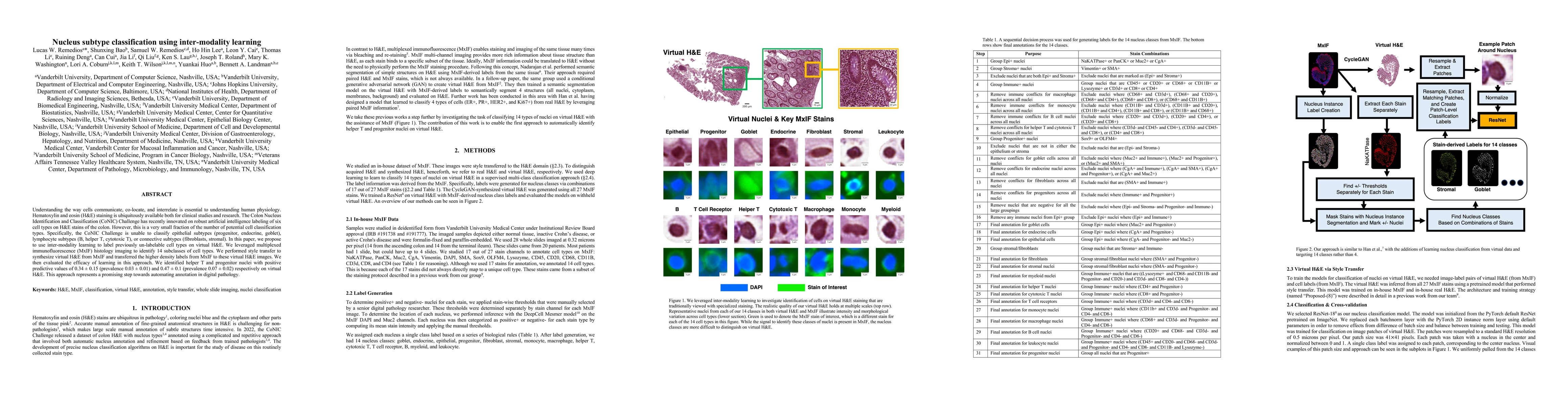
Understanding the way cells communicate, co-locate, and interrelate is essential to understanding human physiology. Hematoxylin and eosin (H&E) staining is ubiquitously available both for clinical s...
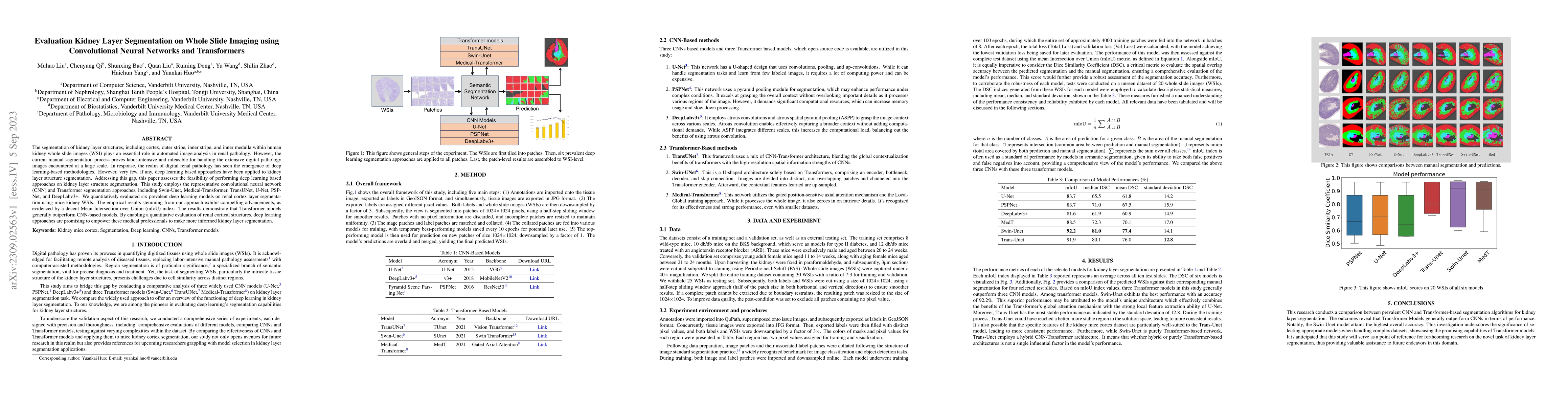
The segmentation of kidney layer structures, including cortex, outer stripe, inner stripe, and inner medulla within human kidney whole slide images (WSI) plays an essential role in automated image a...
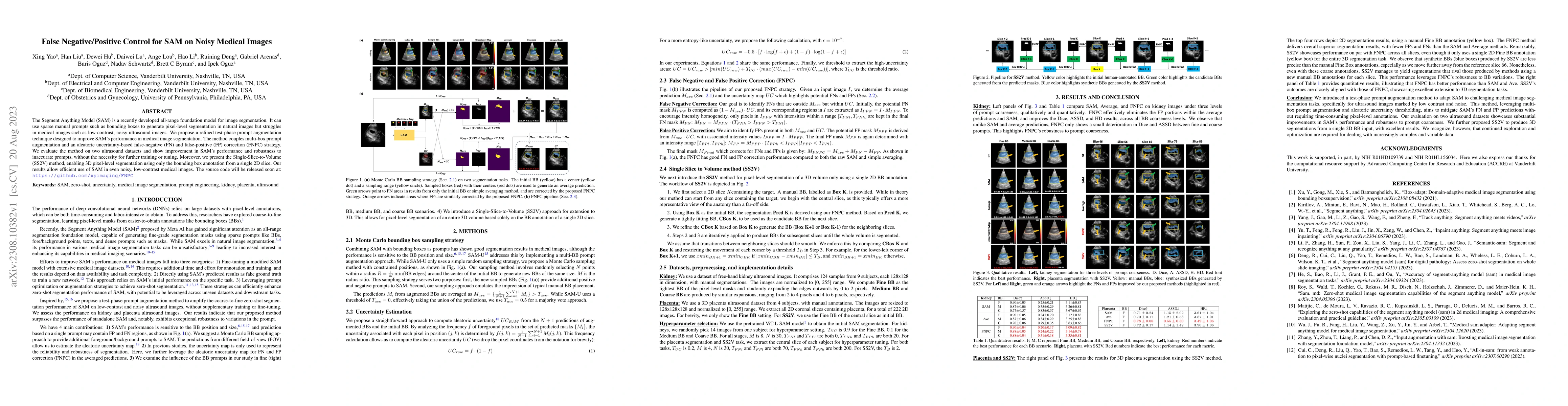
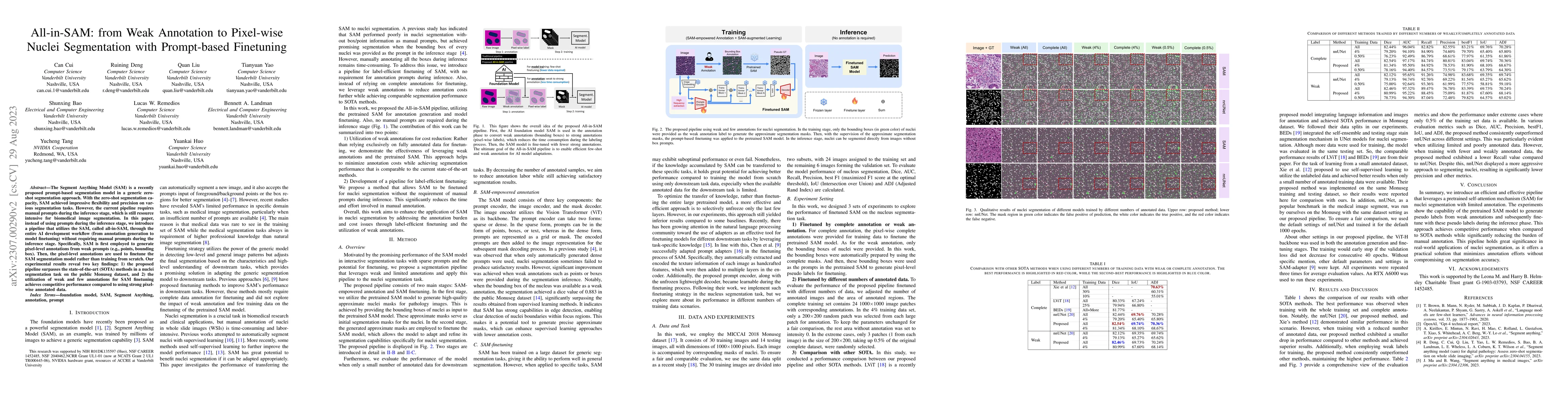
The Segment Anything Model (SAM) is a recently developed all-range foundation model for image segmentation. It can use sparse manual prompts such as bounding boxes to generate pixel-level segmentati...
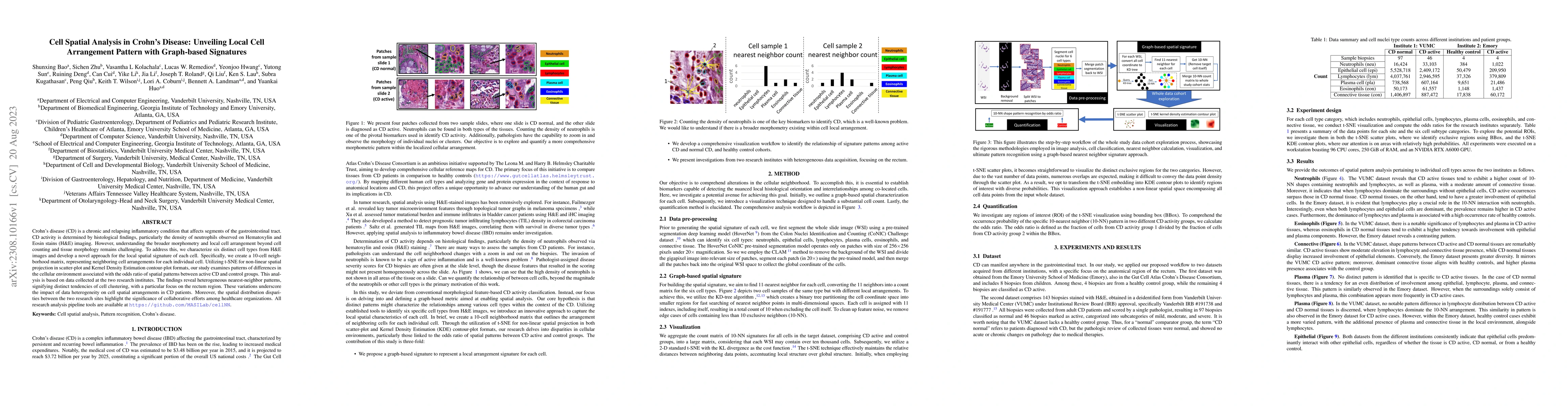
Crohn's disease (CD) is a chronic and relapsing inflammatory condition that affects segments of the gastrointestinal tract. CD activity is determined by histological findings, particularly the densi...
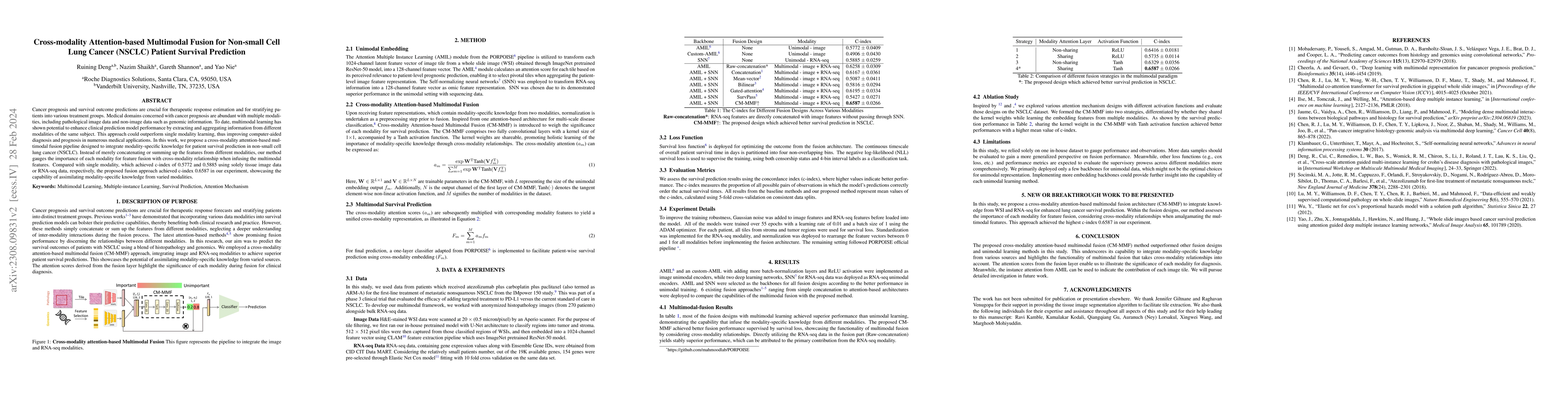
Cancer prognosis and survival outcome predictions are crucial for therapeutic response estimation and for stratifying patients into various treatment groups. Medical domains concerned with cancer pr...
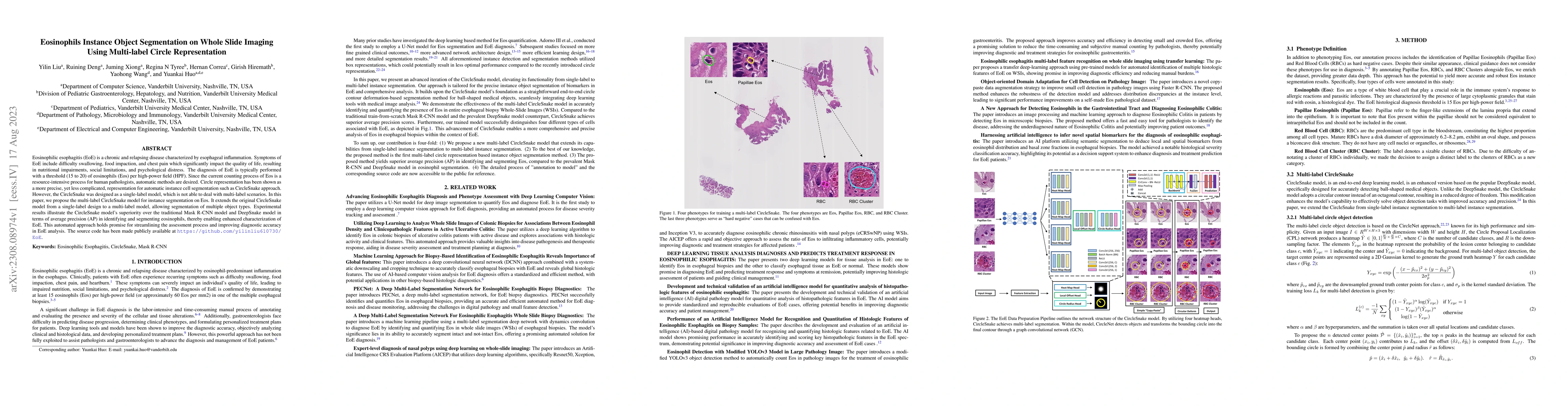
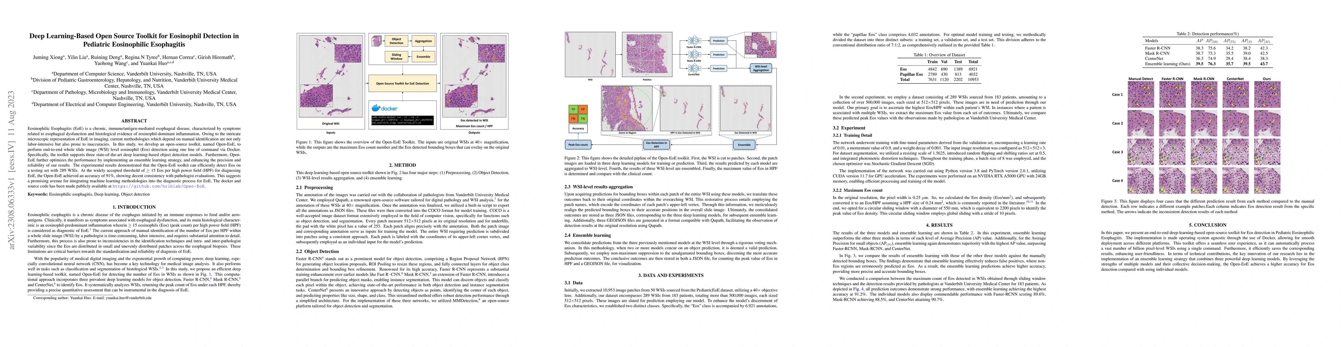
Eosinophilic esophagitis (EoE) is a chronic and relapsing disease characterized by esophageal inflammation. Symptoms of EoE include difficulty swallowing, food impaction, and chest pain which signif...
Eosinophilic Esophagitis (EoE) is a chronic, immune/antigen-mediated esophageal disease, characterized by symptoms related to esophageal dysfunction and histological evidence of eosinophil-dominant ...
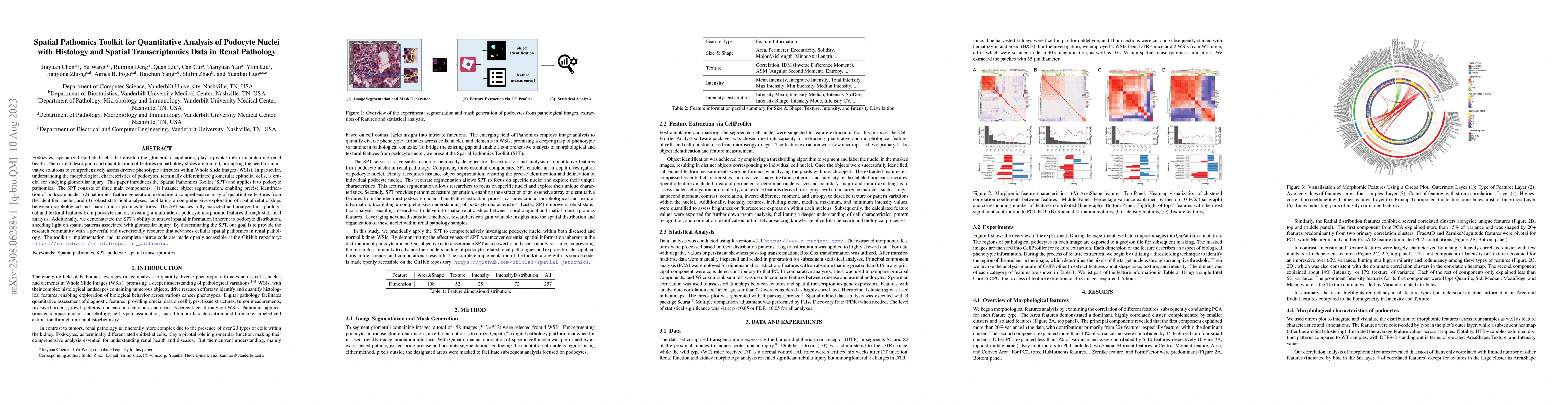
Podocytes, specialized epithelial cells that envelop the glomerular capillaries, play a pivotal role in maintaining renal health. The current description and quantification of features on pathology ...
Precise identification of multiple cell classes in high-resolution Giga-pixel whole slide imaging (WSI) is critical for various clinical scenarios. Building an AI model for this purpose typically re...
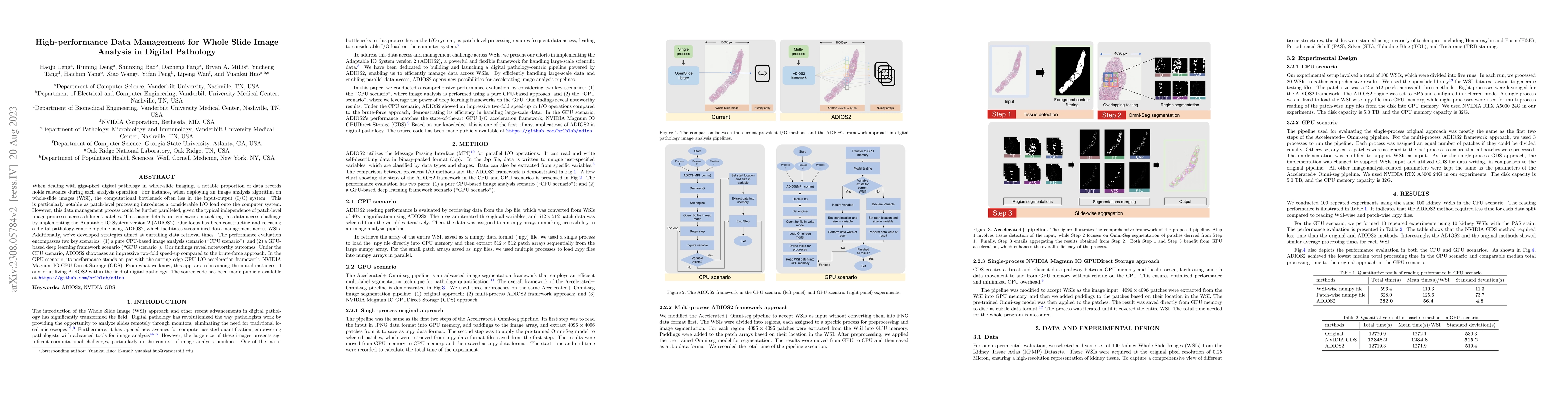
When dealing with giga-pixel digital pathology in whole-slide imaging, a notable proportion of data records holds relevance during each analysis operation. For instance, when deploying an image anal...
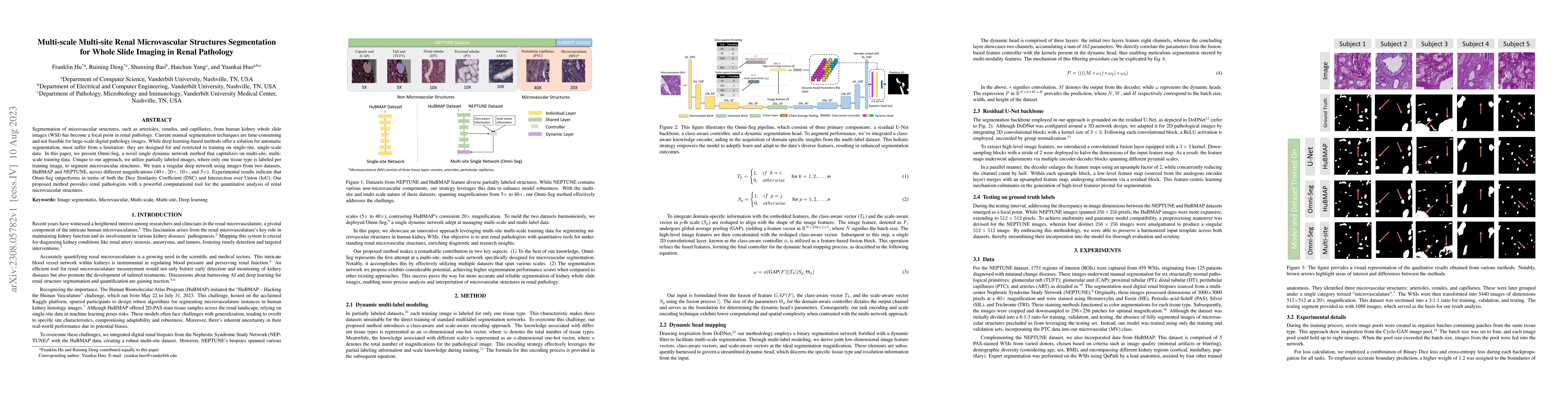
Segmentation of microvascular structures, such as arterioles, venules, and capillaries, from human kidney whole slide images (WSI) has become a focal point in renal pathology. Current manual segment...
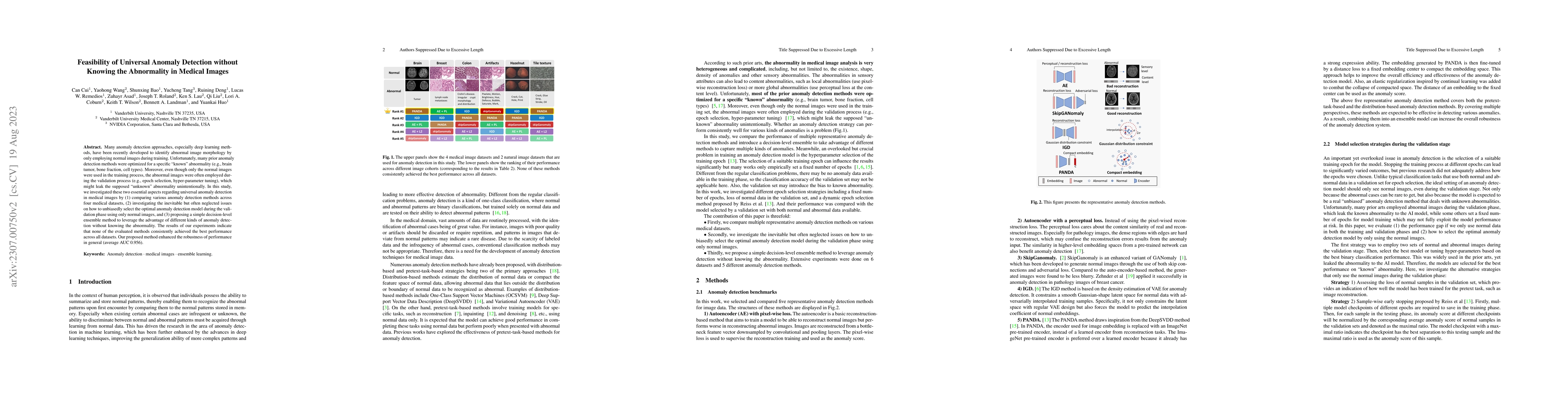
Many anomaly detection approaches, especially deep learning methods, have been recently developed to identify abnormal image morphology by only employing normal images during training. Unfortunately...
The Segment Anything Model (SAM) is a recently proposed prompt-based segmentation model in a generic zero-shot segmentation approach. With the zero-shot segmentation capacity, SAM achieved impressiv...
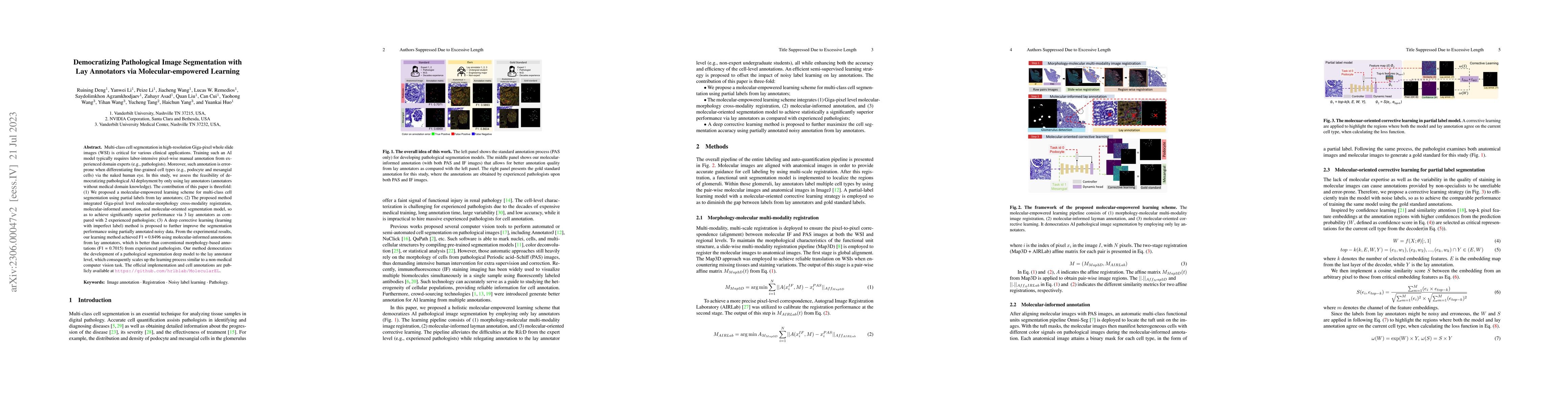
Multi-class cell segmentation in high-resolution Giga-pixel whole slide images (WSI) is critical for various clinical applications. Training such an AI model typically requires labor-intensive pixel...
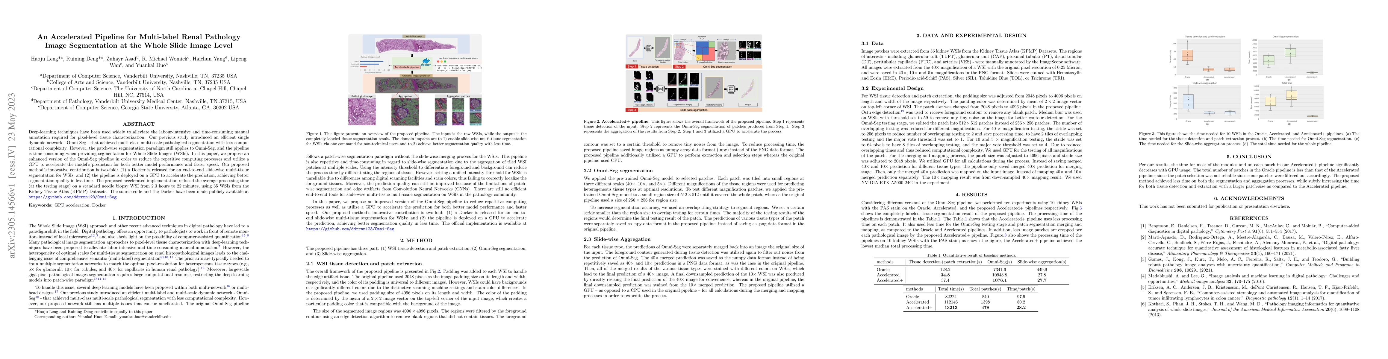
Deep-learning techniques have been used widely to alleviate the labour-intensive and time-consuming manual annotation required for pixel-level tissue characterization. Our previous study introduced ...
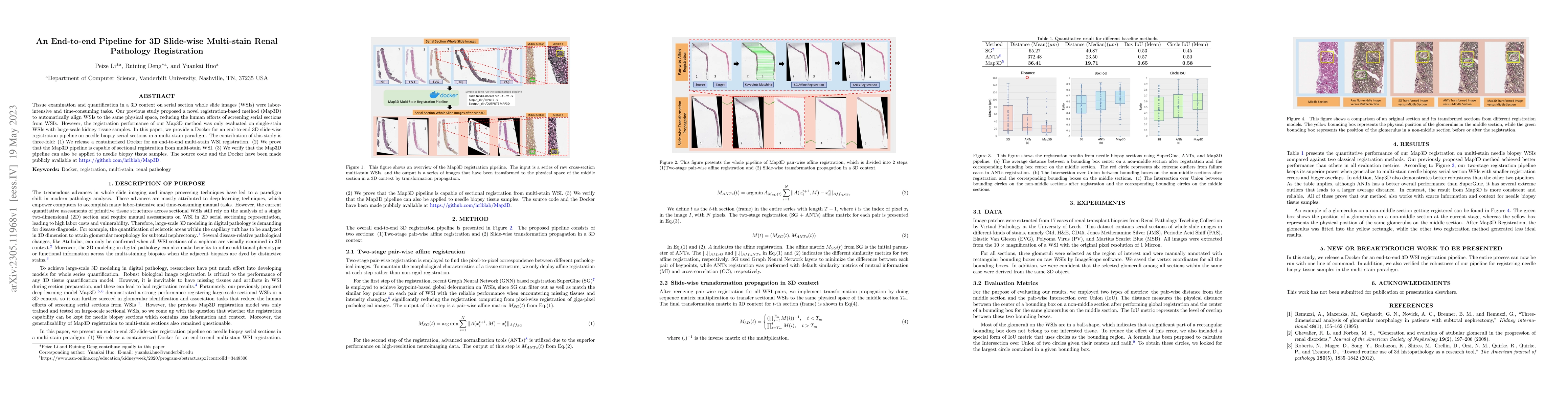
Tissue examination and quantification in a 3D context on serial section whole slide images (WSIs) were laborintensive and time-consuming tasks. Our previous study proposed a novel registration-based...
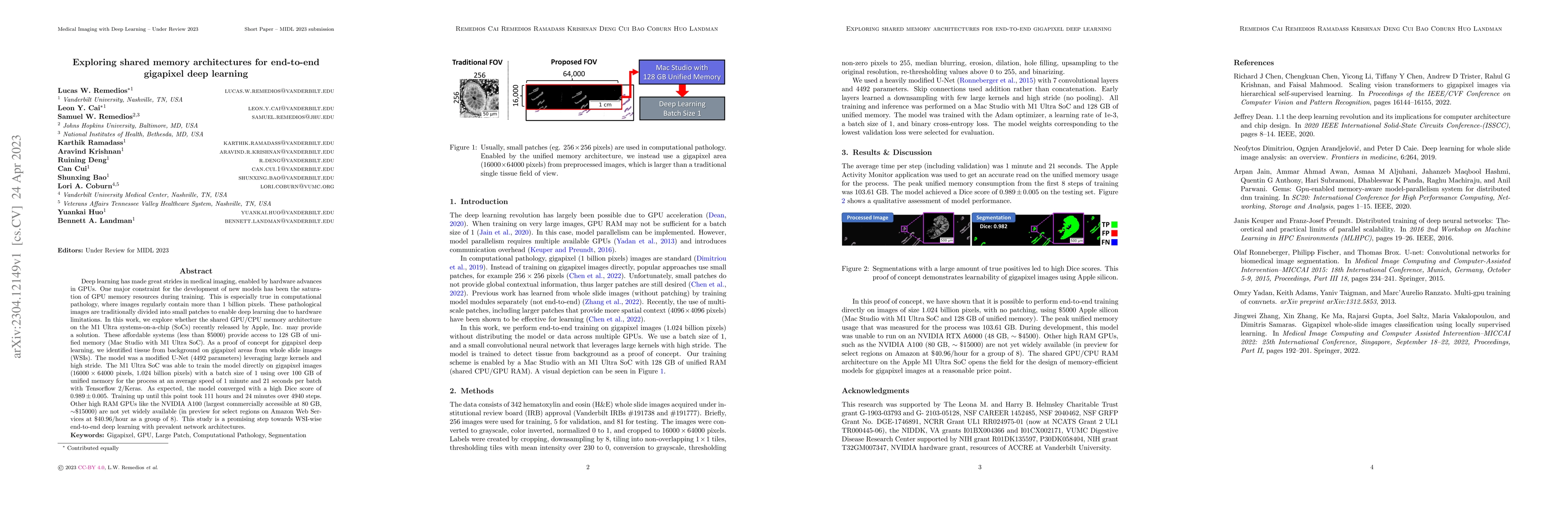
Deep learning has made great strides in medical imaging, enabled by hardware advances in GPUs. One major constraint for the development of new models has been the saturation of GPU memory resources ...
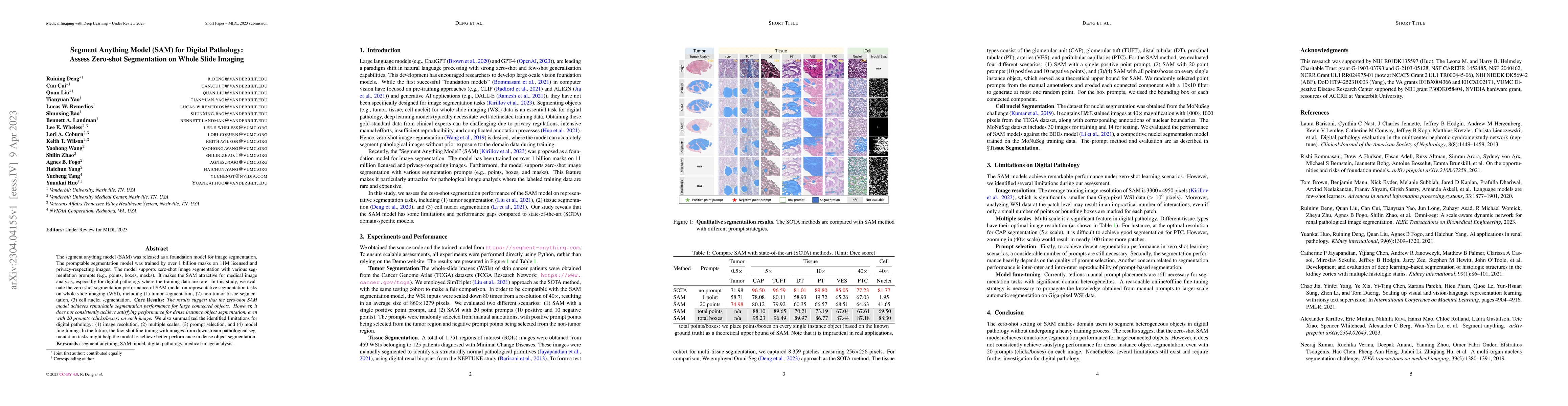
The segment anything model (SAM) was released as a foundation model for image segmentation. The promptable segmentation model was trained by over 1 billion masks on 11M licensed and privacy-respecti...
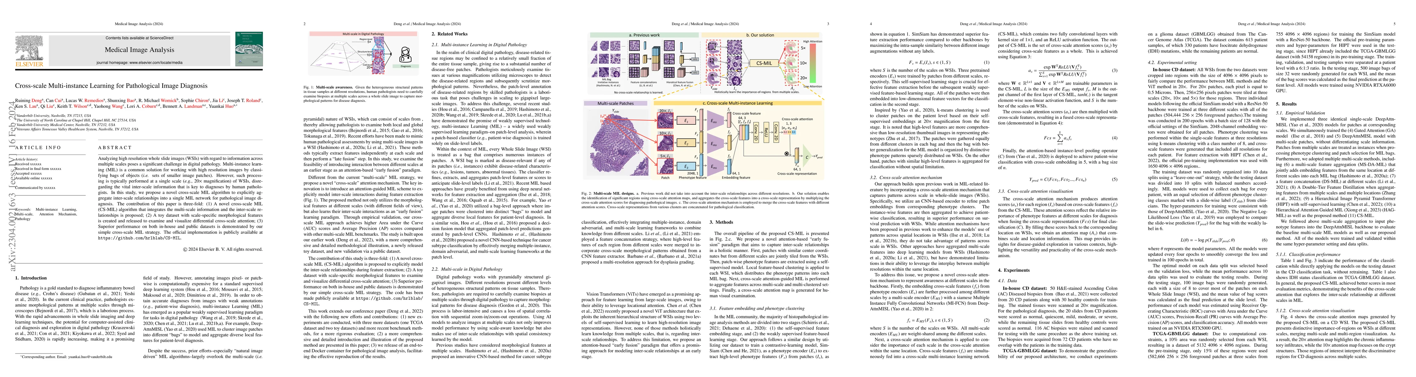
Analyzing high resolution whole slide images (WSIs) with regard to information across multiple scales poses a significant challenge in digital pathology. Multi-instance learning (MIL) is a common so...
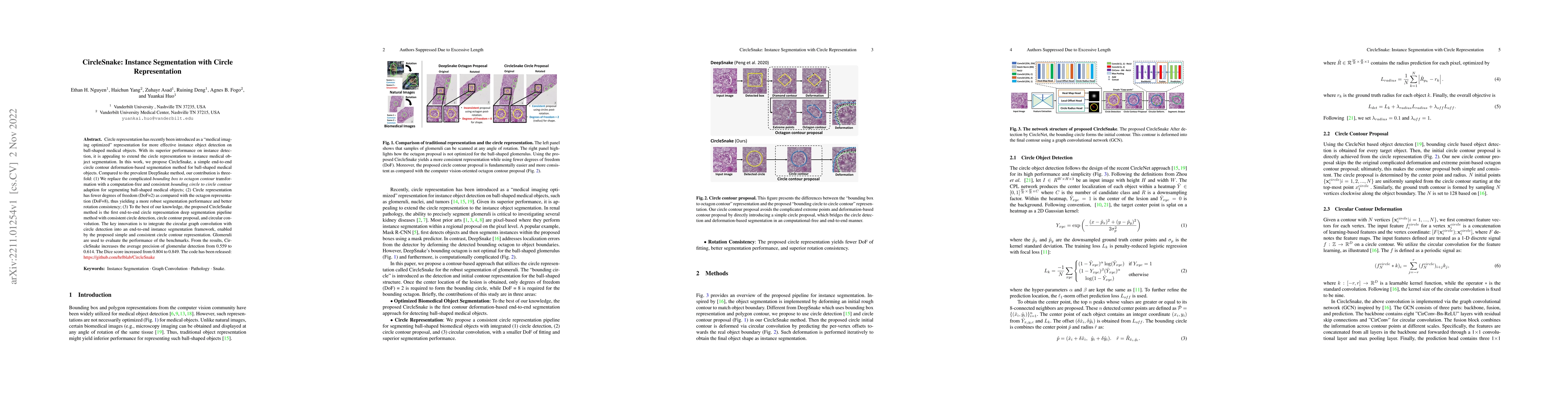
Circle representation has recently been introduced as a medical imaging optimized representation for more effective instance object detection on ball-shaped medical objects. With its superior perfor...
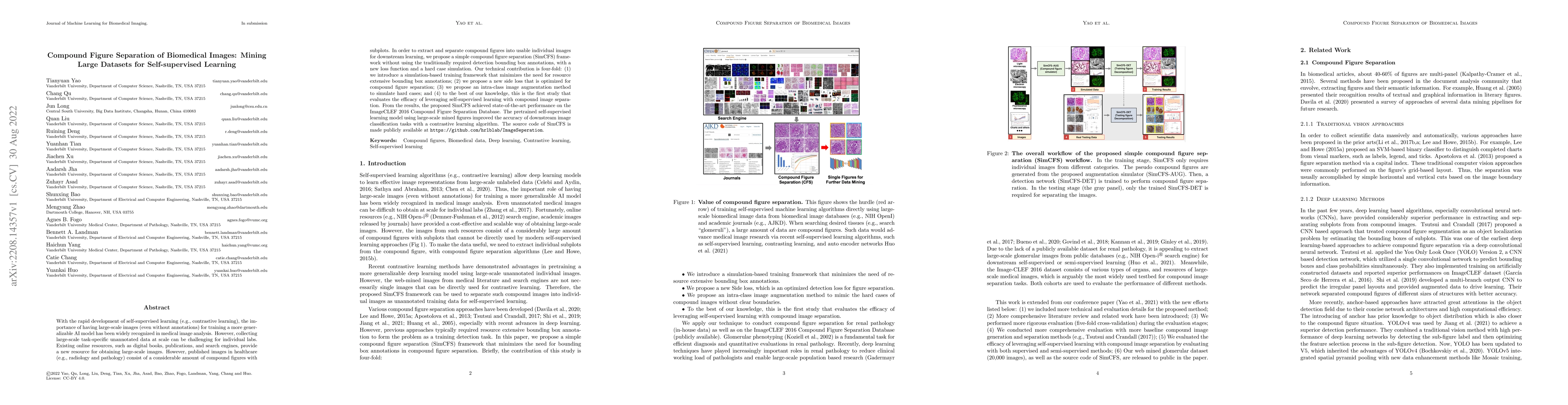
With the rapid development of self-supervised learning (e.g., contrastive learning), the importance of having large-scale images (even without annotations) for training a more generalizable AI model...
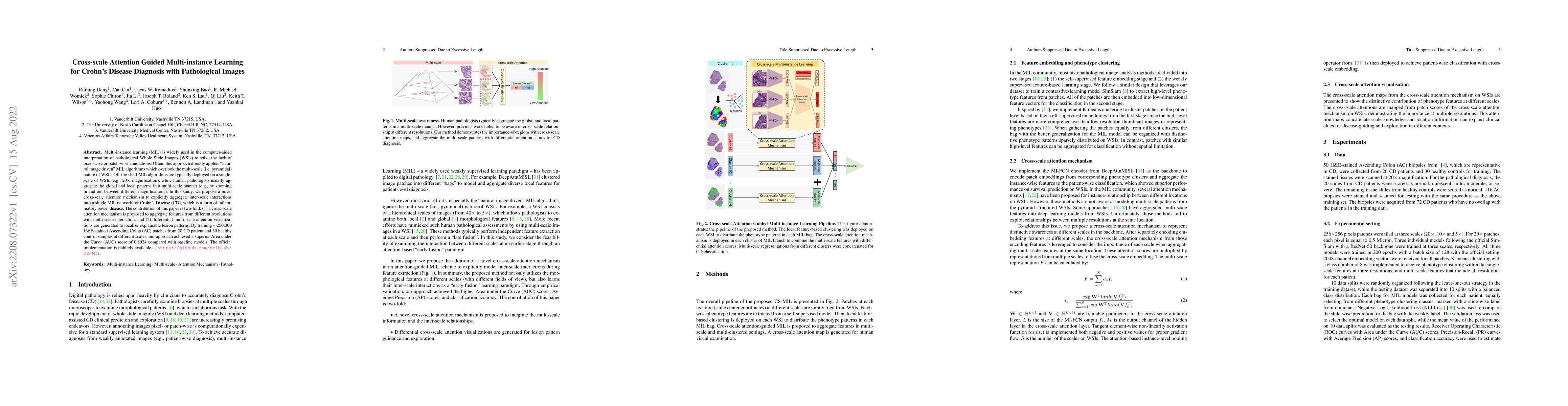
Multi-instance learning (MIL) is widely used in the computer-aided interpretation of pathological Whole Slide Images (WSIs) to solve the lack of pixel-wise or patch-wise annotations. Often, this app...
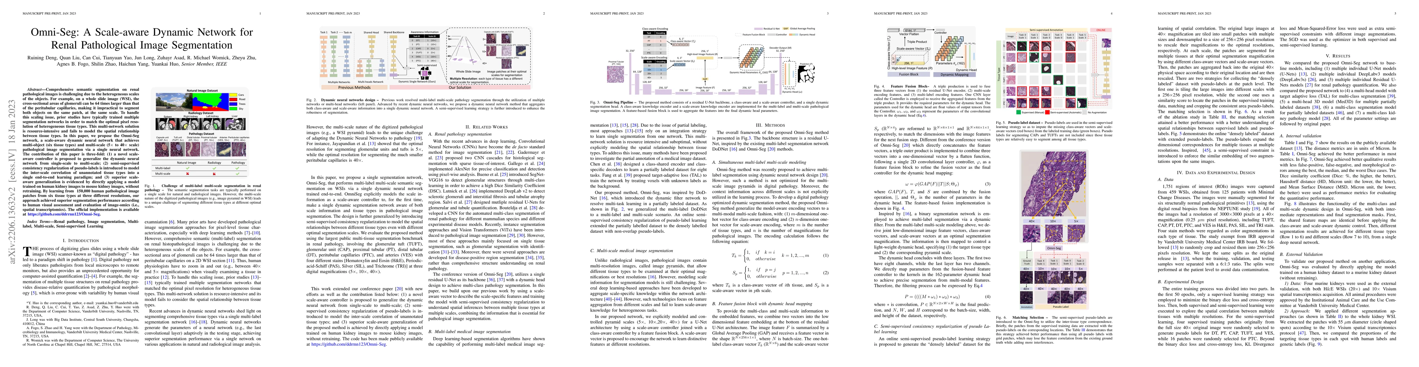
Comprehensive semantic segmentation on renal pathological images is challenging due to the heterogeneous scales of the objects. For example, on a whole slide image (WSI), the cross-sectional areas o...

Integrating cross-department multi-modal data (e.g., radiological, pathological, genomic, and clinical data) is ubiquitous in brain cancer diagnosis and survival prediction. To date, such an integra...
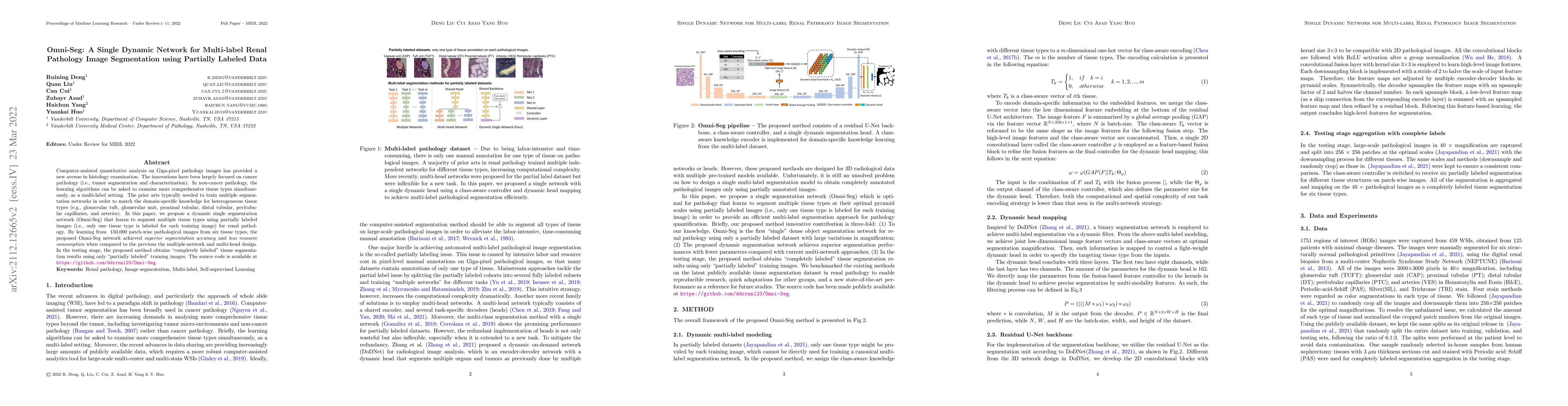
Computer-assisted quantitative analysis on Giga-pixel pathology images has provided a new avenue in histology examination. The innovations have been largely focused on cancer pathology (i.e., tumor ...
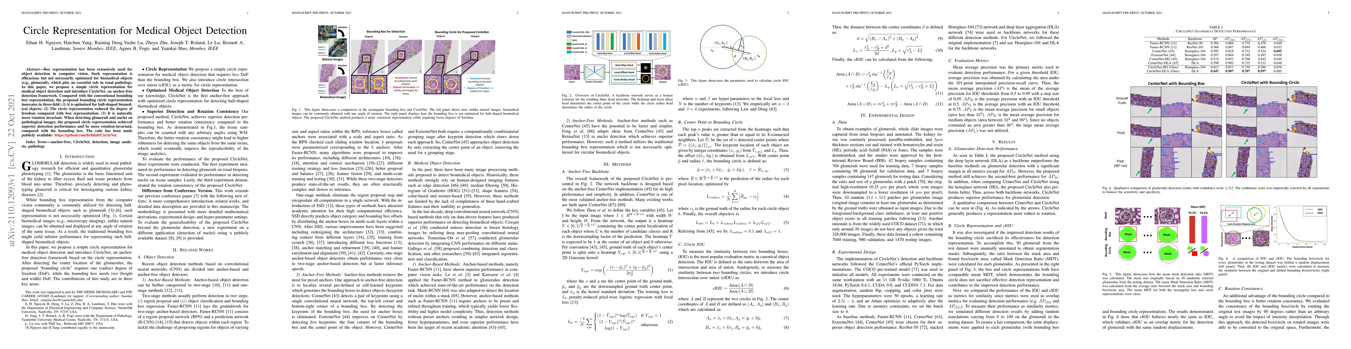
Box representation has been extensively used for object detection in computer vision. Such representation is efficacious but not necessarily optimized for biomedical objects (e.g., glomeruli), which...
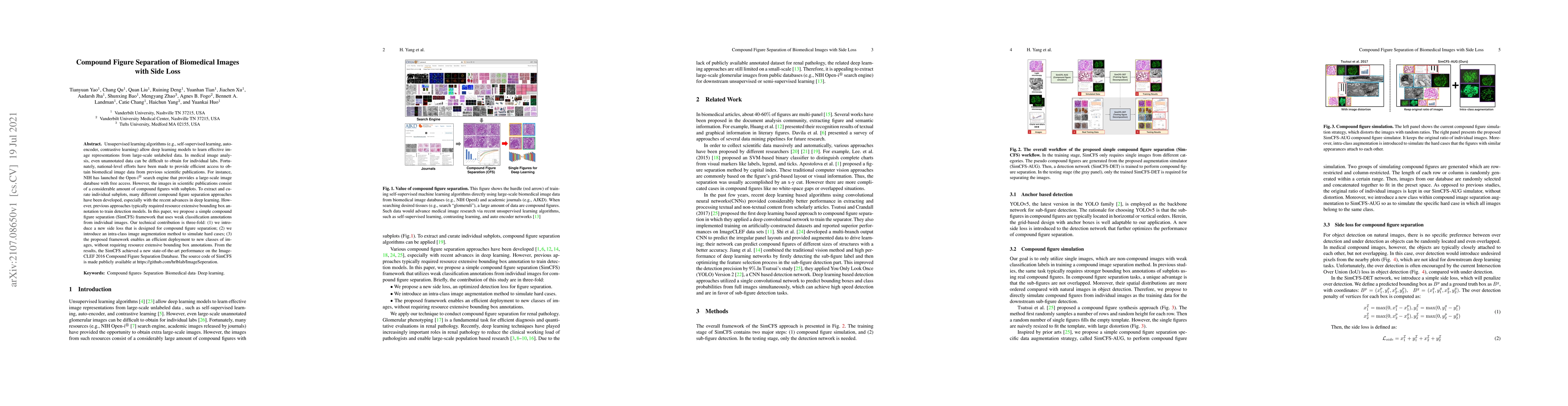
Unsupervised learning algorithms (e.g., self-supervised learning, auto-encoder, contrastive learning) allow deep learning models to learn effective image representations from large-scale unlabeled d...
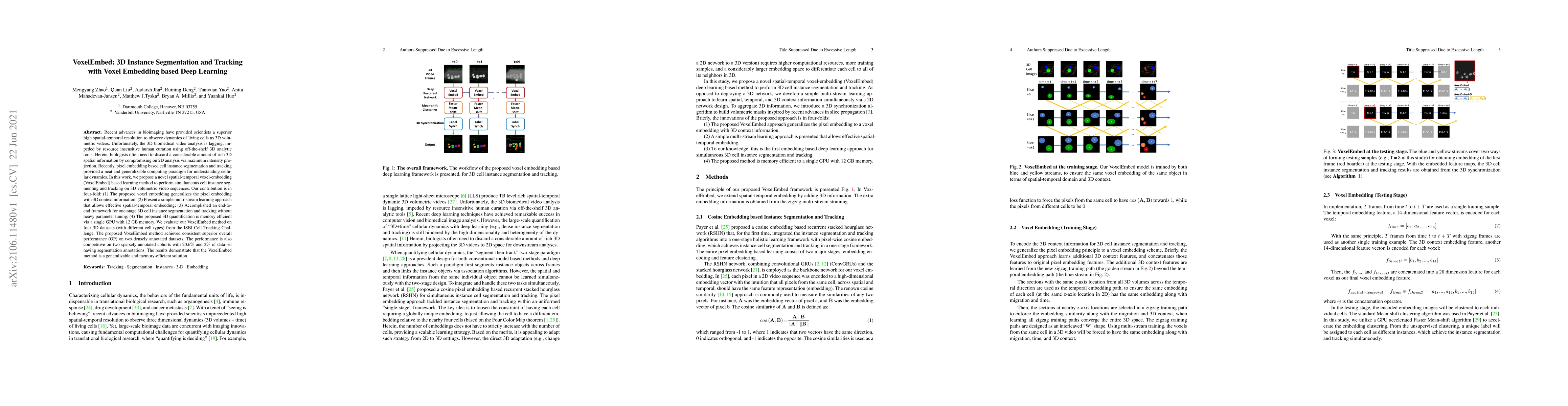
Recent advances in bioimaging have provided scientists a superior high spatial-temporal resolution to observe dynamics of living cells as 3D volumetric videos. Unfortunately, the 3D biomedical video...
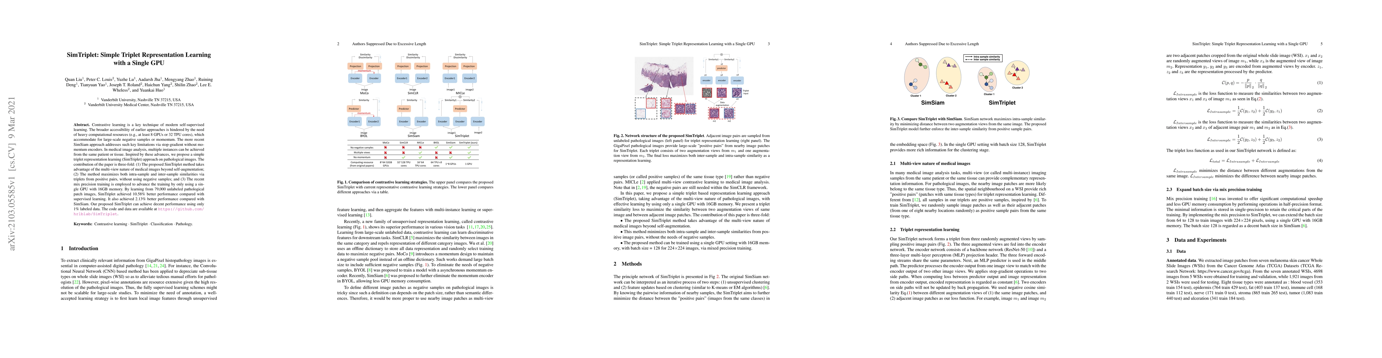
Contrastive learning is a key technique of modern self-supervised learning. The broader accessibility of earlier approaches is hindered by the need of heavy computational resources (e.g., at least 8...
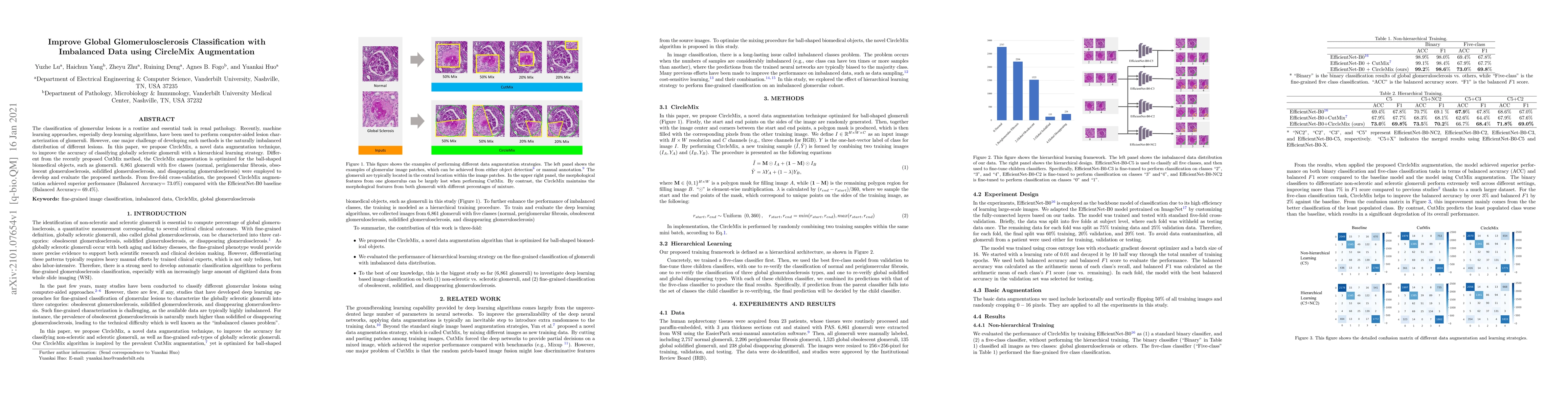
The classification of glomerular lesions is a routine and essential task in renal pathology. Recently, machine learning approaches, especially deep learning algorithms, have been used to perform com...
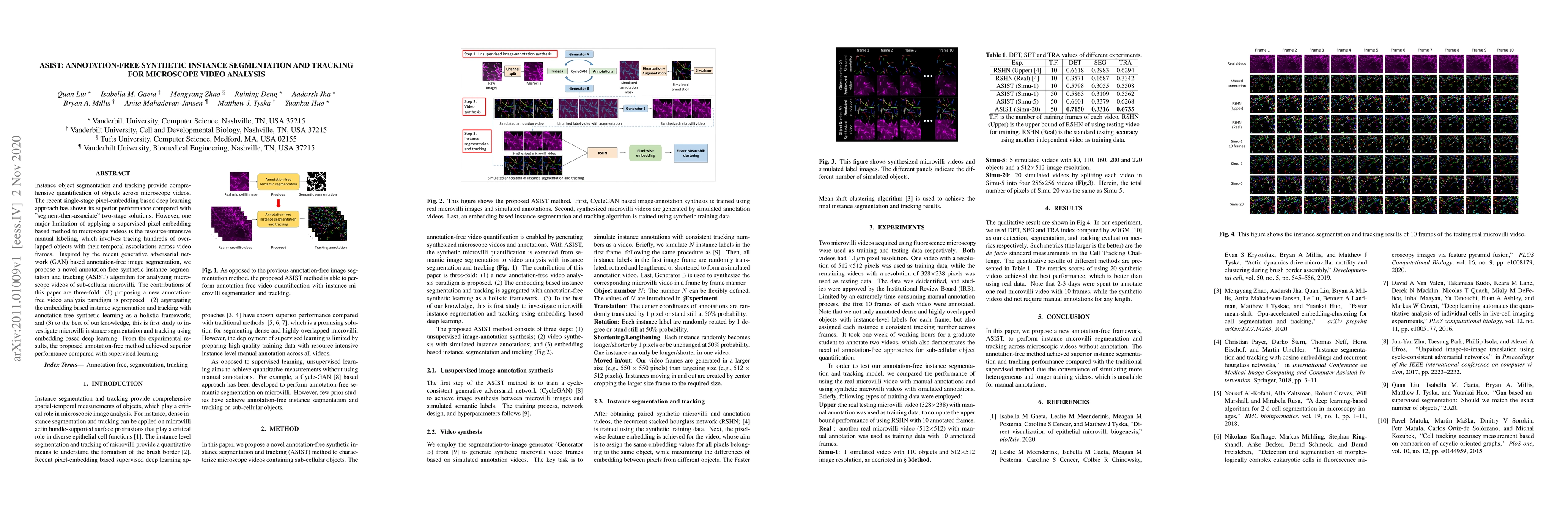
Instance object segmentation and tracking provide comprehensive quantification of objects across microscope videos. The recent single-stage pixel-embedding based deep learning approach has shown its...
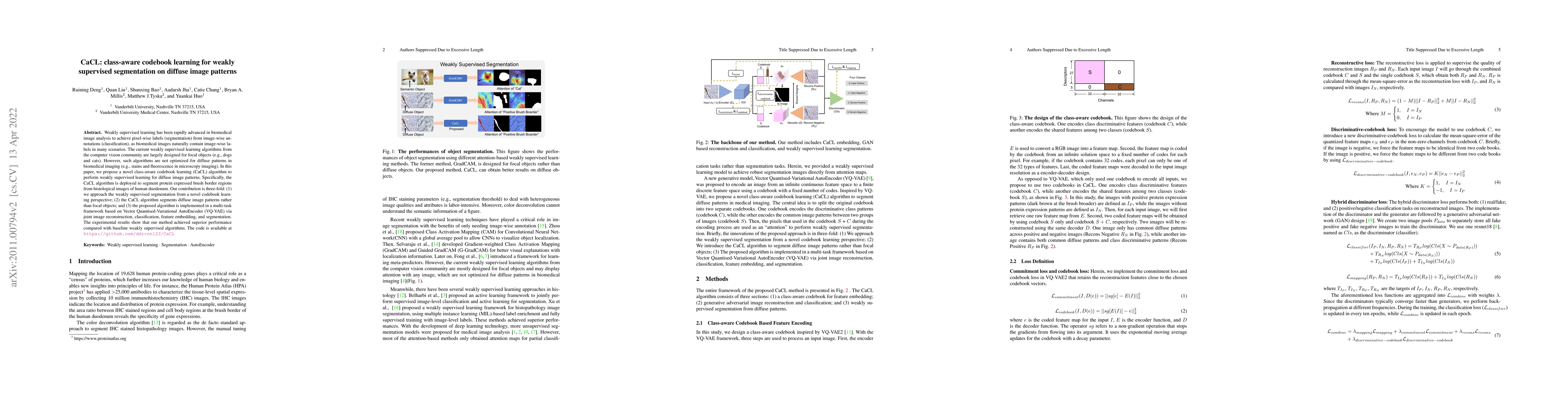
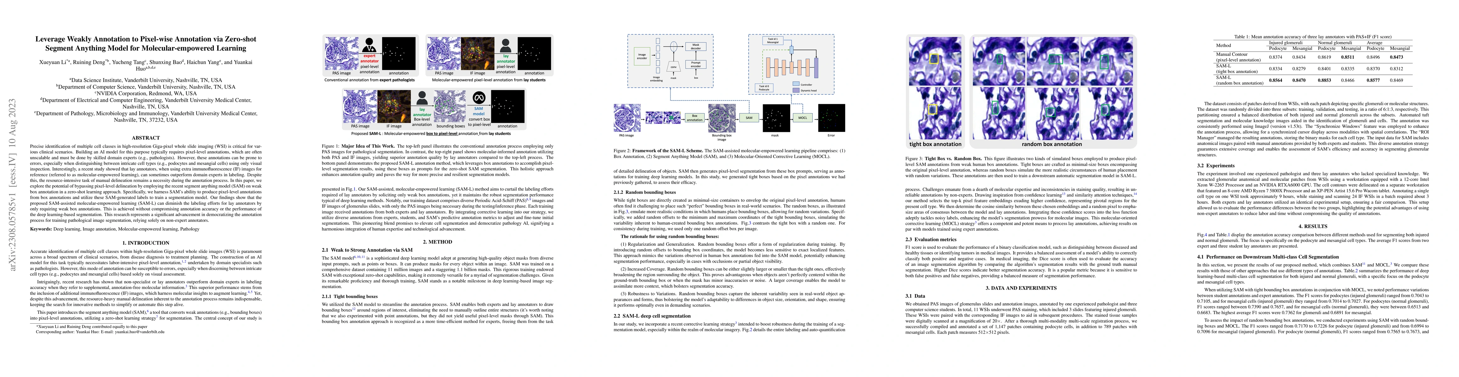
Weakly supervised learning has been rapidly advanced in biomedical image analysis to achieve pixel-wise labels (segmentation) from image-wise annotations (classification), as biomedical images natur...
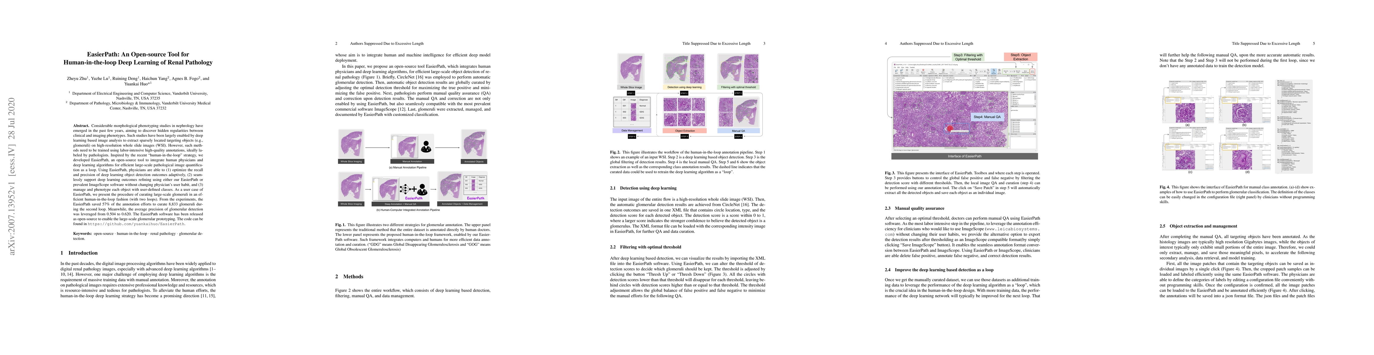
Considerable morphological phenotyping studies in nephrology have emerged in the past few years, aiming to discover hidden regularities between clinical and imaging phenotypes. Such studies have bee...
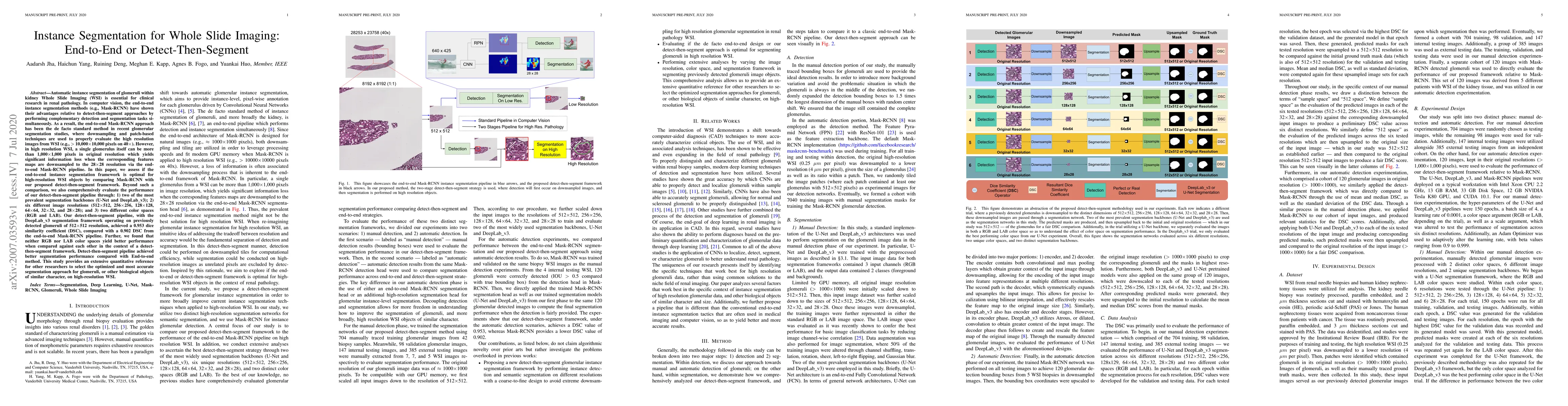
Automatic instance segmentation of glomeruli within kidney Whole Slide Imaging (WSI) is essential for clinical research in renal pathology. In computer vision, the end-to-end instance segmentation m...
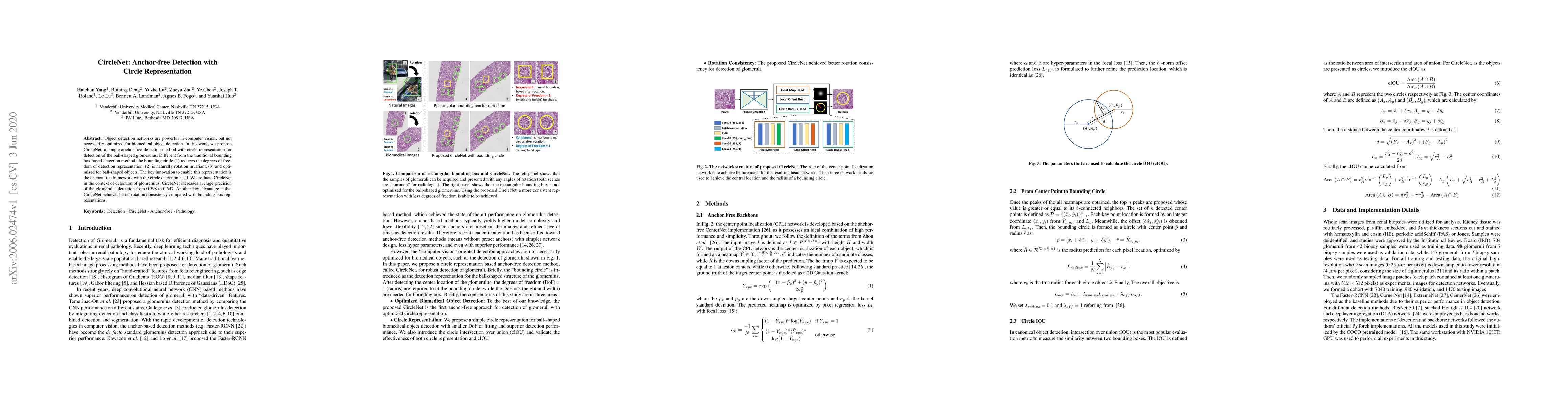
Object detection networks are powerful in computer vision, but not necessarily optimized for biomedical object detection. In this work, we propose CircleNet, a simple anchor-free detection method wi...
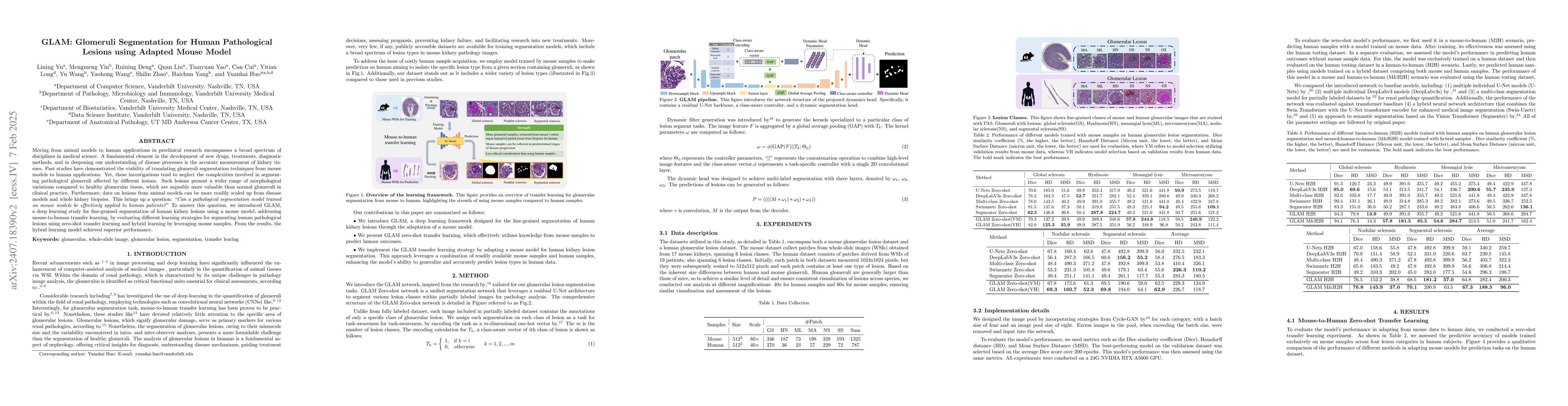
Moving from animal models to human applications in preclinical research encompasses a broad spectrum of disciplines in medical science. A fundamental element in the development of new drugs, treatment...
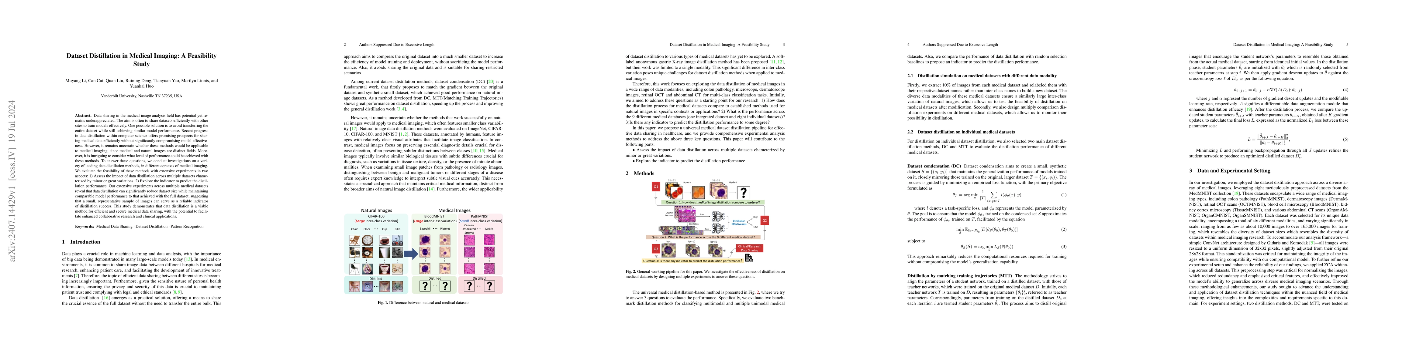
Data sharing in the medical image analysis field has potential yet remains underappreciated. The aim is often to share datasets efficiently with other sites to train models effectively. One possible s...
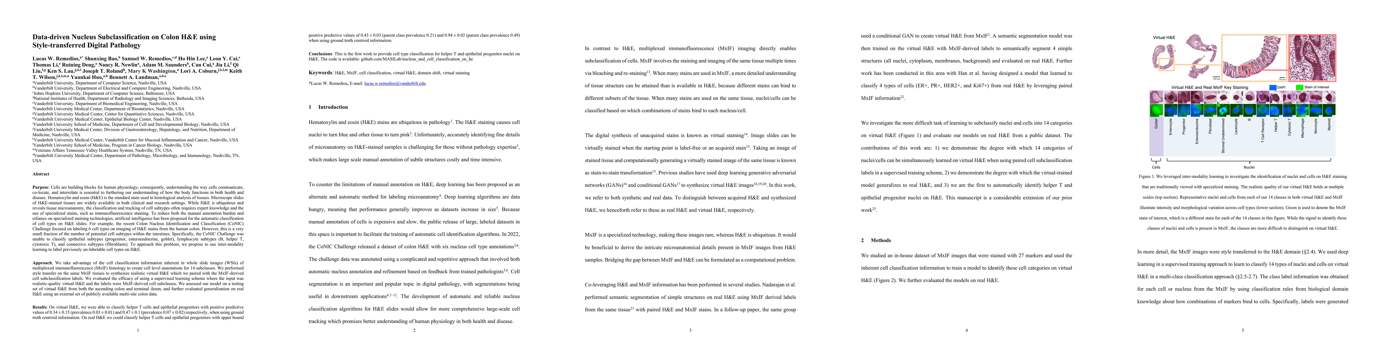
Understanding the way cells communicate, co-locate, and interrelate is essential to furthering our understanding of how the body functions. H&E is widely available, however, cell subtyping often requi...
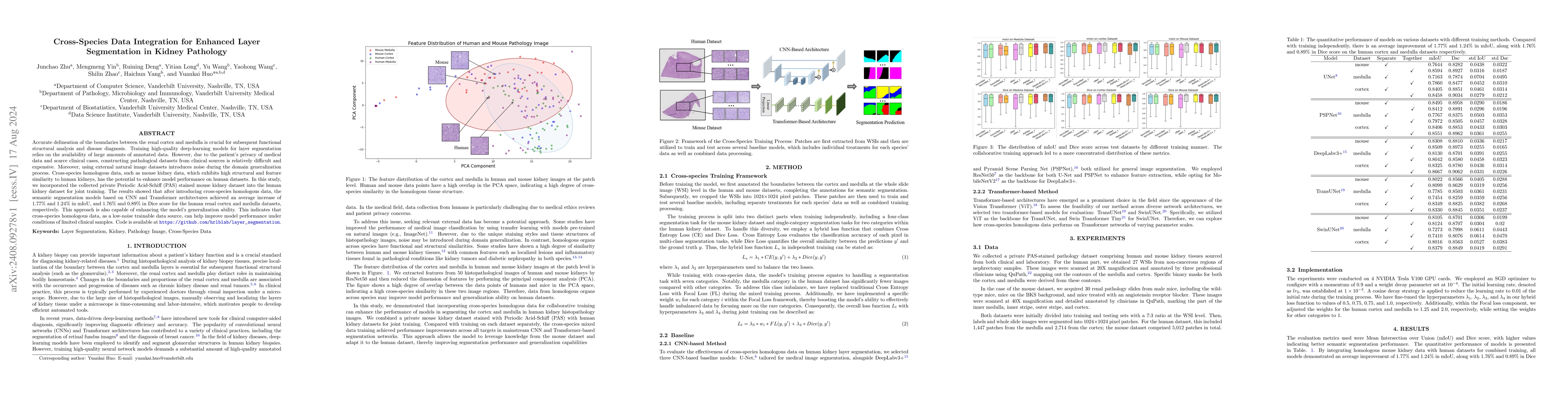
Accurate delineation of the boundaries between the renal cortex and medulla is crucial for subsequent functional structural analysis and disease diagnosis. Training high-quality deep-learning models f...
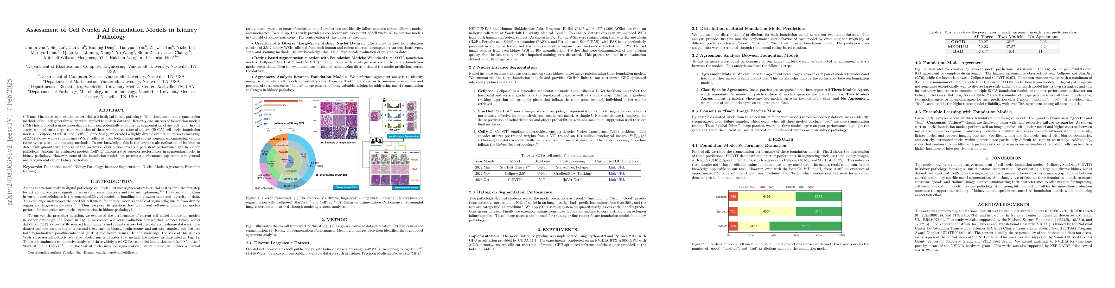
Cell nuclei instance segmentation is a crucial task in digital kidney pathology. Traditional automatic segmentation methods often lack generalizability when applied to unseen datasets. Recently, the s...
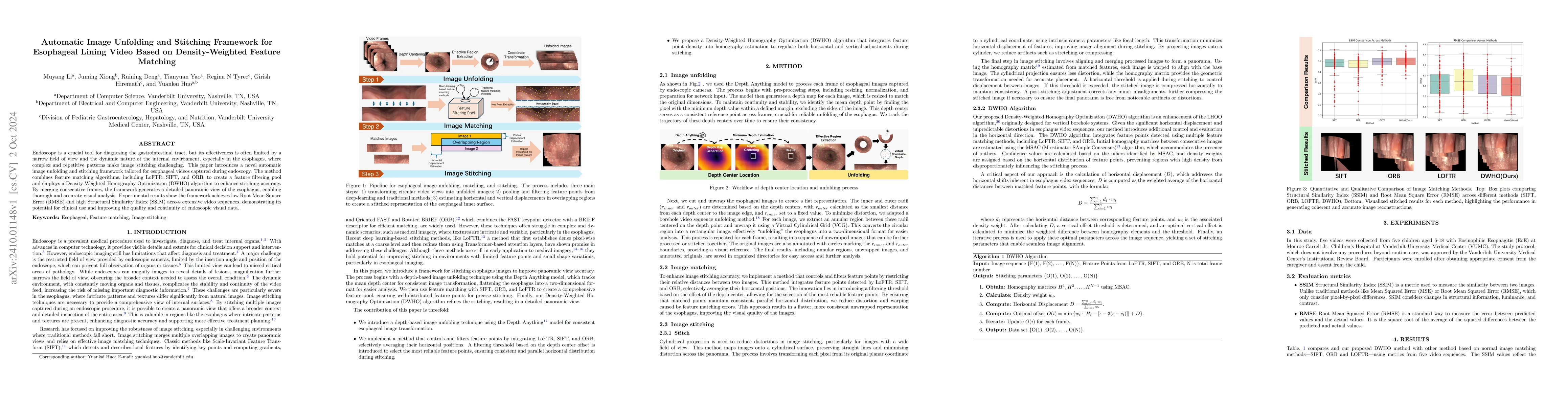
Endoscopy is a crucial tool for diagnosing the gastrointestinal tract, but its effectiveness is often limited by a narrow field of view and the dynamic nature of the internal environment, especially i...
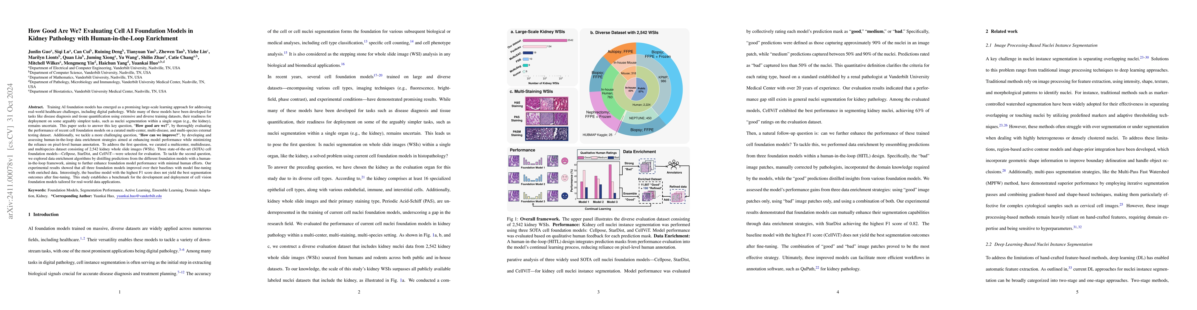
Training AI foundation models has emerged as a promising large-scale learning approach for addressing real-world healthcare challenges, including digital pathology. While many of these models have bee...
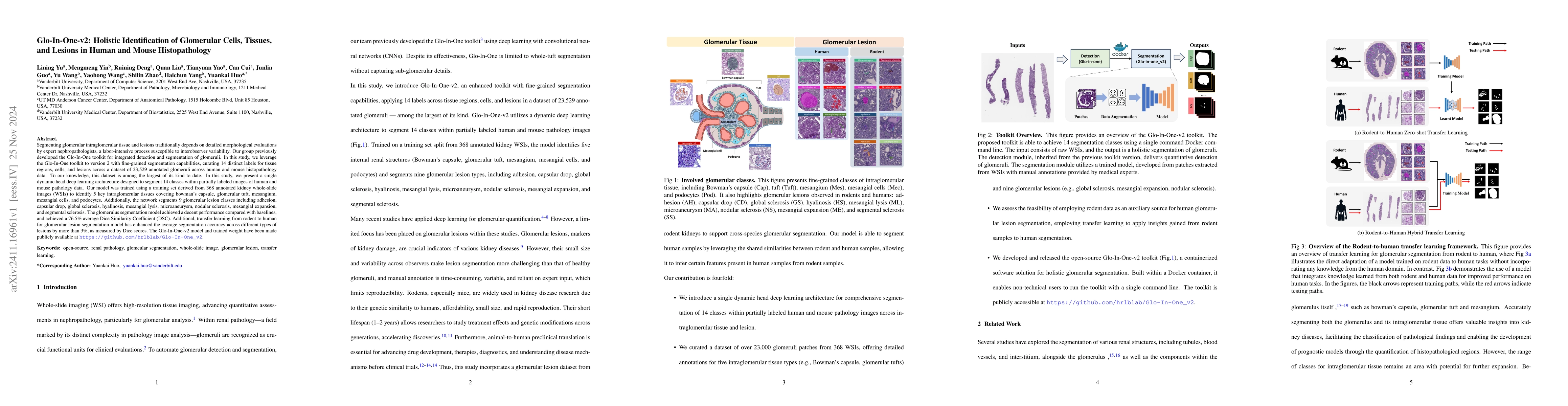
Segmenting glomerular intraglomerular tissue and lesions traditionally depends on detailed morphological evaluations by expert nephropathologists, a labor-intensive process susceptible to interobserve...
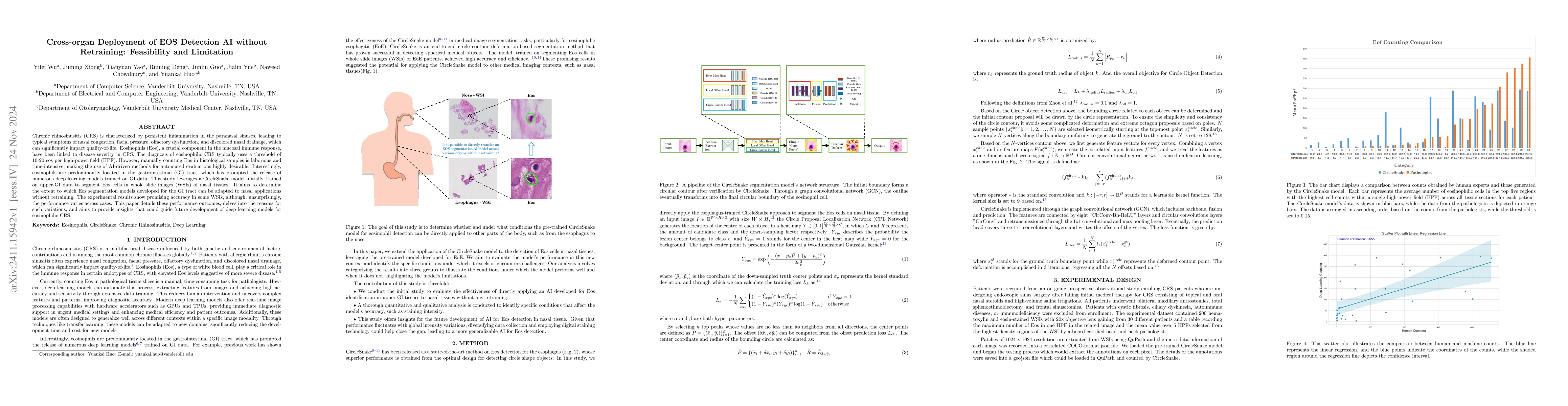
Chronic rhinosinusitis (CRS) is characterized by persistent inflammation in the paranasal sinuses, leading to typical symptoms of nasal congestion, facial pressure, olfactory dysfunction, and discolor...
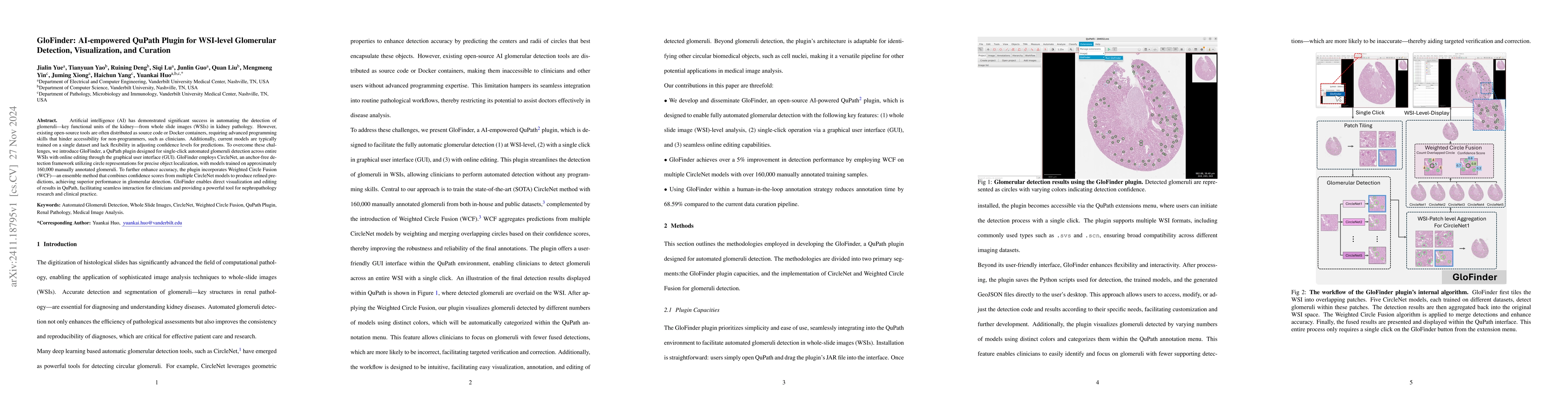
Artificial intelligence (AI) has demonstrated significant success in automating the detection of glomeruli, the key functional units of the kidney, from whole slide images (WSIs) in kidney pathology. ...
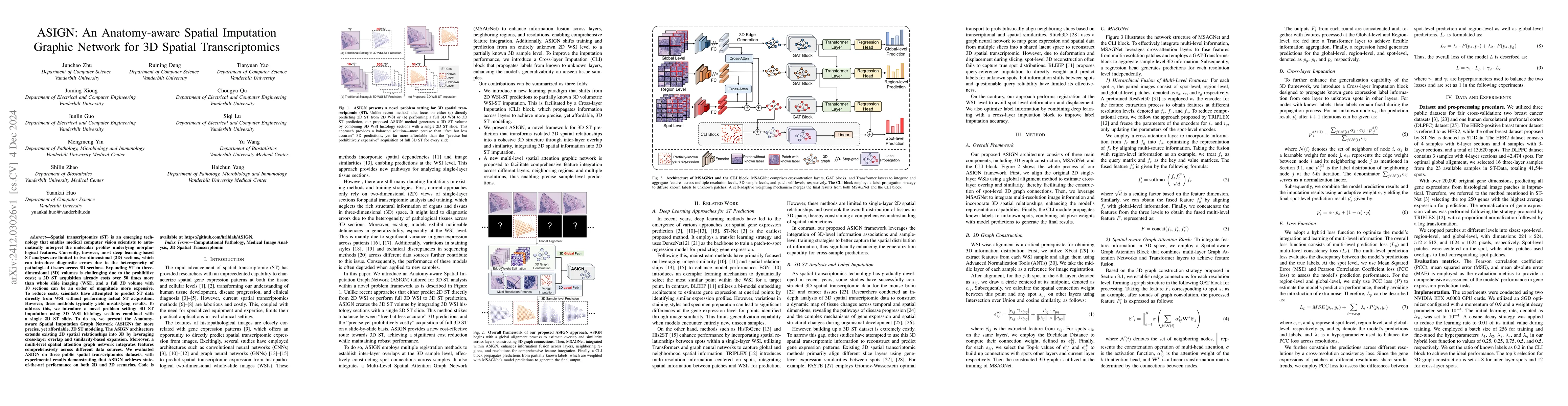
Spatial transcriptomics (ST) is an emerging technology that enables medical computer vision scientists to automatically interpret the molecular profiles underlying morphological features. Currently, h...
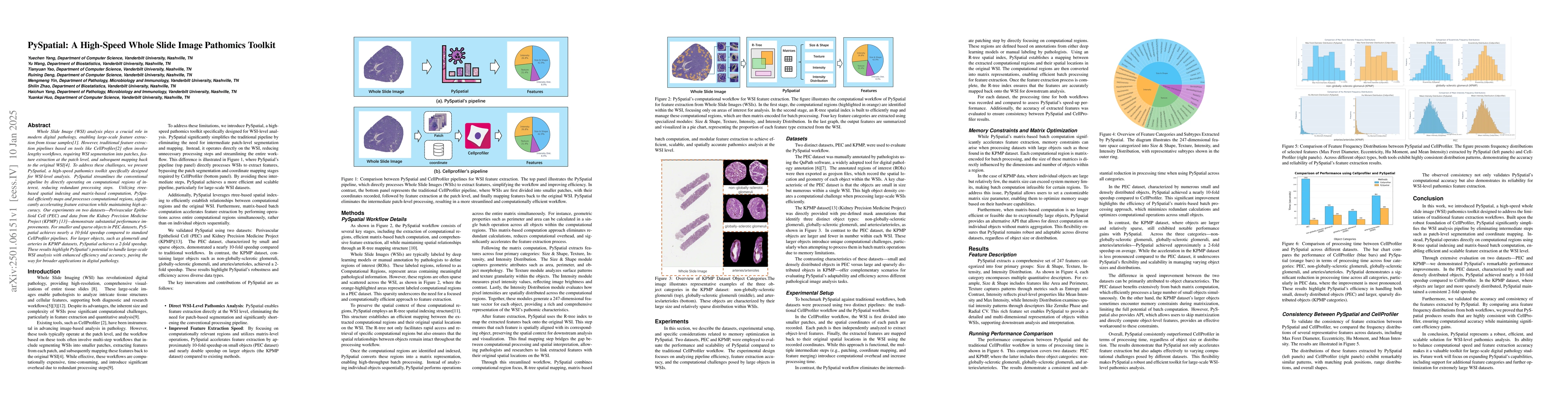
Whole Slide Image (WSI) analysis plays a crucial role in modern digital pathology, enabling large-scale feature extraction from tissue samples. However, traditional feature extraction pipelines based ...
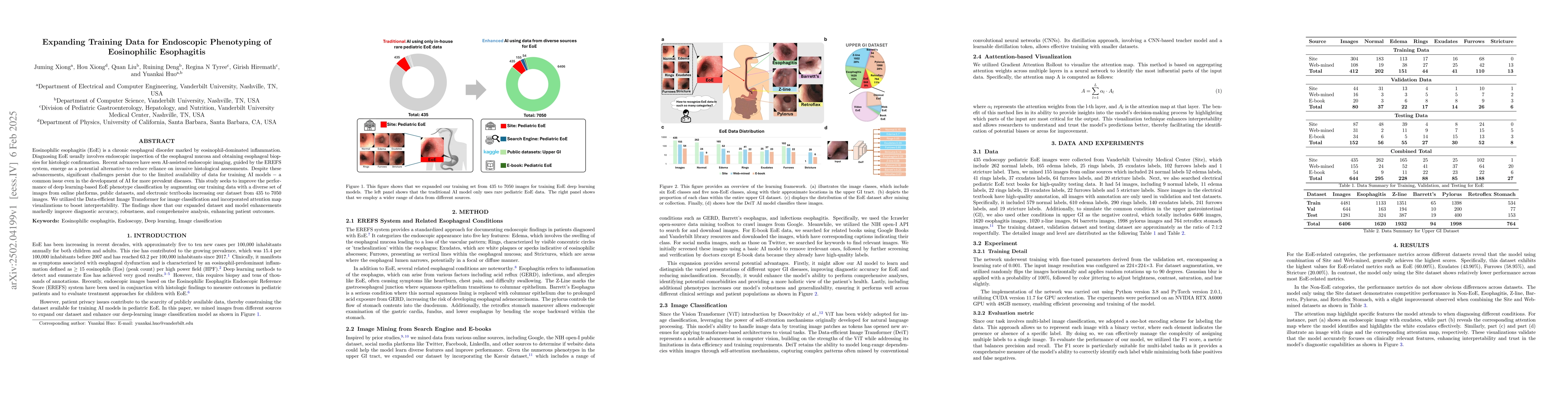
Eosinophilic esophagitis (EoE) is a chronic esophageal disorder marked by eosinophil-dominated inflammation. Diagnosing EoE usually involves endoscopic inspection of the esophageal mucosa and obtainin...
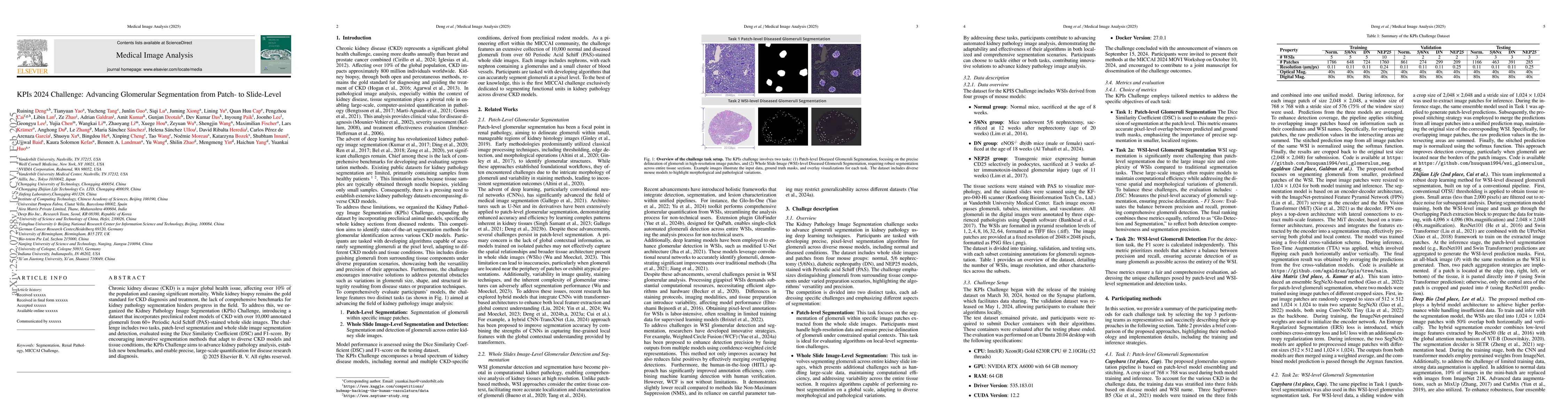
Chronic kidney disease (CKD) is a major global health issue, affecting over 10% of the population and causing significant mortality. While kidney biopsy remains the gold standard for CKD diagnosis and...
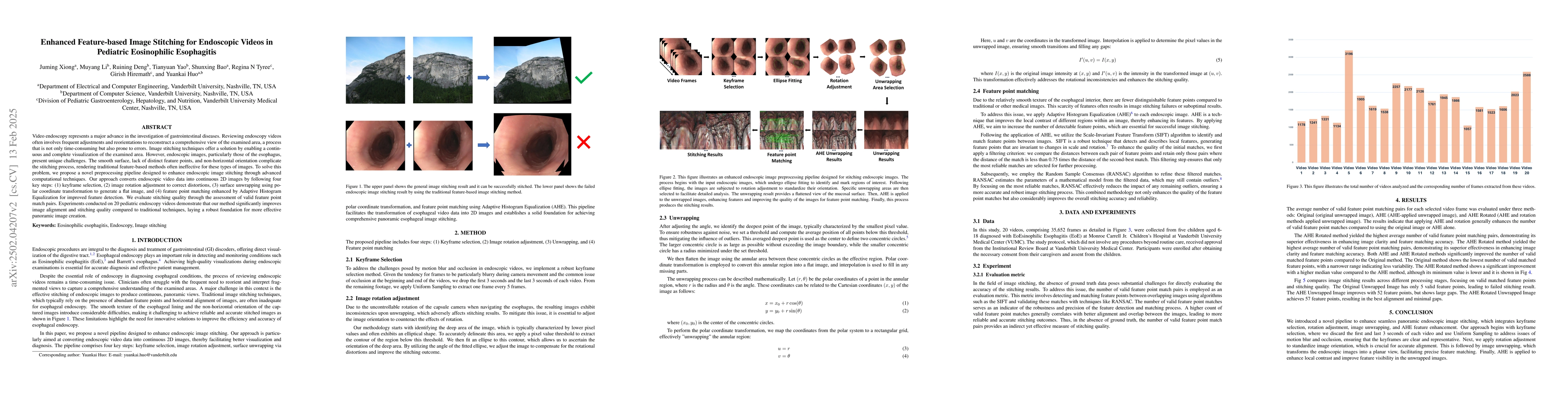
Video endoscopy represents a major advance in the investigation of gastrointestinal diseases. Reviewing endoscopy videos often involves frequent adjustments and reorientations to piece together a comp...
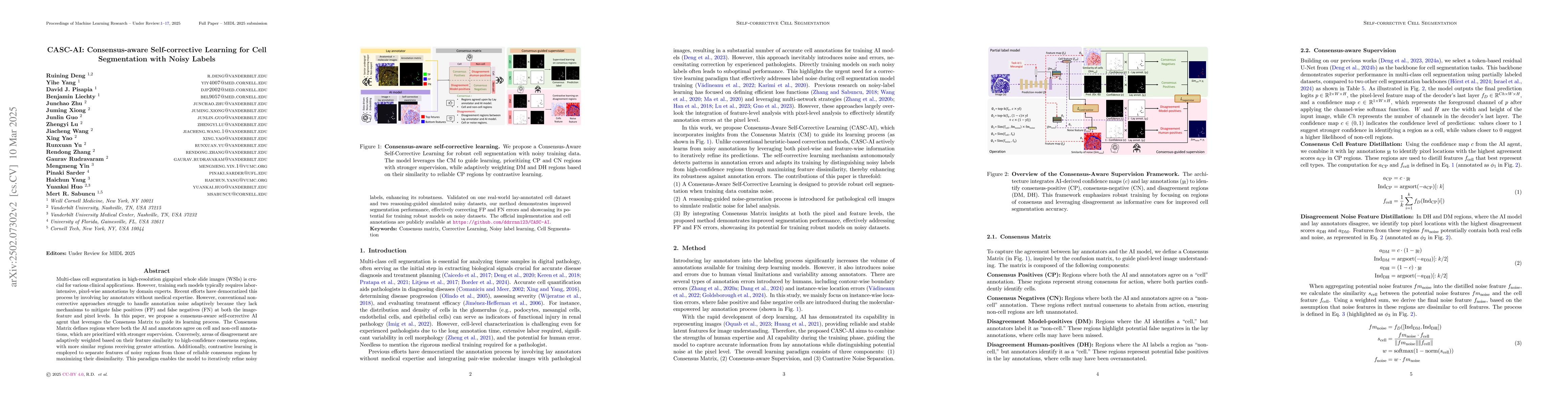
Multi-class cell segmentation in high-resolution gigapixel whole slide images (WSI) is crucial for various clinical applications. However, training such models typically requires labor-intensive, pixe...
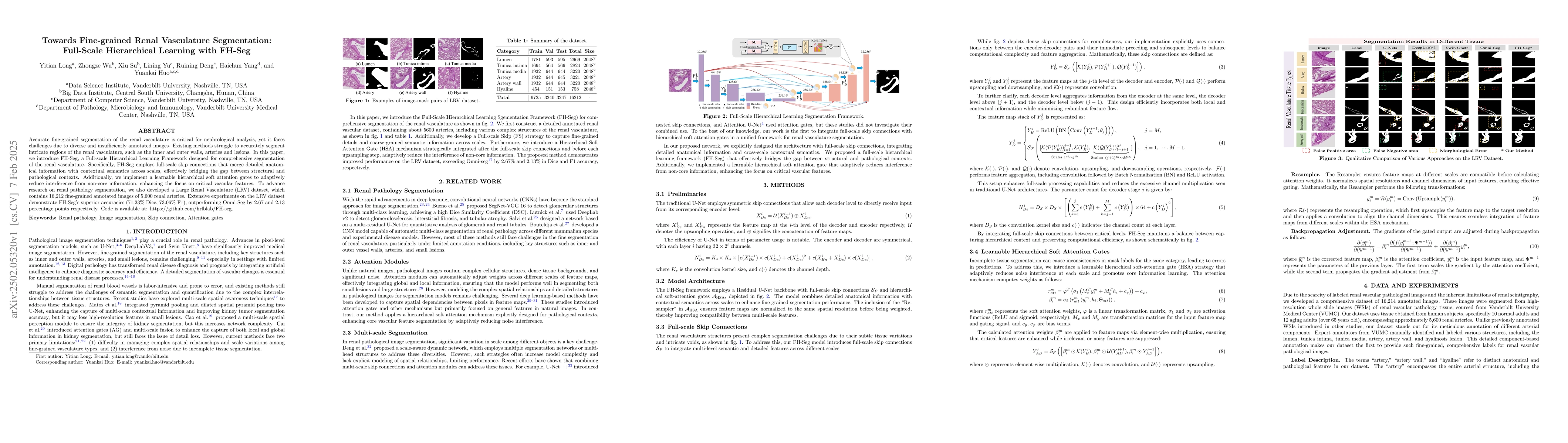
Accurate fine-grained segmentation of the renal vasculature is critical for nephrological analysis, yet it faces challenges due to diverse and insufficiently annotated images. Existing methods struggl...
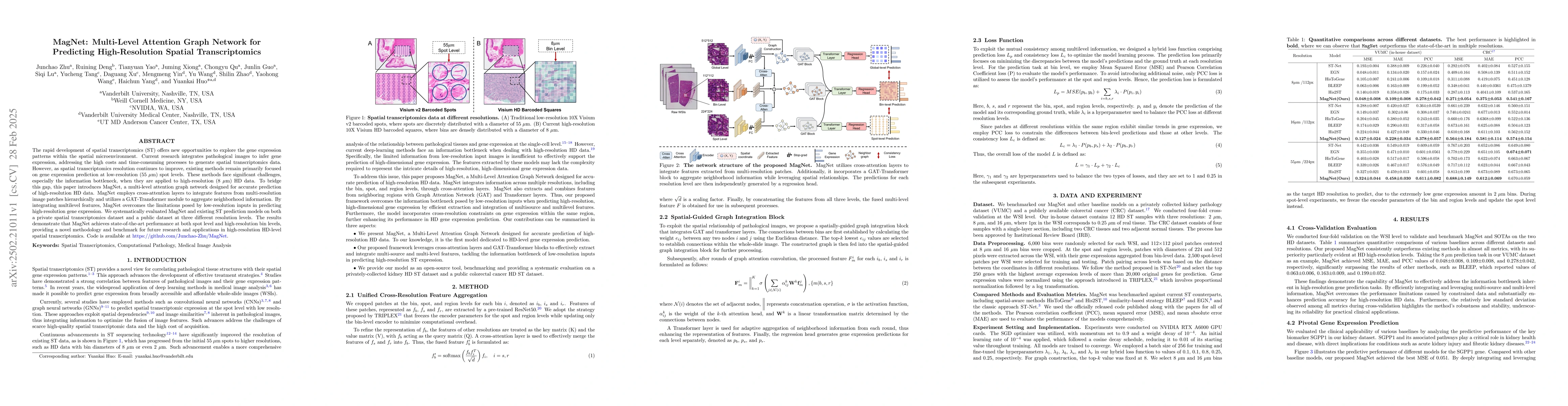
The rapid development of spatial transcriptomics (ST) offers new opportunities to explore the gene expression patterns within the spatial microenvironment. Current research integrates pathological ima...
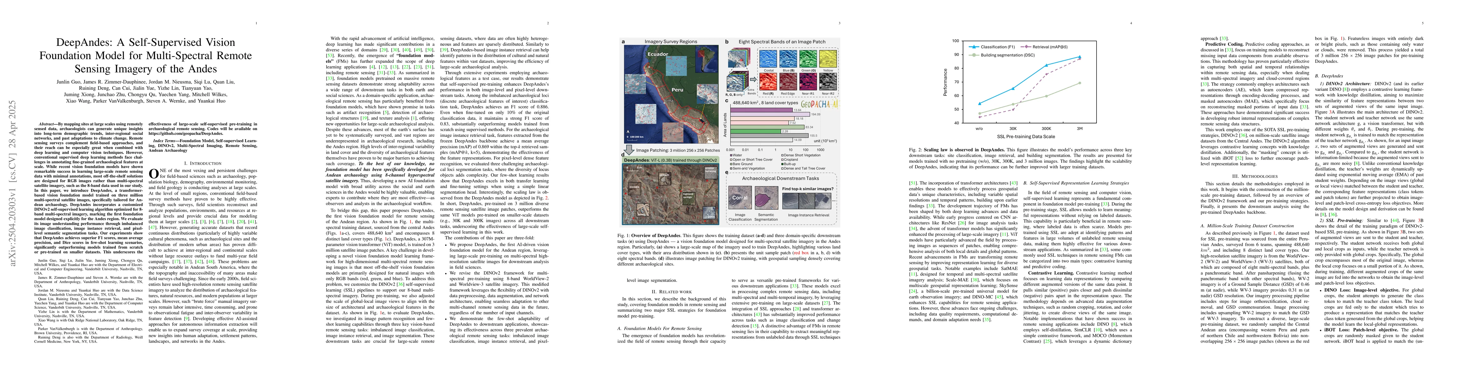
By mapping sites at large scales using remotely sensed data, archaeologists can generate unique insights into long-term demographic trends, inter-regional social networks, and past adaptations to clim...
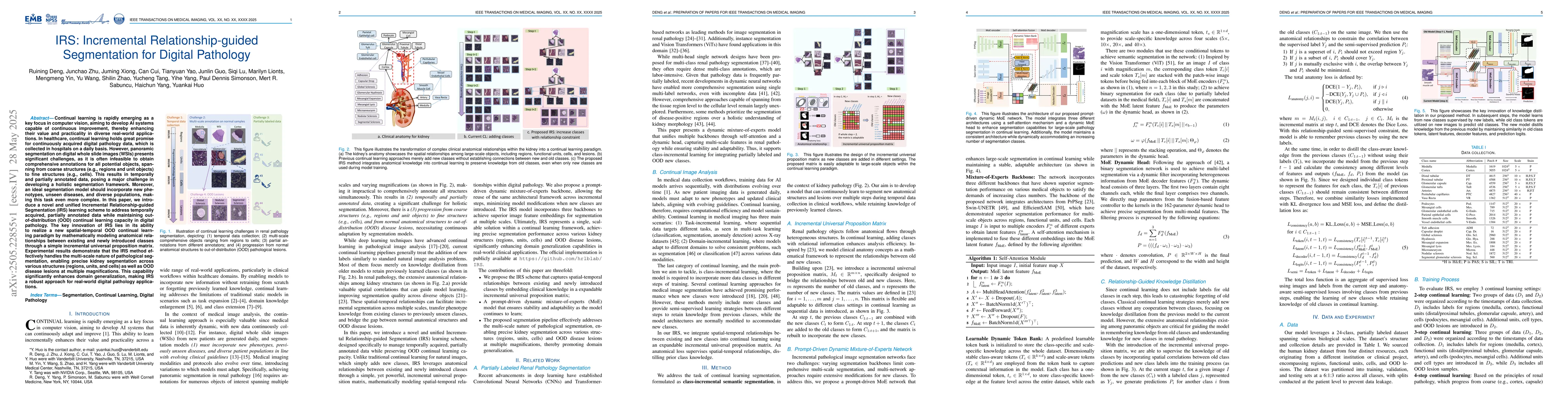
Continual learning is rapidly emerging as a key focus in computer vision, aiming to develop AI systems capable of continuous improvement, thereby enhancing their value and practicality in diverse real...
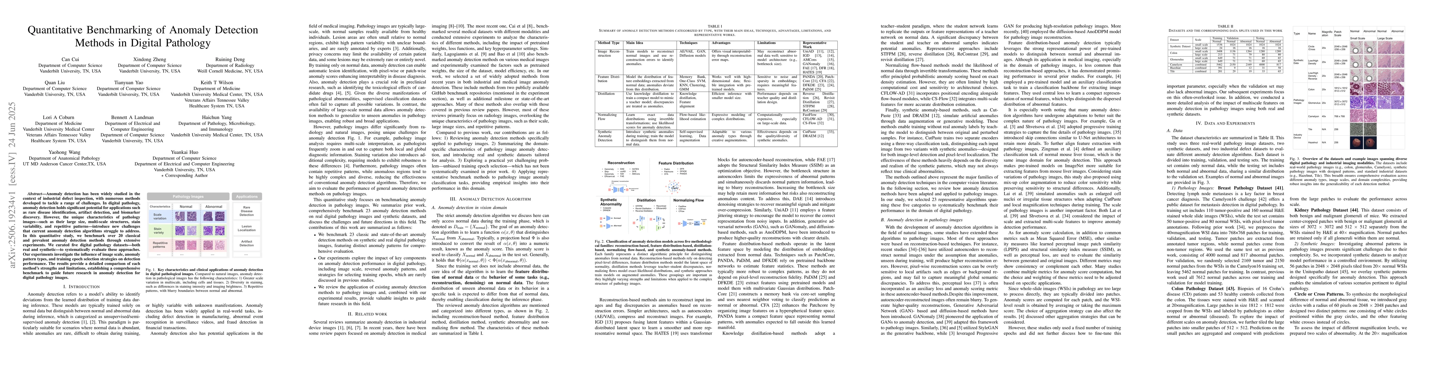
Anomaly detection has been widely studied in the context of industrial defect inspection, with numerous methods developed to tackle a range of challenges. In digital pathology, anomaly detection holds...
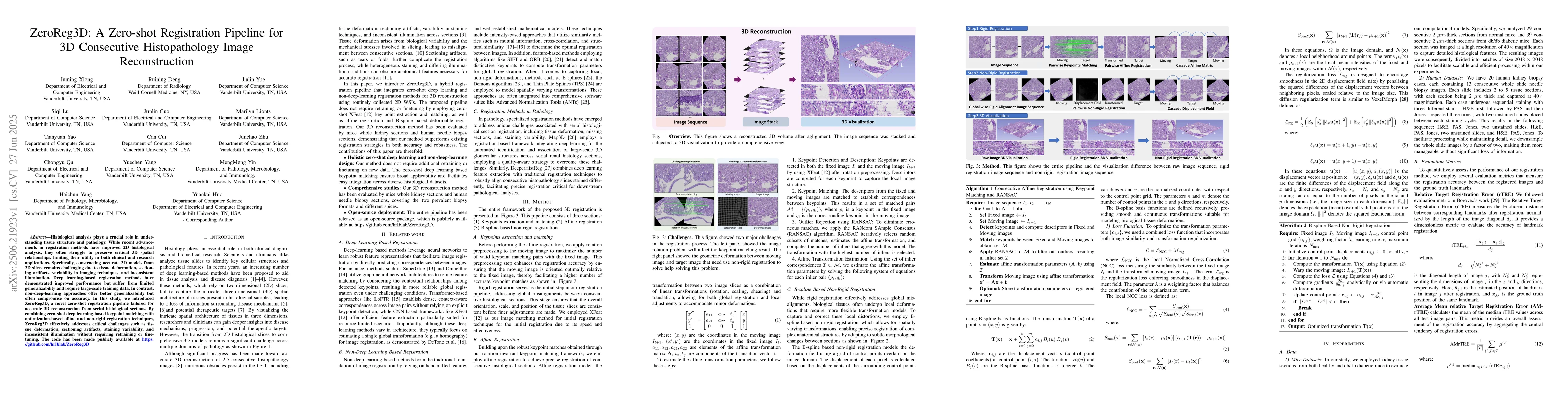
Histological analysis plays a crucial role in understanding tissue structure and pathology. While recent advancements in registration methods have improved 2D histological analysis, they often struggl...
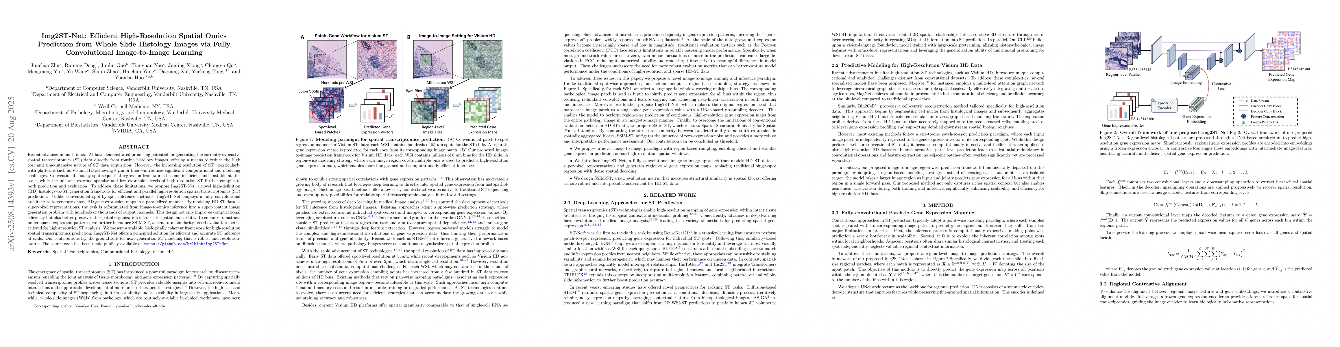
Recent advances in multi-modal AI have demonstrated promising potential for generating the currently expensive spatial transcriptomics (ST) data directly from routine histology images, offering a mean...
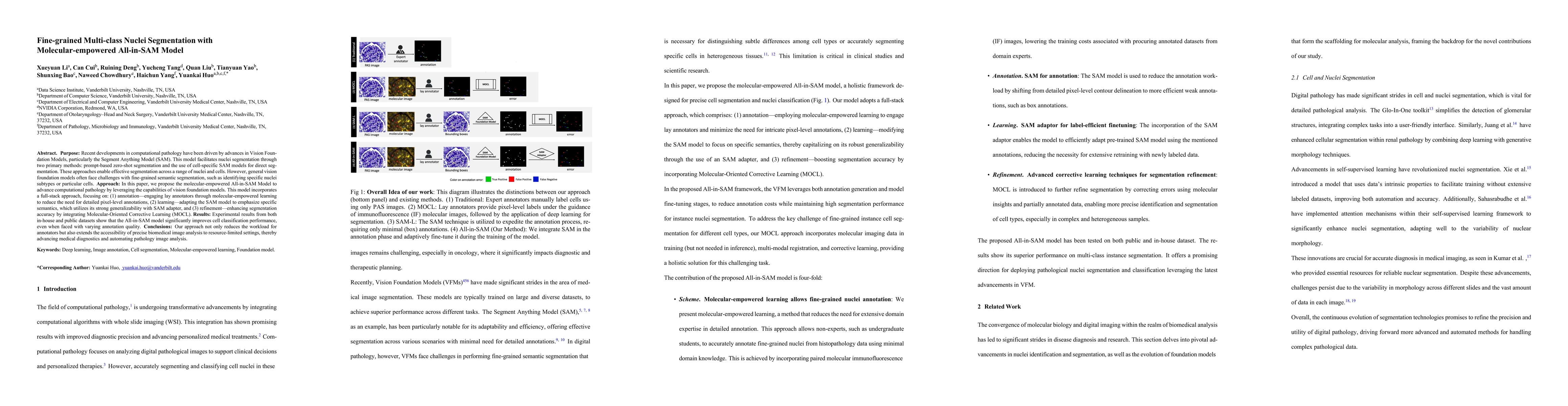
Purpose: Recent developments in computational pathology have been driven by advances in Vision Foundation Models, particularly the Segment Anything Model (SAM). This model facilitates nuclei segmentat...
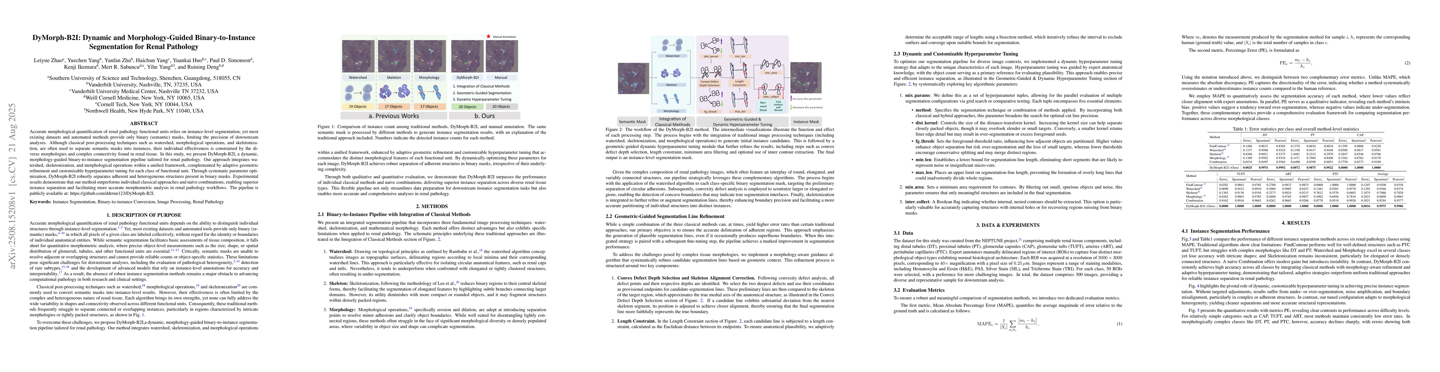
Accurate morphological quantification of renal pathology functional units relies on instance-level segmentation, yet most existing datasets and automated methods provide only binary (semantic) masks, ...
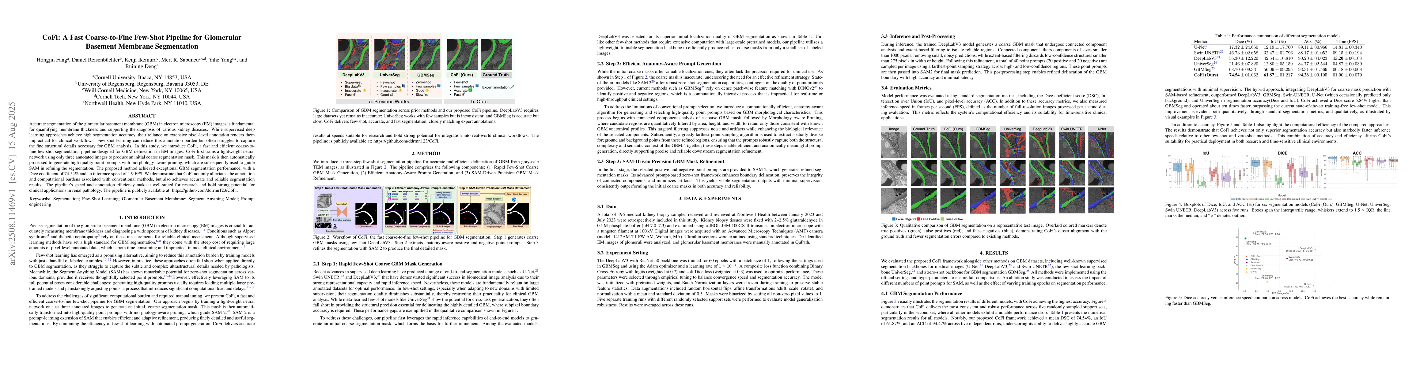
Accurate segmentation of the glomerular basement membrane (GBM) in electron microscopy (EM) images is fundamental for quantifying membrane thickness and supporting the diagnosis of various kidney dise...
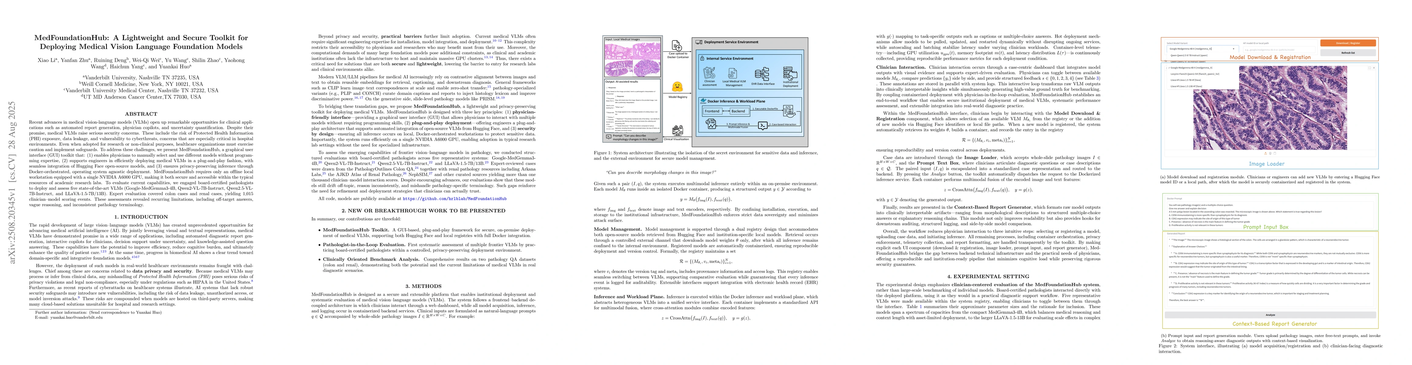
Recent advances in medical vision-language models (VLMs) open up remarkable opportunities for clinical applications such as automated report generation, copilots for physicians, and uncertainty quanti...
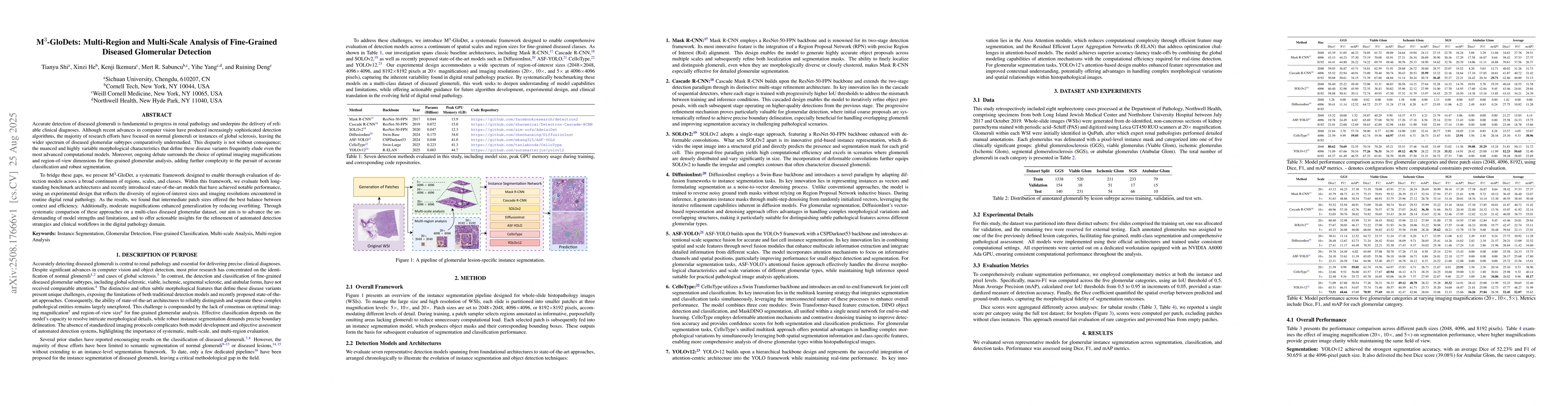
Accurate detection of diseased glomeruli is fundamental to progress in renal pathology and underpins the delivery of reliable clinical diagnoses. Although recent advances in computer vision have produ...
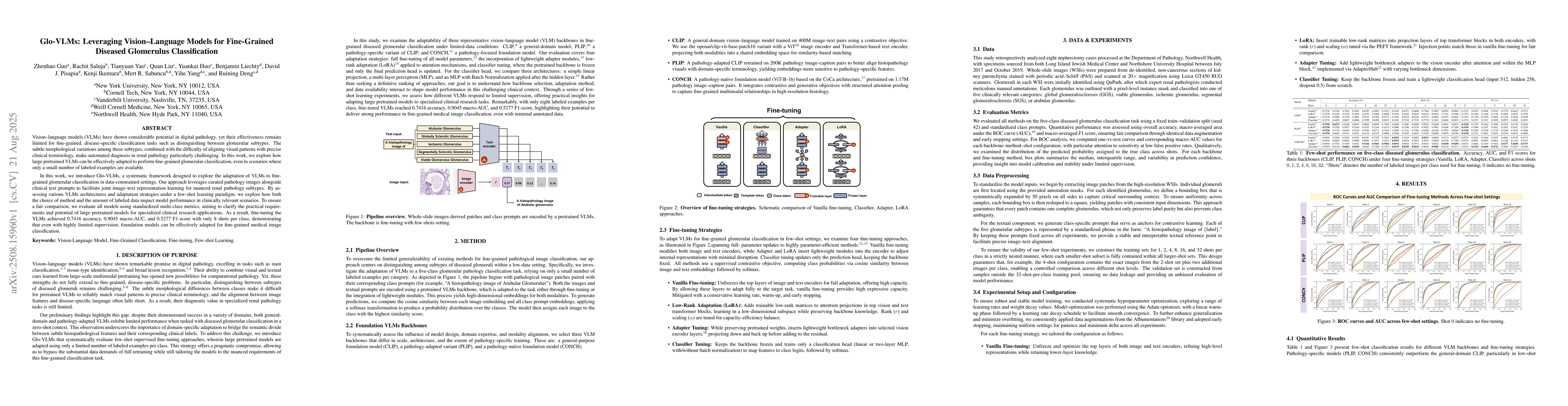
Vision-language models (VLMs) have shown considerable potential in digital pathology, yet their effectiveness remains limited for fine-grained, disease-specific classification tasks such as distinguis...
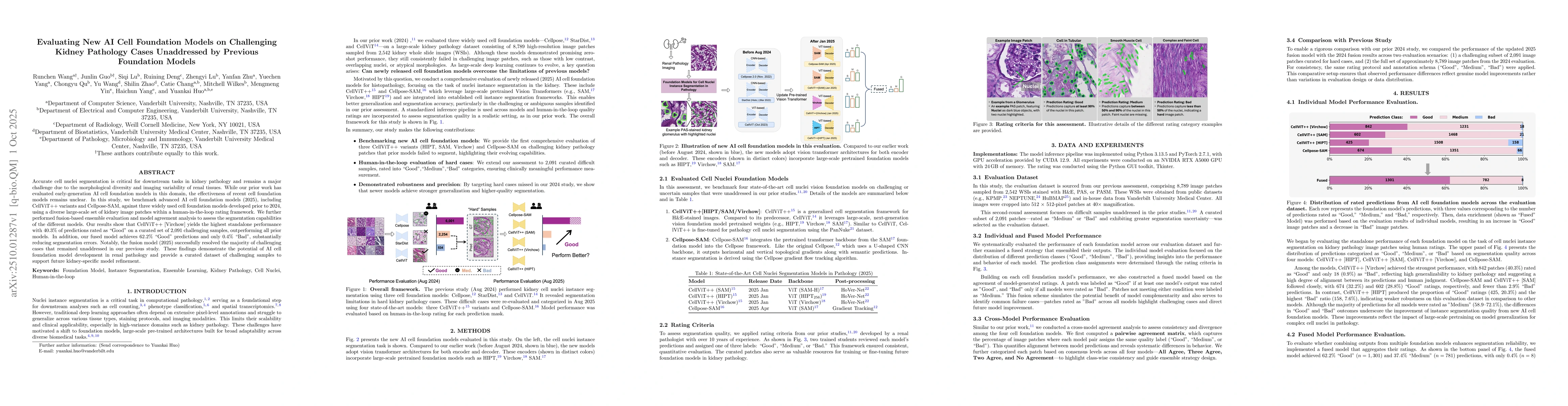
Accurate cell nuclei segmentation is critical for downstream tasks in kidney pathology and remains a major challenge due to the morphological diversity and imaging variability of renal tissues. While ...
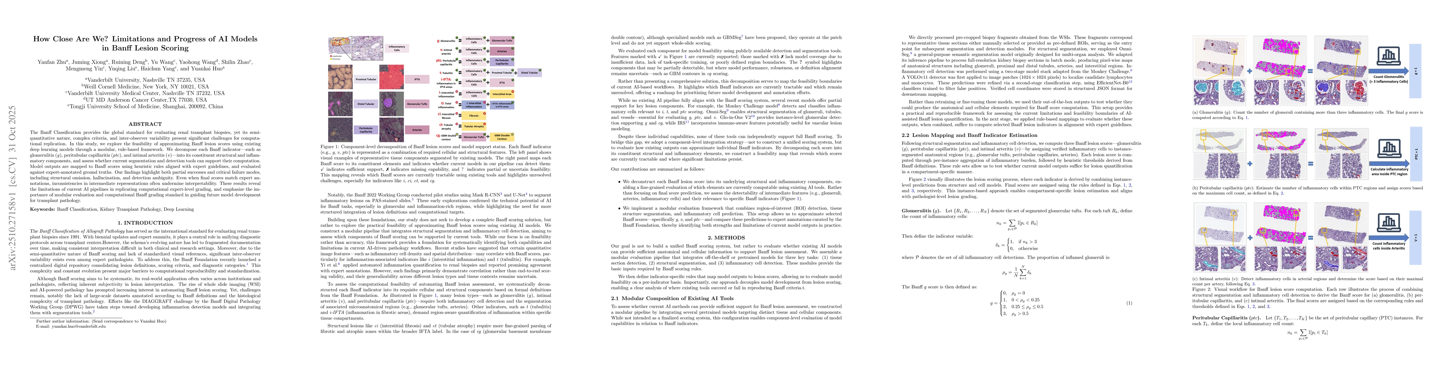
The Banff Classification provides the global standard for evaluating renal transplant biopsies, yet its semi-quantitative nature, complex criteria, and inter-observer variability present significant c...
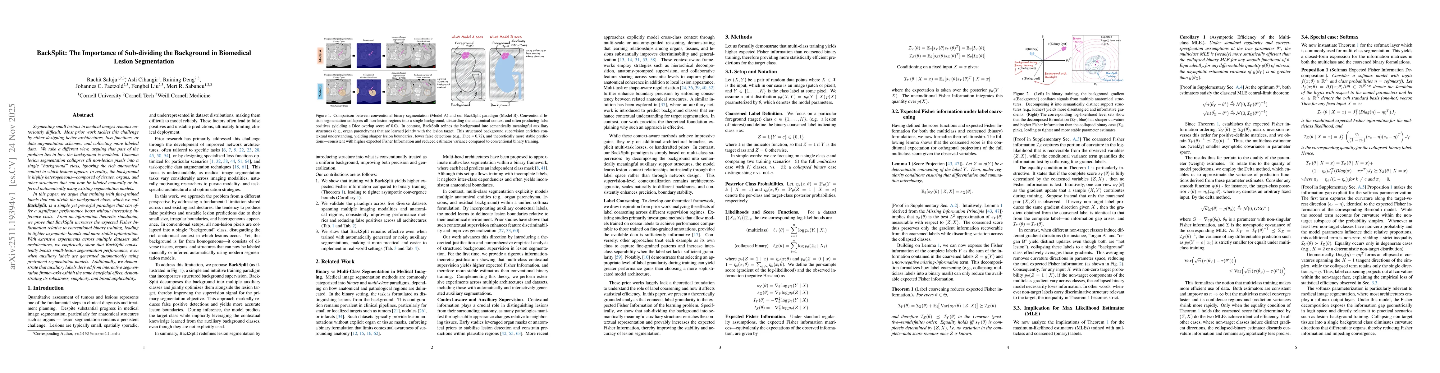
Segmenting small lesions in medical images remains notoriously difficult. Most prior work tackles this challenge by either designing better architectures, loss functions, or data augmentation schemes;...
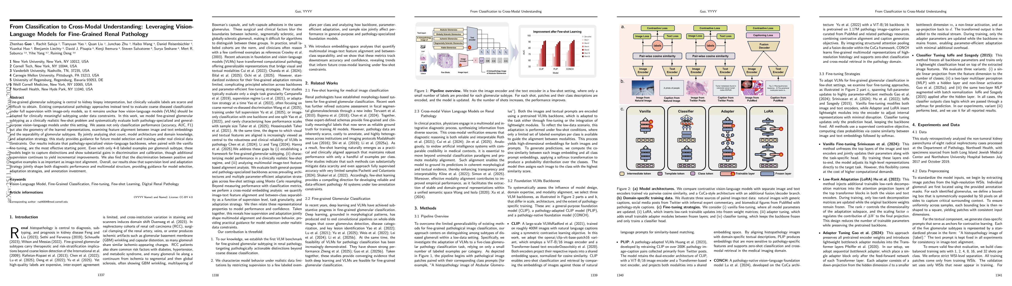
Fine-grained glomerular subtyping is central to kidney biopsy interpretation, but clinically valuable labels are scarce and difficult to obtain. Existing computational pathology approaches instead ten...
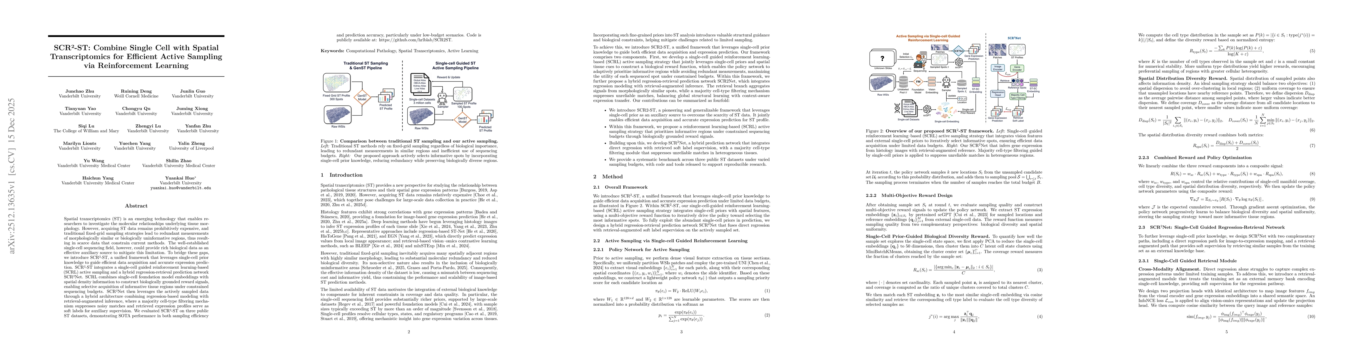
Spatial transcriptomics (ST) is an emerging technology that enables researchers to investigate the molecular relationships underlying tissue morphology. However, acquiring ST data remains prohibitivel...
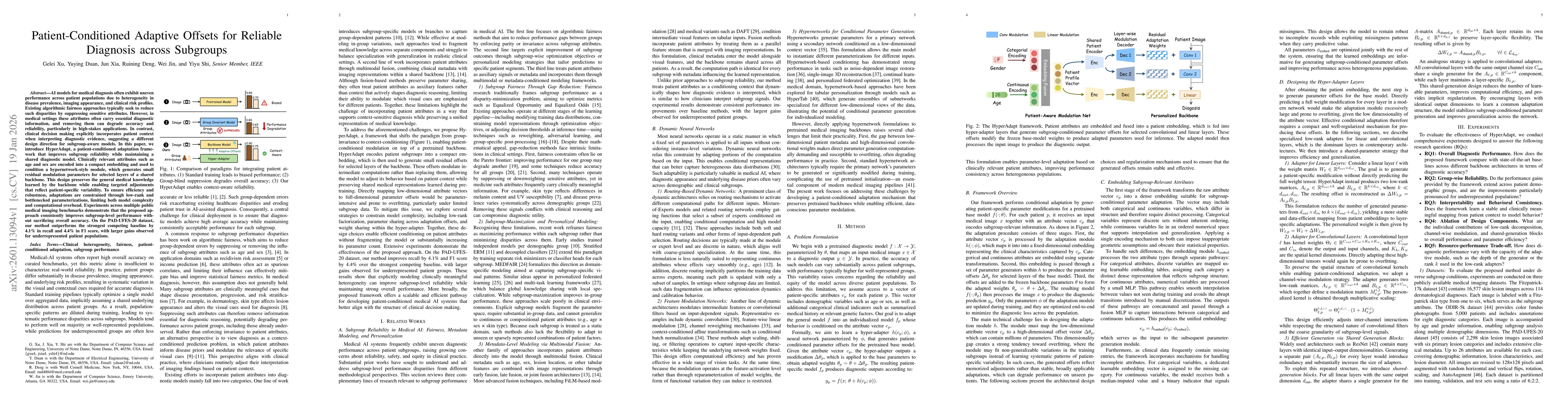
AI models for medical diagnosis often exhibit uneven performance across patient populations due to heterogeneity in disease prevalence, imaging appearance, and clinical risk profiles. Existing algorit...
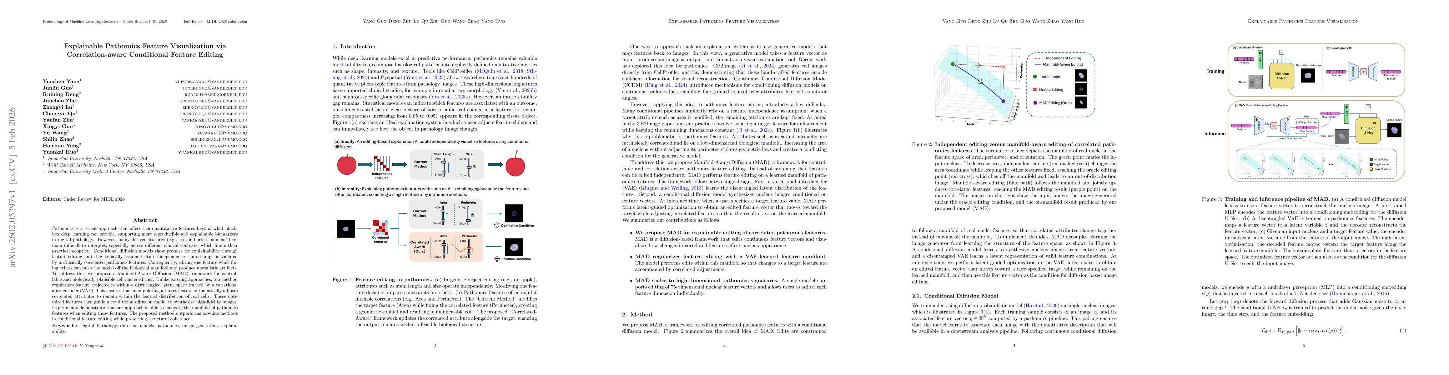
Pathomics is a recent approach that offers rich quantitative features beyond what black-box deep learning can provide, supporting more reproducible and explainable biomarkers in digital pathology. How...
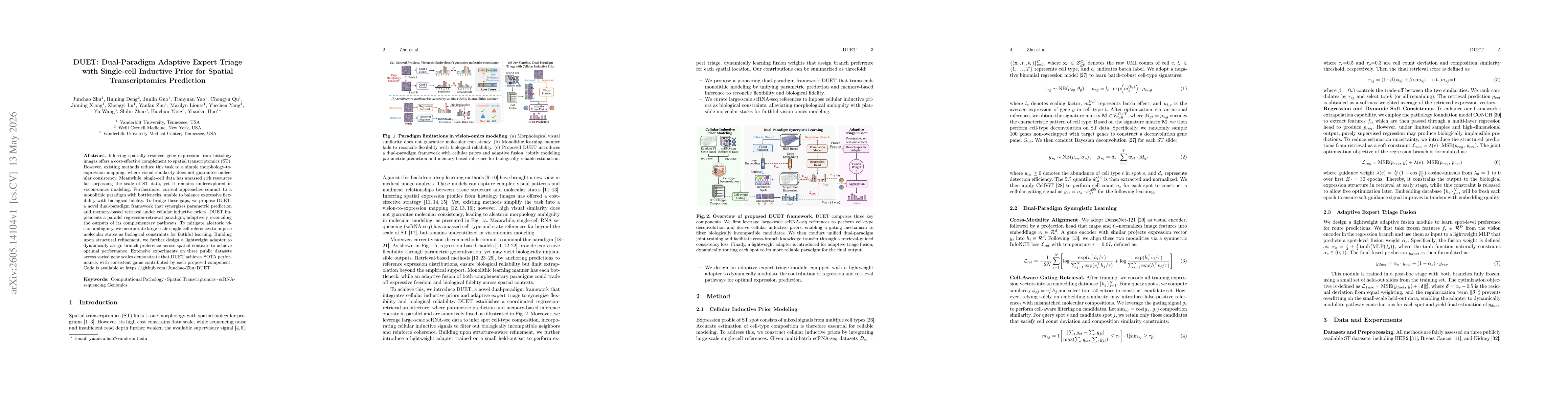
Inferring spatially resolved gene expression from histology images offers a cost-effective complement to spatial transcriptomics (ST). However, existing methods reduce this task to a simple morphology...
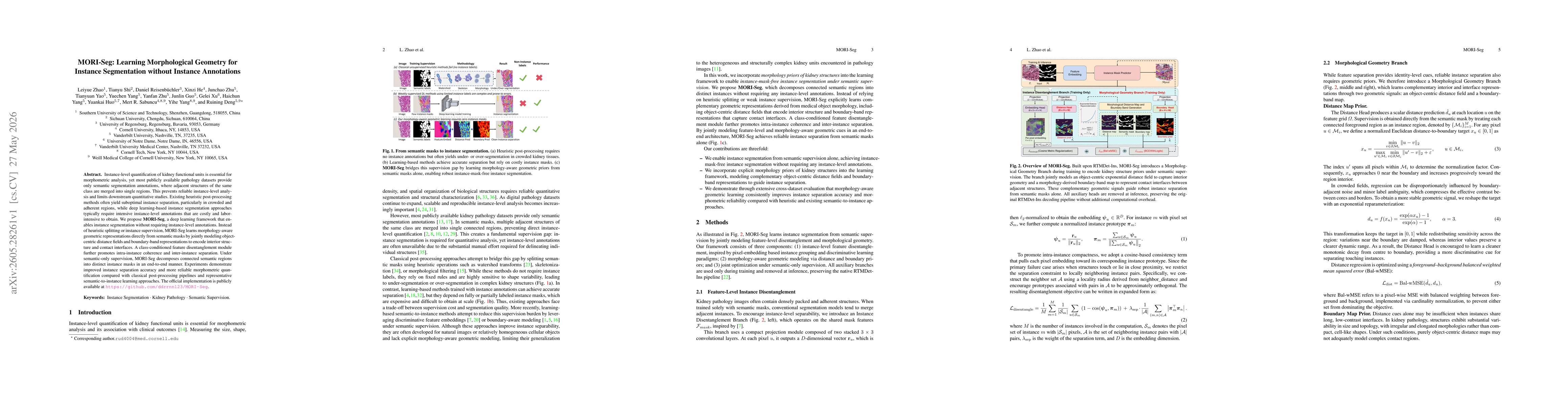
Instance-level quantification of kidney functional units is essential for morphometric analysis, yet most publicly available pathology datasets provide only semantic segmentation annotations, where ad...
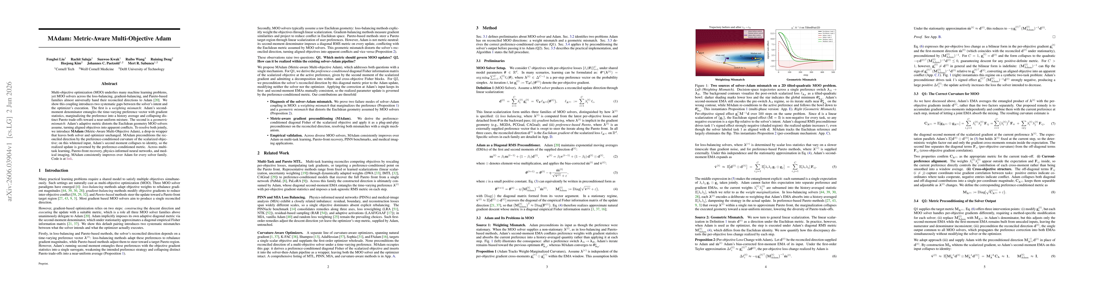
Multi-objective optimization (MOO) underlies many machine learning problems, yet MOO solvers across the loss-balancing, gradient-balancing, and Pareto-based families almost universally hand their reco...
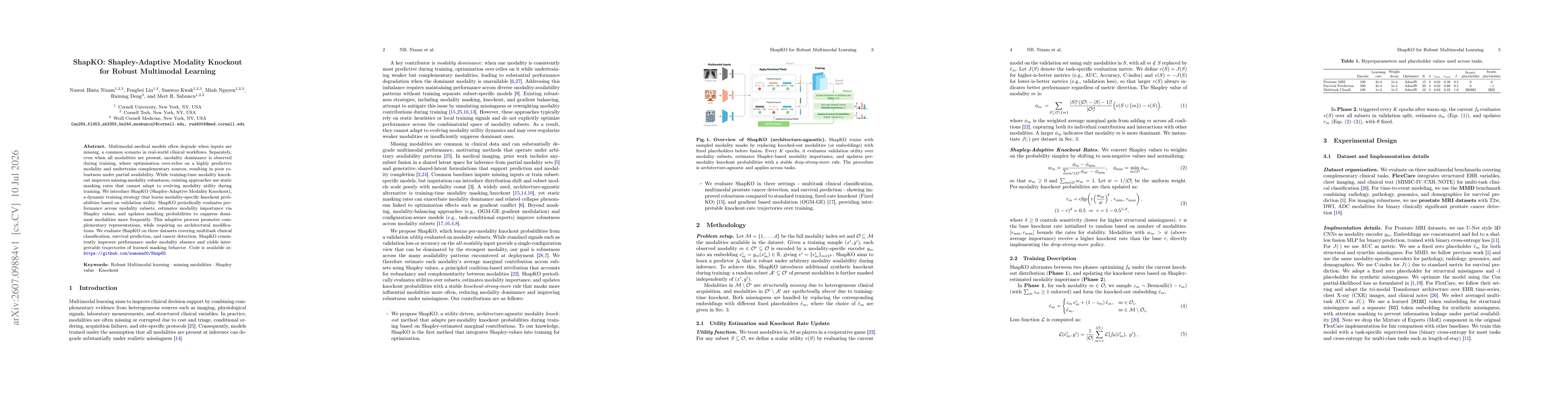
Multimodal medical models often degrade when inputs are missing, a common scenario in real-world clinical workflows. Separately, even when all modalities are present, modality dominance is observed du...