This paper evaluates the performance of three state-of-the-art cell foundation models on a large, curated kidney pathology dataset, finding variability in their outcomes after fine-tuning. It also explores human-in-the-loop data enrichment strategies to enhance model performance with minimal human annotation, demonstrating improved results across all models.
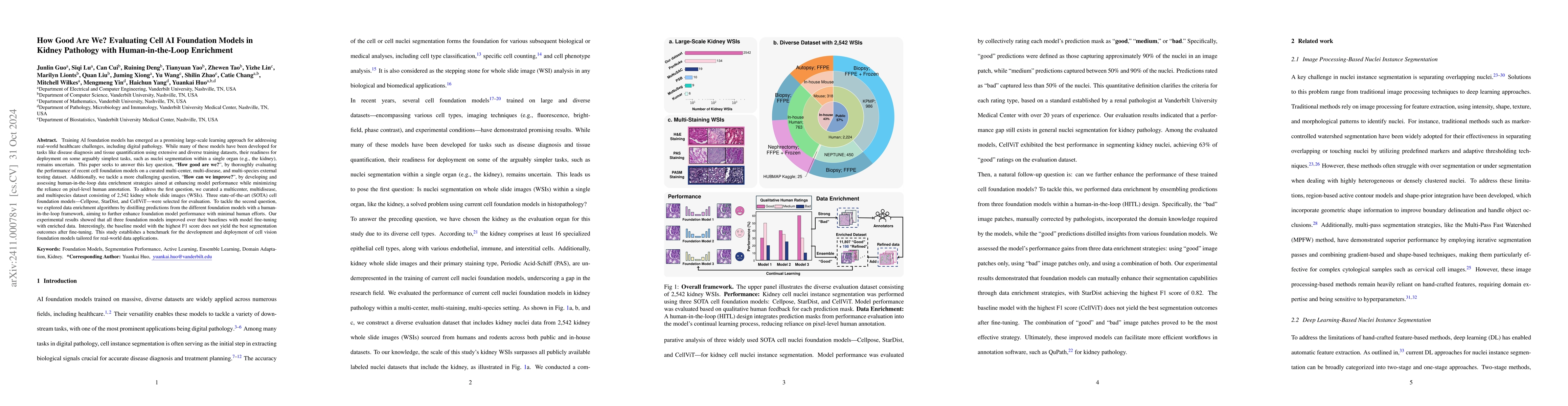
Training AI foundation models has emerged as a promising large-scale learning approach for addressing real-world healthcare challenges, including digital pathology. While many of these models have been developed for tasks like disease diagnosis and tissue quantification using extensive and diverse training datasets, their readiness for deployment on some arguably simplest tasks, such as nuclei segmentation within a single organ (e.g., the kidney), remains uncertain. This paper seeks to answer this key question, "How good are we?", by thoroughly evaluating the performance of recent cell foundation models on a curated multi-center, multi-disease, and multi-species external testing dataset. Additionally, we tackle a more challenging question, "How can we improve?", by developing and assessing human-in-the-loop data enrichment strategies aimed at enhancing model performance while minimizing the reliance on pixel-level human annotation. To address the first question, we curated a multicenter, multidisease, and multispecies dataset consisting of 2,542 kidney whole slide images (WSIs). Three state-of-the-art (SOTA) cell foundation models-Cellpose, StarDist, and CellViT-were selected for evaluation. To tackle the second question, we explored data enrichment algorithms by distilling predictions from the different foundation models with a human-in-the-loop framework, aiming to further enhance foundation model performance with minimal human efforts. Our experimental results showed that all three foundation models improved over their baselines with model fine-tuning with enriched data. Interestingly, the baseline model with the highest F1 score does not yield the best segmentation outcomes after fine-tuning. This study establishes a benchmark for the development and deployment of cell vision foundation models tailored for real-world data applications.
Get AI-generated insights about this paper's methodology, results, and significance.
Current paper (gray), citations (green), references (blue)
No similar papers found for this research.
No citations found for this paper.
Title | Authors | Year | Actions |
---|---|---|---|
CellSAM: A Foundation Model for Cell Segmentation
|
Uriah Israel, Markus Marks, Rohit Dilip, Qilin Li, Changhua Yu, Emily Laubscher, A. Iqbal, Elora Pradhan, Ada Ates, Martin Abt, Caitlin Brown, Edward Pao, Shenyi Li, Alexander Pearson-Goulart, Pietro Perona, Georgia Gkioxari, Ross Barnowski, Yisong Yue, David Van Valen
|
2023
|
View Paper |
CellViT: Vision Transformers for Precise Cell Segmentation and Classification
|
Fabian Hörst, Moritz Rempe, Lukas Heine, Constantin Seibold, J. Keyl, G. Baldini, S. Ugurel, J. Siveke, Barbara Grünwald, J. Egger, J. Kleesiek
|
2023
|
View Paper |
Medical SAM Adapter: Adapting Segment Anything Model for Medical Image Segmentation
|
Junde Wu, Rao Fu, Huihui Fang, Yuanpei Liu, Zhao-Yang Wang, Yanwu Xu, Yueming Jin, T. Arbel
|
2023
|
View Paper |
Segment Anything Model for Medical Image Analysis: an Experimental Study
|
M. Mazurowski, Haoyu Dong, Han Gu, Jichen Yang, N. Konz, Yixin Zhang
|
2023
|
View Paper |
Segment Anything Model (SAM) for Digital Pathology: Assess Zero-shot Segmentation on Whole Slide Imaging
|
Ruining Deng, C. Cui, Quan Liu, Tianyuan Yao, Lucas W. Remedios, Shunxing Bao, Bennett A. Landman, Lee E. Wheless, Lori A. Coburn, K. Wilson, Yaohong Wang, Shilin Zhao, A. Fogo, Haichun Yang, Yucheng Tang, Yuankai Huo
|
2023
|
View Paper |
Nuclei and glands instance segmentation in histology images: a narrative review
|
Esha Sadia Nasir, Arshi Parvaiz, M. Fraz
|
2022
|
View Paper |
Scaling Vision Transformers to Gigapixel Images via Hierarchical Self-Supervised Learning
|
Richard J. Chen, Chengkuan Chen, Yicong Li, Tiffany Y. Chen, A. Trister, R. G. Krishnan, Faisal Mahmood
|
2022
|
View Paper |
Cellpose 2.0: how to train your own model
|
C. Stringer, Marius Pachitariu
|
2022
|
View Paper |
Nuclei Instance Segmentation and Classification in Histopathology Images with Stardist
|
Martin Weigert, Uwe Schmidt
|
2022
|
View Paper |
TSFD-Net: Tissue specific feature distillation network for nuclei segmentation and classification
|
Talha Ilyas, Zubaer Ibna Mannan, Abbas Khan, S. Azam, Hyongsuk Kim, F. D. Boer
|
2022
|
View Paper |
How Many Cell Types Are in the Kidney and What Do They Do?
|
Michael S. Balzer, T. Rohacs, K. Suszták
|
2021
|
View Paper |
Lizard: A Large-Scale Dataset for Colonic Nuclear Instance Segmentation and Classification
|
S. Graham, M. Jahanifar, A. Azam, M. Nimir, Y. Tsang, K. Dodd, E. Hero, H. Sahota, Atisha Tank, K. Benes, N. Wahab, F. Minhas, S. Raza, H. Eldaly, K. Gopalakrishnan, D. Snead, N. Rajpoot
|
2021
|
View Paper |
Image-based cell phenotyping with deep learning.
|
Aditya Pratapa, Michael Doron, Juan C. Caicedo
|
2021
|
View Paper |
Whole-cell segmentation of tissue images with human-level performance using large-scale data annotation and deep learning
|
N. Greenwald, G. Miller, Erick Moen, A. Kong, Adam Kagel, Thomas Dougherty, C. Fullaway, Brianna J. McIntosh, K. Leow, Morgan Schwartz, Cole Pavelchek, Sunny Cui, I. Camplisson, Omer Bar-Tal, Jaiveer Singh, Mara Fong, Gautam Chaudhry, Zion Abraham, J. Moseley, S. Warshawsky, Erin Soon, Shirley Greenbaum, Tyler T. Risom, T. Hollmann, S. Bendall, Leeat Keren, William Graf, Michael Angelo, David Van Valen
|
2021
|
View Paper |
CPP-Net: Context-Aware Polygon Proposal Network for Nucleus Segmentation
|
Shengcong Chen, Changxing Ding, Minfeng Liu, Jun Cheng, D. Tao
|
2021
|
View Paper |
An Image is Worth 16x16 Words: Transformers for Image Recognition at Scale
|
Alexey Dosovitskiy, Lucas Beyer, Alexander Kolesnikov, Dirk Weissenborn, Xiaohua Zhai, Thomas Unterthiner, Mostafa Dehghani, Matthias Minderer, G. Heigold, S. Gelly, Jakob Uszkoreit, N. Houlsby
|
2020
|
View Paper |
Deep learning in digital pathology image analysis: a survey
|
Shujian Deng, Xin Zhang, Wen Yan, E. Chang, Yubo Fan, M. Lai, Yan Xu
|
2020
|
View Paper |
Cellpose: a generalist algorithm for cellular segmentation
|
C. Stringer, Tim Wang, Michalis Michaelos, Marius Pachitariu
|
2020
|
View Paper |
Caliban: Accurate cell tracking and lineage construction in live-cell imaging experiments with deep learning
|
Morgan Schwartz, Erick Moen, G. Miller, Tom Dougherty, Enrico Borba, Rachel Ding, William Graf, Edward Pao, David Van Valen
|
2019
|
View Paper |
PanNuke: An Open Pan-Cancer Histology Dataset for Nuclei Instance Segmentation and Classification
|
Jevgenij Gamper, Navid Alemi Koohbanani, Ksenija Benet, A. Khuram, N. Rajpoot
|
2019
|
View Paper |
Acute lymphoblastic leukemia cells image analysis with deep bagging ensemble learning
|
Y. Liu, Feixiao Long
|
2019
|
View Paper |
Segmentation of Nuclei in Histopathology Images by Deep Regression of the Distance Map
|
Peter Naylor, M. Laé, F. Reyal, Thomas Walter
|
2019
|
View Paper |
Hover-Net: Simultaneous segmentation and classification of nuclei in multi-tissue histology images
|
S. Graham, Q. Vu, S. Raza, A. Azam, Y. Tsang, J. T. Kwak, N. Rajpoot
|
2018
|
View Paper |
Blood Cell Images Segmentation using Deep Learning Semantic Segmentation
|
Thanh Tran, Oh-Heum Kwon, Ki-Ryong Kwon, Suk-Hwan Lee, Kyung-Won Kang
|
2018
|
View Paper |
A Structured Tumor-Immune Microenvironment in Triple Negative Breast Cancer Revealed by Multiplexed Ion Beam Imaging
|
Leeat Keren, M. Bosse, Diana Marquez, Roshan Angoshtari, Samir Jain, S. Varma, Soo-Ryum Yang, A. Kurian, D. V. Valen, Robert West, S. Bendall, Michael Angelo
|
2018
|
View Paper |
Micro‐Net: A unified model for segmentation of various objects in microscopy images
|
S. Raza, Linda Cheung, M. Shaban, S. Graham, David B. A. Epstein, S. Pelengaris, Michael Khan, N. Rajpoot
|
2018
|
View Paper |
Multi-Pass Fast Watershed for Accurate Segmentation of Overlapping Cervical Cells
|
Afaf Tareef, Yang Song, Heng Huang, D. Feng, Mei Chen, Y. Wang, Weidong (Tom) Cai
|
2018
|
View Paper |
Data-analysis strategies for image-based cell profiling
|
Juan C. Caicedo, Sam Cooper, Florian Heigwer, Scott Warchal, P. Qiu, Csaba Molnar, A. Vasilevich, Joseph D Barry, Harmanjit Singh Bansal, Oren Z. Kraus, Mathias Wawer, L. Paavolainen, M. Herrmann, M. Rohban, Jane Hung, H. Hennig, J. Concannon, Ian Smith, P. Clemons, Shantanu Singh, P. Rees, P. Horváth, Roger G. Linington, Anne E Carpenter
|
2017
|
View Paper |
Mask R-CNN
|
Kaiming He, Georgia Gkioxari, Piotr Dollár, Ross B. Girshick
|
2017
|
View Paper |
A Dataset and a Technique for Generalized Nuclear Segmentation for Computational Pathology
|
Neeraj Kumar, R. Verma, Sanuj Sharma, S. Bhargava, Abhishek Vahadane, A. Sethi
|
2017
|
View Paper |
A survey on deep learning in medical image analysis
|
G. Litjens, Thijs Kooi, B. Bejnordi, A. Setio, F. Ciompi, Mohsen Ghafoorian, J. Laak, B. Ginneken, C. I. Sánchez
|
2017
|
View Paper |
Large‐scale image‐based screening and profiling of cellular phenotypes
|
Nicola Bougen-Zhukov, Sheng Yang Michael Loh, H. Lee, L. Loo
|
2017
|
View Paper |
QuPath: Open source software for digital pathology image analysis
|
P. Bankhead, M. Loughrey, José A Fernández, Yvonne Dombrowski, D. McArt, P. Dunne, S. McQuaid, R. Gray, L. Murray, H. Coleman, J. James, M. Salto‐Tellez, P. Hamilton
|
2017
|
View Paper |
Deep Learning Automates the Quantitative Analysis of Individual Cells in Live-Cell Imaging Experiments
|
D. V. Valen, Takamasa Kudo, Keara M. Lane, Derek N. Macklin, N. Quach, Mialy Defelice, Inbal Maayan, Yu Tanouchi, E. Ashley, M. Covert
|
2016
|
View Paper |
Automatic segmentation for cell images based on bottleneck detection and ellipse fitting
|
M. Liao, Yu-qian Zhao, Xiang-hua Li, Peishan Dai, Xiao-wen Xu, Jun-kai Zhang, Beiji Zou
|
2016
|
View Paper |
U-Net: Convolutional Networks for Biomedical Image Segmentation
|
O. Ronneberger, P. Fischer, T. Brox
|
2015
|
View Paper |
Automatic Nuclei Segmentation in H&E Stained Breast Cancer Histopathology Images
|
M. Veta, P. V. van Diest, R. Kornegoor, A. Huisman, M. Viergever, J. Pluim
|
2013
|
View Paper |
Detection and Segmentation of Cell Nuclei in Virtual Microscopy Images: A Minimum-Model Approach
|
S. Wienert, Daniel Heim, K. Saeger, A. Stenzinger, M. Beil, P. Hufnagl, M. Dietel, C. Denkert, F. Klauschen
|
2012
|
View Paper |
Segmentation of Clustered Nuclei With Shape Markers and Marking Function
|
Jierong Cheng, Jagath Rajapakse
|
2009
|
View Paper |
Nuclei Segmentation Using Marker-Controlled Watershed, Tracking Using Mean-Shift, and Kalman Filter in Time-Lapse Microscopy
|
Xiaodong Yang, Houqiang Li, Xiaobo Zhou
|
2006
|
View Paper |
Applying watershed algorithms to the segmentation of clustered nuclei.
|
N. Malpica, C. O. Solórzano, J. Vaquero, Andrés Santos, I. Vallcorba, J. M. García-Sagredo, F. D. Pozo
|
1998
|
View Paper |
Segment Anything in Medical Images
|
Jun Ma, Bo Wang
|
2023
|
View Paper |
Accurate Cervical Cell Segmentation from Overlapping Clumps in Pap Smear Images
|
Youyi Song, Ee-Leng Tan, Xudong Jiang, Jie-Zhi Cheng, Dong Ni, Siping Chen, B. Lei, Tianfu Wang
|
2017
|
View Paper |
“An integrated region, boundary, shape-based active contour 39
|
|||
its fine-tuned counterparts
|
Showing first page preview. Click "Open Full PDF" to view the complete paper.
Your comment contains inappropriate language and has been rejected.
Are you sure you want to remove this tag? This action cannot be undone.
Someone just favorited a paper!
Comments (0)