Summary

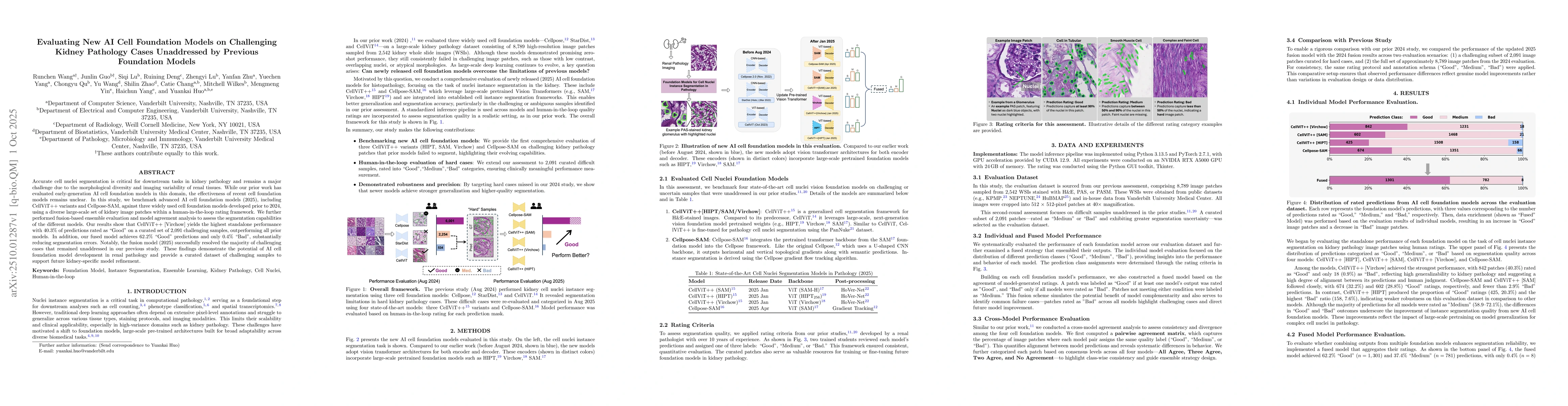
Accurate cell nuclei segmentation is critical for downstream tasks in kidney pathology and remains a major challenge due to the morphological diversity and imaging variability of renal tissues. While our prior work has evaluated early-generation AI cell foundation models in this domain, the effectiveness of recent cell foundation models remains unclear. In this study, we benchmark advanced AI cell foundation models (2025), including CellViT++ variants and Cellpose-SAM, against three widely used cell foundation models developed prior to 2024, using a diverse large-scale set of kidney image patches within a human-in-the-loop rating framework. We further performed fusion-based ensemble evaluation and model agreement analysis to assess the segmentation capabilities of the different models. Our results show that CellViT++ [Virchow] yields the highest standalone performance with 40.3% of predictions rated as "Good" on a curated set of 2,091 challenging samples, outperforming all prior models. In addition, our fused model achieves 62.2% "Good" predictions and only 0.4% "Bad", substantially reducing segmentation errors. Notably, the fusion model (2025) successfully resolved the majority of challenging cases that remained unaddressed in our previous study. These findings demonstrate the potential of AI cell foundation model development in renal pathology and provide a curated dataset of challenging samples to support future kidney-specific model refinement.

AI Key Findings

Generated Oct 05, 2025

Methodology

The study benchmarks advanced AI cell foundation models (2025) against prior models using a curated dataset of 2,091 challenging kidney image patches. Human-in-the-loop ratings and fusion-based ensemble evaluation were employed to assess segmentation performance.

Key Results

  • CellViT++ [Virchow] achieved the highest standalone performance with 40.3% of predictions rated as 'Good'.
  • The fused model achieved 62.2% 'Good' predictions and only 0.4% 'Bad', significantly reducing segmentation errors.
  • The fusion model resolved most challenging cases previously unaddressed by earlier models.

Significance

This research demonstrates significant improvements in kidney pathology cell segmentation, offering a curated dataset for future model refinement and enhancing diagnostic accuracy in renal histopathology.

Technical Contribution

Introduction of a fusion strategy that combines multiple AI cell foundation models to enhance segmentation reliability and reduce prediction uncertainty.

Novelty

The study introduces a fusion-based ensemble approach and evaluates it on a newly curated set of challenging kidney pathology cases, demonstrating substantial improvements over previous models.

Limitations

  • The study focuses on a specific dataset of kidney images, which may limit generalizability to other tissue types.
  • The human-in-the-loop rating system introduces potential subjectivity in evaluation.

Future Work

  • Expanding the curated dataset to include diverse tissue types and imaging modalities.
  • Investigating the integration of additional models or modalities to further improve segmentation accuracy.
  • Developing automated rating systems to reduce human bias in model evaluation.

Paper Details

PDF Preview

Download PDF

Comments (0)

This website uses cookies to ensure you get the best experience. Learn more